Lab Practical 1 Study Guide
Anatomical Terminology: 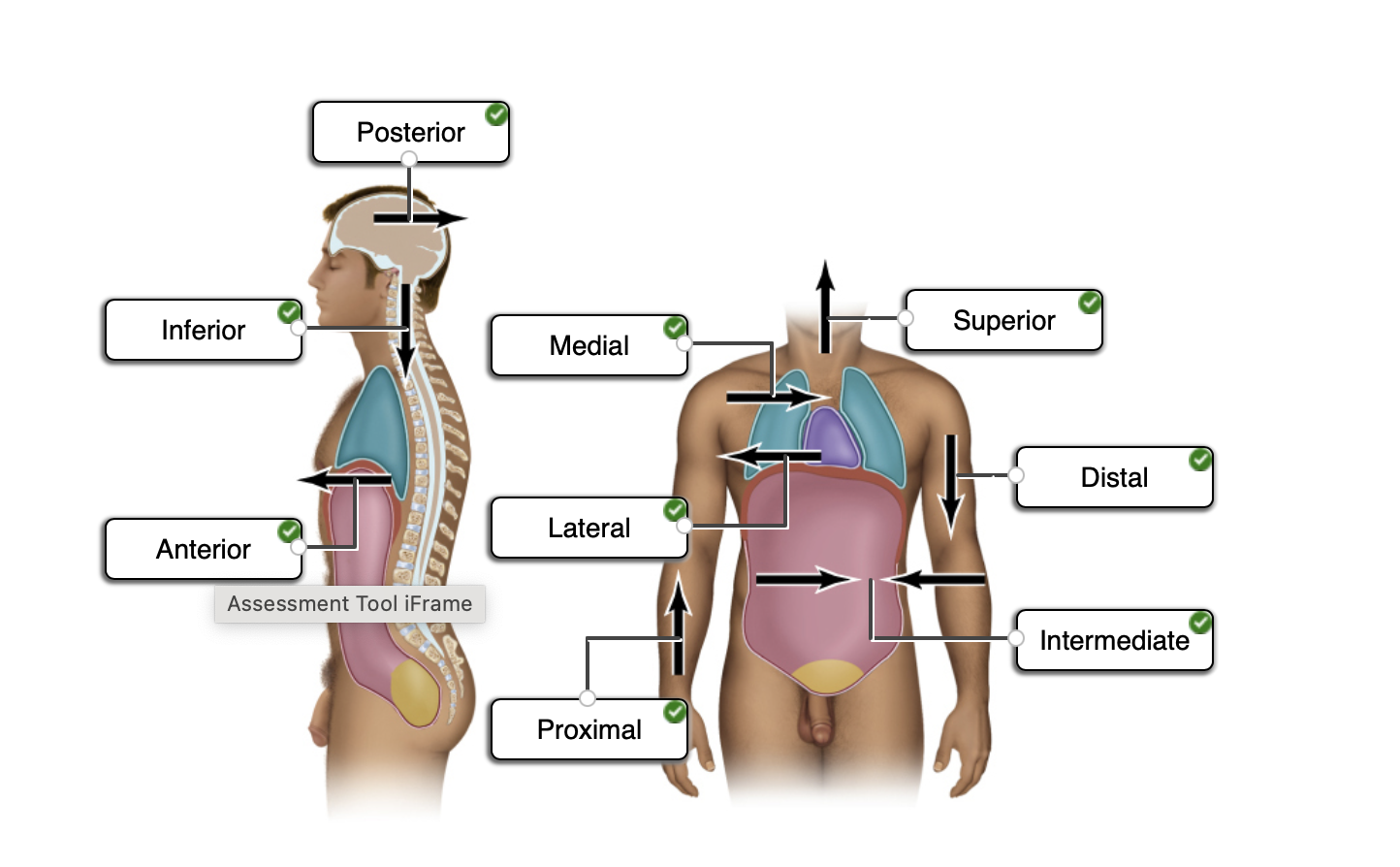
- Posterior: Toward the dorsal (back) side
- Anterior: Toward the ventral (front) side
- Inferior: Toward the tail or feet
- Superior: Toward the head
- Medial: Toward the median plane
- Lateral: Away from the median plane
- Proximal: Toward the site of attachment
- Distal: Away from the site of attachment
- Intermediate: In between
11 Major Organ Systems of the Human Body
Integumentary:
Principal organs: skin, hair, nails, cutaneous glands
Principal functions: Protection, water retention, thermoregulation
Vitamin D synthesis, cutaneous sensation, nonverbal communication
Skeletal:
Principal organs: Bones, cartilages, ligaments
Principal functions: Support, movement, protective enclosure of viscera,
Blood formation, mineral storage, electrolyte and acid-base balance
Muscular:
Principal organs: skeletal muscles
Principle functions: movement, stability, communication, control of body openings, heat production
Lymphatic:
Principal organs: Brain, spinal cord, nerves, ganglia
Principal functions: Rapid internal communication, coordination, motor control and sensation
Endocrine:
Principal organs: Pituitary gland, thyroid gland, pineal glands, thymus, adrenal glands, pancreas, testes, ovaries
Principal functions: Hormone production, internal chemical communication and coordination
Circulatory:
Principal organs: Heart, blood vessels
Principal functions: Distribution of nutrients, oxygen, wastes, hormones, electrolytes, heat, immune cells and antibodies; fluid, electrolyte and acid-base balance
Lymphatic:
Principal organs: Lymph nodes, lymphatic vessels, thymus, spleen, tonsils
Principal functions: recovery of excess tissue fluid, detection of pathogens, production of immune cells, defense against disease
Respiratory:
Principal organs: nose, pharynx, larynx, trachea, bronchi, lungs
Principal functions: absorption of oxygen, discharge of carbon dioxide, acid-base balance, speech
Urinary:
Principal organs: Kidneys, ureters, urinary bladder, urethra
Principal functions: elimination of wastes, regulation of blood volume and pressure, stimulation of red blood cell formation, control of fluid, electrolyte and acid-base balance, detoxification
Digestive:
Principal organs: teeth, tongue, salivary glands, esophagus, small and large intestines, liver, gallbladder, pancreas
Principal functions: nutrient breakdown and absorption. Liver functions include: metabolism of carbohydrates, lipids, proteins, vitamins, and minerals; synthesis of plasma proteins, disposal of drugs, toxins, and hormones; cleansing of blood
Male reproductive:
Principal organs: testes, epididymides, spermatic ducts, seminal vesicles, prostate, bulbourethral glands, penis
Principal functions: production and delivery of sperm; secretion of sex hormones
Female reproductive:
Principal organs: ovaries, uterine tubes, uterus, vagina, mammary glands
Principal functions: production of eggs; site of fertilization and fetal development; fetal nourishment, birth, lactation, secretion of sex hormones
Microscopy
- Goal: create a magnified image that is too small to be seen with the naked eye
- Allows visualization of cell morphology and their organization into a tissue
- Three factors that determine the quality of a microscopic image
- Magnification: increase in image size
- Resolution: ability to distinguish detail
- Contrast: ability to distinguish objects from the background
- Brightfield microscopes use a combination of glass lenses and light to view a specimen
- Fields of study where microscopy is necessary:
- Cytology: the study of cells
- Histology: the study of tissues
- Pathology: the study of disease
- Four broad categories of tissues:
- Epithelial tissue
- Connective tissue
- Nervous tissue
- Muscle tissue
- Terms to know:
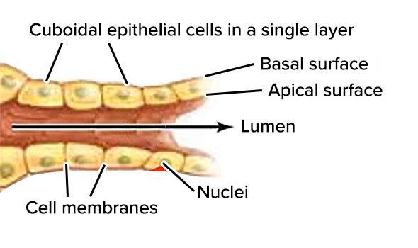
- Apical surface: The surface of an epithelial cell facing the lumen or external environment
- Basal surface: The surface of an epithelial cell facing the basement membrane
- Basement membrane: found underneath the epithelial and underlying connective tissue layers; composed on non-cellular layers of collagen, glycoproteins and proteoglycans. Aids in attachment of epithelial tissue and as a selective molecular barrier to underlying connective tissue
- Lumen: Central cavity or open space within an organ
Epithelial Tissue: 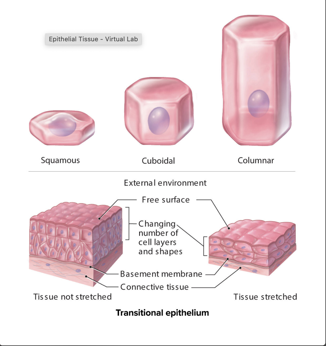
- Covers body surfaces and lines the insides of our hollow organs
- Form in sheets of cells that border a lumen (an open space)
- Cell shapes include:
- Squamous: flat/ scale like
- Cuboidal: cube shaped
- Columnar: column like
- Transitional: changes shape from polyhedral to flat
- Tissue layers include:
- Simple: a single layer of cells
- Stratified: multiple layers of cells
- Pseudostratified: Single layer of cells that appears to be multiple layers
- Naming an epithelial tissue consists of naming the number of layers and the cell shape
Simple Epithelial Tissues
- Simple squamous epithelium
- Structure: single layer of thin, flat cells resembling irregular floor tiles; the single nucleus of each cell bulges at its center
- Function: Thinnest possible barrier to allow for rapid diffusion and filtration; secretion in serous membranes
- Location: Air sacs in lungs (alveoli); lining of lumen of blood vessels and lymph vessels (endothelium) serous membranes of body
- Simple cuboidal epithelium
- Structure: single layer of cells about as tall as they are wide; spherical and centrally located nucleus
- Function: absorption and secretion; forms secretory tissue of most glands and small ducts
- Location: Kidney tubules, thyroid gland follicles; surface of ovary; secretory regions and ducts of most glands
- Simple columnar epithelium
- Structure: single layer of cells taller than they are wide; oval shaped nucleus oriented lengthwise in basal region of cell; apical regions of cell may have microvilli; may contain goblet cells that secrete mucin
- Function: absorption and secretion; secretion of mucin
- Location: inner lining of most digestive tract (stomach, small and large intestine
- Nonciliated Pseudostratified Columnar Epithelium
- Structure: Single layer of cells with varying heights; all cells connect to the basement membrane, but not all cells reach the apical surface; lacks goblet cells and cilia
- Function: Protection
- Location: Rare-lining of part of the male urethra and epididymis
- Ciliated Pseudostratified columnar epithelium
- Structure: single layer of cells with varying heights; all cells connect to the basement membrane, but not all cells reach the apical surface; has goblet cells and cilia
- Function: protection; secretion of mucin and movement of mucus along apical surface of epithelium by cilia
- Location: lining of the larger airways of respiratory tract, including nasal cavity, part of pharynx, parts of larynx, trachea, and bronchi
Stratified epithelial tissues
- Stratified squamous epithelium:
- Keratinized Stratified Squamous Epithelium
- Structure: multiple cell layers; basal cells are cuboidal or polyhedral whereas apical cells are squamous; apical cells are dead and filled with the protein keratin
- Function: protection of underlying tissue from abrasion
- Location: epidermis
- Nonkeratinized stratified squamous epithelium
- Structure: multiple cell layers; basal cells are cuboidal or polyhedral whereas apical (superficial) cells are squamous; cells lack keratin; superficial cells are alive and kept moist
- Function: Protection of underlying tissue from abrasion
- Location: lining of oral cavity, part of pharynx, part of larynx, esophagus, lining of vagina and anus
- Keratinized Stratified Squamous Epithelium
- Stratified cuboidal epithelium
- Structure: two or more layers of cells; cells at the apical surface are about as tall as they are wide
- Function: Protection and secretion
- Location: Ducts of most exocrine glands and ovarian follicles
- Stratified columnar epithelium
- Structure: two or more layers of cells; cells at the apical surface are taller than they are wide
- Function: Protection and secretion
- Location: Large ducts of salivary glands, conjunctiva covering the eye, and membranous part of male urethra
Transitional Epithelium
- Structure: epithelial appearance varies, depending upon whether tissue is relaxed or distended; relaxed epithelium has cuboidal or polyhedral cells and the apical cells are large and rounded. Distended epithelium has flattened cells at the apical surface; some cells are binucleated
- Function: accommodates volume changes by distending or relaxing in the urinary bladder, ureters, and part of urethra
- Location: lining of the urinary bladder, ureter, and part of urethra
Connective Tissue
- Connective tissue is a diverse group of tissues that are found distributed widely throughout the body. The cells are typically separated by significant amounts of extracellular matrix and found deep to epithelial tissue
- Has diverse functions:
- Support, protection, binding of structures, storage, transport, immune
- Terms to know:
- Ground substance: noncellular material produced by the connective tissue; In combination with the protein fibers forms the extracellular matrix
- Protein fibers: Fibers are made of protein and found outside of the cells. The protein fibers may provide strength or stretch for a tissue. In combination with the ground substance forms the extracellular matrix.
- Collagen fibers: very strong unbranched “rope-like” proteins that provide strength and flexibility to a tissue.
- Elastic fibers: composed of the protein elastin and provide stretch to a tissue
- Reticular fibers: thin branched fibers similar to collagen, that provide a tough and flexible scaffold
Loose connective tissue proper
- Areolar tissue
- Structure: abundant viscous ground substance; few collagen and elastic fibers; scattered fibroblasts; many blood vessels
- Function: Protects tissues and organs; binds skin and some epithelia to deeper tissue
- Location: papillary layer of the dermis, subcutaneous layer (deep to skin); surrounds organs, nerve cells, some muscle cells, and blood vessels
- Adipose tissue
- Structure: closely packed adipocytes; nucleus pushed to edge of cell by large fat droplet; contains many blood vessels
- Function: stores energy; insulates, cushions, and protects
- Location: subcutaneous layer; surrounds and covers some organs
- Reticular tissue
- Structure: Viscous ground substance; meshwork of reticular fibers, leukocytes, and some fibroblasts
- Function: provides stroma (supportive framework) to lymphatic organs
- Location: spleen, lymph nodes, and red bone marrow
Dense connective tissue proper
- Dense regular connective tissue
- Structure: densely packed, parallel arrays of collagen fibers; fibroblasts squeezed between layers of fibers; scarce ground substance; limited blood supply
- Function: attaches bone to bone (most ligaments) as well as muscle to bone (tendons); resists stress applied in one direction
- Location: tendons (attach muscle to bone); ligaments (typically attach bone to bone)
- Dense irregular connective tissue
- Structure: collagen fibers randomly arranged and clumped together; fibroblasts in spaces among fibers; more ground substance than in dense regular connective tissue; extensive blood supply
- Function: Withstands stresses applied in all directions; durable
- Location: most of dermis of skin; periosteum covering bone; perichondrium covering cartilage, epineurium covering nerves, epimysium covering skeletal muscle, some organ capsules
- Elastic connective tissue
- Structure: predominantly composed of elastic fibers; fibroblasts occupy some spaces between the fibers
- Function: allows for stretching and recoil
- Location: Walls of elastic arteries (such as the aorta), trachea, vocal chords
Supporting connective tissue
- Cartilage
- Hyaline cartilage
- Structure: glassy appearing matrix; irregular chondrocytes in lacunae
- Function: Provides support; forms most of fetal skeleton
- Location: Tip of nose; trachea; most of larynx, costal cartilage; both the epiphyseal (growth) plates and articular ends of long bones; most of fetal skeleton
- Fibrocartilage
- Structure: readily visible, numerous, parallel collagen fibers with limited ground substance; large chondrocytes in lacunae
- Function: resists compression; acts as shock absorber in some joints
- Location: intervertebral discs; public symphysis; menisci of knee joints
- Elastic cartilage
- Structure: Abundant elastic fibers that form weblike mesh; closely packed chondrocytes in lacunae
- Function: maintains shape while permitting extensive flexibility
- Location: external ear; epiglottis of larynx
- Bone
- Structure: calcified extracellular matrix containing osteocytes trapped in lacunae; compact bone organized in osteons (concentric lamellae arranged around a central canal); spongy bone is a meshwork that has a different organization from compact bone; well vascularized
- Function: provides levers for body movement, supports soft structures, protects organs, stores calcium and phosphorus; spongy bone contains hemopoietic tissue and is the site for hemopoiesis
- Location: bones of the body
- Hyaline cartilage
Fluid connective tissue
- Blood
- Structure: contains formed elements (erythrocytes, leukocytes, and platelets); dissolved protein within a liquid ground substance called plasma
- Function: erythrocytes transport respiratory gasses (oxygen and carbon dioxide); leukocytes help protect the body from infectious agents; and platelets help with blood clotting. Plasma transports nutrients, wastes, and hormones throughout the body
- Location: Primarily within blood vessels and in the heart
- Lymph: lymph fluid is found within lymph vessels and derives from interstitial fluid. It is mostly water and dissolved solutes and typically no cellular components.
Nervous Tissue
- Composed of glial cells and neurons
- Neurons: receive and transmit nerve impulses
- Located in the brain, spinal cord, and nerves
- Structure: cell body and cell processes such as dendrites and an axon
- Dendrites: receive information and transmit it to the cell body
- Axon: transmits the nerve impulse away from the cell body to another cell, such as another neuron, muscle cell, or gland
- Multipolar neurons: most common structural type of neuron; make up motor neurons and interneurons. Contain multiple dendrites and a single axon
- Glial cells: protect and care for the neurons
Muscle Tissue
- Muscle tissue is unique in that its cells have the ability to contract when stimulated
- Muscle tissue allows for both voluntary and involuntary movement and heat generation
- Skeletal muscle
- Attaches to bone or skin
- Cells are long striated cylindrical cells and multinucleate
- Function: voluntary movement of skeletal muscle
- Cardiac muscle
- Found in the wall of the heart
- Cells are short, branched, striated, and uni- or binucleate
- Function: involuntary movement of heart muscle
- Smooth muscle
- Found in the walls of most viscera
- Cells are short with a fusiform shape, lack striations, and are uninucleate
- Function: involuntary movement such as peristalsis of an organ or vasoconstriction of a blood vessel