Biophysics Summarised Notes.docx
- Thermodynamics. Thermodynamic systems and quantities. First law of thermodynamics and bio-systems.
- Thermodynamics is concerned with heat and temperature and their relation to energy and work.
- There are 3 TD systems;
Isolated= no exchange of matter and heat
Closed= no exchange of matter, can exchange heat/work energy or both
Open= can exchange matter and heat/work energy
- Thermodynamic state= it is described by the composition of its variables
TD variables include;
-Temperature= average kinetic energy of particles
-Pressure= number and magnitude of particle strikes (vibrations)
-Internal energy= depends on the type of state (energy of each particle) {Sum of potential and kinetic energy}
-Entropy= measure of order/chaos (particle arrangement)
-Thermal energy/Heat
- First Law of TD= Energy cannot be created or destroyed but only transformed from one form to another.
dE= Q-W dE= heat added to the system – work done by the system
dE= change of internal energy
Q= heat transfer into a system
W=work done by the system
- Entropy. Free energy. Second law of thermodynamics applied to bio-systems. Stationary systems, Prigozhin theorem.
- Entropy= the measure of disorder - the higher the chaos, the bigger the entropy.
- Boltzman’s Entropy: S= k.lnW
S= entropy
k= Ludwig boltzman constant
W= thermodynamical probability
Heat energy increases when chaos increases, while the ability to do work decreases.
- Free energy= the energy in a physical system that can be converted to work
- Second Law of TD= the entropy of an isolated system never decreases, because isolated systems spontaneously evolve towards TD equilibrium- state of maximal entropy
dQ=T.dS – entropy and heat energy evolve in the same direction, i.e. they both move in the same direction, either both increases/both decrease
dQ= change of heat energy
T= temperature
dS= change of entropy
- Biothermodynamics;
- Biological systems are open
- Bioprocesses are irreversible
- Biosystems are non-equilibrium but are stationary systems
Biorganisms are characterized by;
- permanent inflow of substance, outflow of products
- permanent use of energy to maintain gradients
- stability of TD variables; internal energy and entropy
- Prigozhin theorem= a stationary state of TD system (obstructs reaching equilibrium) corresponds to minimal rise of entropy. No obstructions= entropy rise reaches zero
- Bio-membranes: structure, dynamics, phase transitions, functions.
Biological membrane- A complex dynamic structure consisting of lipids and proteins organized in a double layer.
Lipids (phospholipids)- form hydrocarbons chains with hydrophilic heads and hydrophobic tails
There are 2 structures; monolayer- ball-shaped (micelle) and bilayer
Membrane proteins:
-There are 2 main types of membrane proteins- peripheral and integrated.
-They have various forms; single (integral) or multipass (trans-membrane proteins), tunnel-shaped and those attached to one side of the membrane are called peripheral proteins (on a transmembrane protein/on the
Dynamics of membranes: (variety of motions)
- Lateral diffusion= diffusion with neighboring molecules
- Rotation= lipid rotates on own axis
-Swing= side-to-side
-Flexion= contraction
-Transverse Diffusion (flip flop)= movement of molecules from one half of monolayer to the other
Main properties of membranes:
- Membrane fluidity= maintains cell function and allows small molecules
to rapidly diffuse through
- Barrier function and selective transport= small non-polar molecules
like oxygen and carbon dioxide. Whereas, glucose and other large
polar, water-soluble molecules, ions and proteins do not pass. I.e.
selective permeability
- Phase transition= an increases of temperature results in bilayer
variation- thickness and flexibility. Membrane becomes more permeable
thus increases rate of diffusion.
- Passive membrane transport. Gradients. Equilibrium potentials. Diffusion types- nonspecific-, facilitated-, and exchange diffusion.
Passive Membrane transport- the movement of molecules from high conc. to low conc. across a partially permeable membrane with FREE energy (without input of ATP).
Concentration gradient= the graduated difference in concentration of a solute per unit distance through a solution.
C2 – C1 = Grad C
Equilibrium= the net flow of molecules through a channel/ across membrane is zero
Diffusion types;
Facilitated diffusion= diffusion carried out by transport proteins.
Transport proteins are penetrated in the bilayer. It enables soluble molecules to move across the membrane from high to low concentration. Diffusion rate depends on conc. grad. and the number of transport proteins.
Exchange diffusion= carrier transport mechanism across a membrane. The transport is fast but does not result in net transport.
Non-specific diffusion= from high to low concentration across a semi-permeable membrane without the use of ion channels or integral proteins.
- Ionophores and ion channels.
Ionophores: are lipid soluble peptide chains which mediate the passage of ions across the membrane.
E.g. Valinomycin – carrier ionophore that can freely move across a lipid bilayer due to its chemical structure and spatial organization. It transports extracellular Potassium (K) into the cell.
Gramicidin- channel-former ionopher. It forms a tunnel through the membrane which allows Sodium (Na) ions to pass. It is formed by 2 parts of the gramicidin helix.
Ion channels: are pore forming membrane proteins which allow water and water-soluble substances to pass across the membrane.
There are several mechanisms;
- Voltage-gated – open and close in response to membrane potential
- Ligand-gated – opens when neurotransmitter attaches to its receptor
a) Extracellular b) Intracellular
Types of ion channels include; Selective, Nonselective, Cationic, Anionic
It is dependent on the ions passed through.
- Active membrane transport. Primary and Secondary active transport. Na-K pump, Ca pump, Ca-Na exchanger.
Active membrane transport – the movement of molecules across a membrane with the help of ATP.
Protein pump properties: they have a specific affinity for ion binding and the ability to dissociate ATP (which provides the energy for transport) .
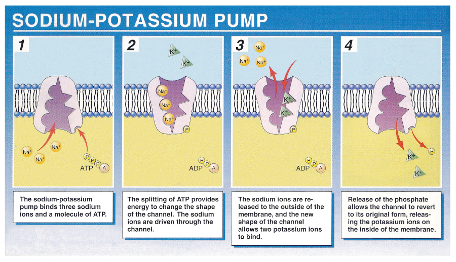
Primary AT: the transport of ions against electro-concentration forces. Conc. gradients are provided by ion pumps. The pump protein use the energy stored in a cell to carry ions in and out simultaneously. Eg. Na (out) and K (in) ions
Secondary AT (Ion-coupling): the transport of molecules across the cell membrane using energy in other forms than ATP. The energy is obtained from the electrochemical gradient created by pumping ions out of the cell.
(A special protein carrier where ion gradient forces are conjugated and organic molecules are transferred. No chemical energy is consumed.)
Process: Ions move down the electrochemical gradient; driving ion, which drives the uphill movement of another ion/molecule; driven ion/molecule. Coupling between the driven and driving ion is obligatory.
Sodium-Potassium Pump:
Calcium Pump: group of ion transporters found in the cell membrane and are responsible for active transport of calcium out of the cell and thus maintain a steep Ca2+ electrochemical gradient across the membrane.
Sodium-Calcium Pump: a membrane protein that removes calcium from cells. It uses the energy stored in the electrochemical gradient of Sodium by allowing sodium to flow down the gradient across the membrane in exchange for the calcium ions.
- Electric membrane potential. Equilibrium potential. Resting potential
- Nernst-Bernstein equation. Goldman equation. Membrane ion permeability.
Electric membrane potential/ Membrane voltage: difference in electrical potential between the interior and exterior biological cell due to the presence of varying concentrations of ions. Typical values range between 40 to -80mV.
Equilibrium potential: the electrical driving force at which it equals to the concentration driving force so that the net ion flow is zero. It is measured in volts.
Nernst Equation: equilibrium potential of ion X
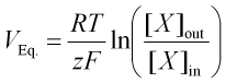
Veq= equilibrium potential in volts
R= universal gas constant
T= temperature in Kelvins
z= valency of ionic species
F= Faraday’s constant
Xout=conc. of ionic species X, extracellular
Xin= concc. Of ionic species X, intracellular
E.g. EK= -90mV
ENa= 52mV
ECa= 134mV
Bernstein Hypothesis: membrane potential of bio cells is based completely on potassium equilibrium potential, as the cell membrane is permeable to potassium ions mainly.
Goldman-Hodgkin-Katz model: Membrane potential is not at equilibrium but is stationary. It is formed by complex participation of all ions presented in cellular and outer compartments. Calculating membrane potential for membranes permeable to multiple ions.
Relative permeability P acts as a weighing factor. The greater the ions permeability, the greater the presence it holds in the equation. Membrane potential is therefore closer to the equilibrium potential of that ion.
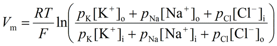
Vm= membrane potential
R= universal gas constant
T= temperature in Kelvins
F= Faraday’s constant
PK/PNa/PCl= relative membrane permeability of ions
[K]/ [Na]/ [Cl]0/i= conc. of ion in extracellular/intracellular fluid
Resting Potential: equilibrium of all electro-concentration forces at which polarity of membrane is stable. The parameters which maintain stability include; net flow of ions, cell permeability and ion-pump activity.
E.g. Skeletal muscle cells= -95mV
Smooth muscle cells= -60mV
Neurons= -60 to -70mV
Membrane Ion Potential: the ease of an ion to cross the cell membrane.
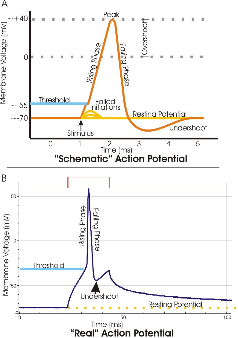
- Action potential. Alterations in the membrane permeability during an action potential.
Action potential: external stimulus -> sharp change of membrane electric potential, which has a short duration and represents membrane response to over-threshold (-55mV) irritation
Depolarization: increase of membrane potential from the resting potential
Repolarization: immediate decrease of membrane potential to negative
Alterations in the membrane permeability during action potential:
1) Na+ channels open, Na+ enters cell (beginning of Depolarization)
2) K+ channels open, K+ leaves the cell
3) Na+ channels close (in Overshoot)
4) Repolarization begins (Na+ channels closed)
5) Extra K+ leaves cell
6) K+ channels close (when equilibrium potential is reached)
- Ion theory of excitation (Hodgkin & Huxley theory) - Ion currents measurement by fixed voltage scheme
Ion theory of excitation:
The magnitude of the ion current depends on the membrane conductance and the moment value of membrane potential. For potassium and sodium currents:
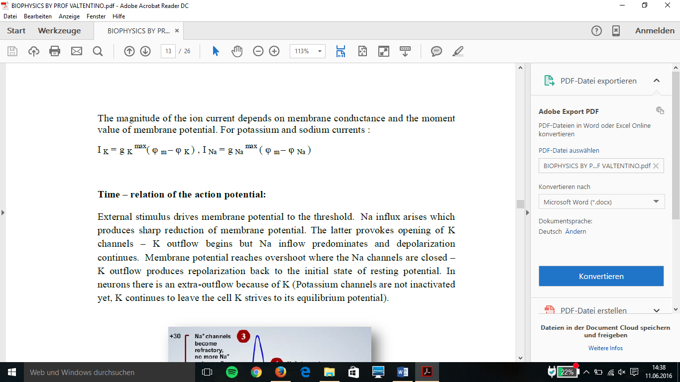
IK or INa = ion current of potassium or sodium
g = electric conductance of membrane
φm = membrane potential
φK or φNa = equilibrium potential of ion
Ion currents measurement by fixed voltage scheme:

A) Depolarizing voltage jump from -65 mV to 0mV
B) Total current due to the movement of Na+ (sodium) and K+ (potassium) ions
C) Outward (out of cell) K+ (potassium) current after TTX blocks the Na+ (sodium) channels
D) Inward (inside cell) Na+ (sodium) current after TEA blocks the K+ (potassium) channels
- Action potential propagation. Electrotone potential. Blockage of AP spreading.
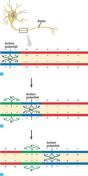
Action potential propagation:
1) AP passes along the membrane provided by so called local currents or transverse currents– these currents are the movement of sodium (Na+) and potassium (K+) ions.
2) The neighbouring cell which is in the resting state is irritated by these currents, provoking depolarization of resting membrane.
3)Potential reaches threshold – open the ion channels and start of a new AP.
-> Action potential will propagate from one cell to its neighbouring till it reaches the terminal synapse.
Gradient maintaining the stability of AP:
DEF: Concentration gradient
C2 – C1 or grad C is
-> The graduated difference in concentration of a solute per unit distance through a solution.
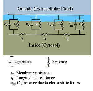
The evaluation of AP time propagation is measured by cable model and this formula:
t = df.C.R/U
t = time
df = depolarization threshold
C = capacitance
R = Resistance
U = voltage between excited and neighboring area
-> the area of initial AP undergoes “refractory state”, during such state the membrane is not excitable, therefore AP propagation has a certain direction only
Electrotonic Potential
Electrotonic potential is a non-propagated local potential, resulting from a local change in ionic conductance.
Electrotonic potential triggers Action potential in long neurons. In some short neurons you only have electrotonic potential, which is conducted faster, but not suitable for long distance signaling.
Cable Model/Theory
Cable model is used for calculation of time propagation of AP.
Blockage of AP spreading – Interruption of AP propagation
Example: Local anesthetics
Local anesthetic drugs act by inhibiting Na+ (sodium) influx through voltage gated sodium channels. When influx interrupted, action potential cannot rise and signal conduction is inhibited.
Examples of anesthetics:
- Lidocaine
- Articaine
- Septocaine
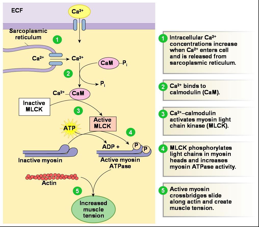
- Bioelectrical activity, structure and contractile type of smooth muscle. Contractile mechanisms of smooth muscles.
Bioelectric activity: electrical conductivity and impulses in living organisms. It is based on charged ions such as Na+, K+ and Cl-, Ca2+
Smooth Muscle
Structure:
- SM are networks of contractile fibers (protein fibers): actin and myosin bounded by dense bodies – for contraction, temporary contact between actin and myosin is required
Contractile Type of SM
- SM possessing pacemaker cells are called phasic – this type of tissue makes spontaneous contractions
- SM may contract phasically (rapid contraction, relaxation), or tonically (slow, continued contraction)
- Action potentials of phasic SM are generated by Cajal cells – they generate recurring AP with specific time-shape
Steps:
- Bioelectrical activity, structure and properties of skeletal and cardiac muscles. Contractile mechanisms of striated muscles.
Skeletal Muscle:
Structure:
- Cylinder-shape - cells organized in parallel manner to form single fiber groups which form muscle
- Contractive filaments are actin and myosin, strongly ordered in sacromeres – each thick-myosin filament is surrounded by 6 thin-actin filaments
Contraction:
- No pacemaker activity – activity regulated by CNS through motor neurons – contraction starts when neuron stimulates muscle bundle
Contraction of Striated Muscle through Electro-mechanical coupling – Skeletal AND Cardiac:
Steps:
1) ACh- Acetylcholine is released and diffuses across synaptic cleft and binds to receptor proteins on muscle fibers membrane triggering AP
2) AP travels along the membrane down T tubules
3) AP triggers Ca2+ release from SR-sarcoplasmic reticulum
4) Ca2+ binds to troponin in thin filament, myosin binding sites are exposed
5) ATP activates myosin cross-bridge, causing the thin filament to slide toward center of sarcomere
6) cytosolic Ca2+ is removed by active transport into SR after AP finishes
7) tropomyosin blockage is restored, contraction ends and fiber relaxes
Cardiac Muscles:
- Are myogenic cells
- Can contract without nervous stimulation, like SM
- Long, cylindrical cells
- Sarcomere is the contractile unit of myocardial cell
- Source of initial AP is sinus node
- Depending on region of the heart, AP waveforms can be different
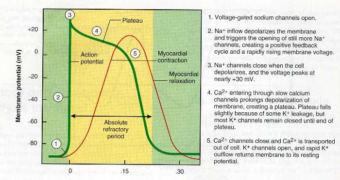
Ventricular Muscle Action Potential- time relation (AP of Cardiac muscle):
Summary:
Resting membrane potential- Na+ channels open- K+ channels open- Na+ channels close- Ca2+ channels open- Ca2+ and K+ generate plateau- Ca2+ and K+ channels close returning to resting membrane potential.
- Intercellular communications. Types of information systems: electrical and chemical information translation.
There are two main types of intracellular communication,
1) Electrical
2) Chemical
Neuronal transmission- high velocity communication
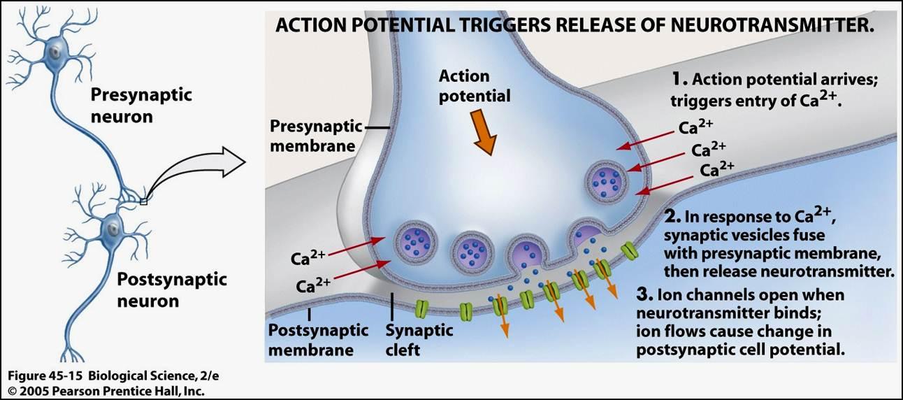
Combination of chemical and electrical transduction. Electrical signals are carried along the neuron by action potential, reaching synapse signal is transferred to next neuron by chemical transduction;
Synapse= junction between nerve cell and another cell
Synaptic cleft= space between two cells
Gap junction/Nexus= specialized intracellular connection – connecting cytoplasm of two cells allowing molecules and ions to pass freely. It is composed of two connexons allowing electrical and chemical communication
Neurotransmitter(e.g. Hormones)= required to cross synaptic cleft – nts diffuse across the cleft and bind to receptors on other cell membrane causing ion channels on the other cell to open They are active for a short time - enzymes in cleft inactivate the nts – which are taken back into axon and recycled
Chemical signaling system:
Intracellular communication by hormones secreted by cells
1) Endocrine= hormone distributed in blood binds to distant target cell
2) Paracrine= acts locally by diffusing from source to target cell
3) Autocrine= hormone acts on same cell that produced it
4) Contact dependent= communication through membrane to membrane interface by signal-receptor complex
-> The main difference in chemical intercellular communication are in the speed and selectivity with which signals are delivered to their targets!
- Receptors- receiving and translation of information. Intracellular system of signal spreading.
2nd stage of cell signaling is translation of info. into target cell.
Receptors= complex proteins integrated in membrane
= they; identify informer molecules,
bind ligands
transmit signal
= one or more binding domains identify a specific ligand and binds it
* Ligand=ion or molecule that reacts to form a complex with another molecule
There are 4 groups=
1) Channel linked receptors- signal binds, channel opens, ion flows through membrane e.g. Acetylcholine N type receptor
2) Enzyme linked receptors- signal binds, enzyme activated, enzyme generates product
3) G-protein coupled receptors- signal binds, G-protein binds and is activated e.g. Acetylcholine M type receptor
4) Intracellular receptors- signal binds to intracellular receptor and activated receptor regulates transcription
- Electrokinetic phenomena. Biological significance of electrokinetic properties.
Electrokinetic Phenomena: different effects that occur in heterogeneous fluids, or in porous bodies filled with fluid, or in a fast flow over a flat surface.
Electrokinetic potential= Zeta potential; a potential difference between the dispersion medium and the stationary layer of fluid attached to the dispersed particle.
Zeta potential value can be related to the stability of colloidal dispersion. It indicates the degree of repulsion between adjacent or similarly charged particles in a dispersion.
High zeta value means more repulsion between particles than attraction therefore colloids are stable and resist aggregation where as low zeta potential colloids will aggregate so colloids are unstable.
Zeta potential can be estimated by measuring the speed of movement of a disperse phase in an electric field by Smoluchowski’s formula:
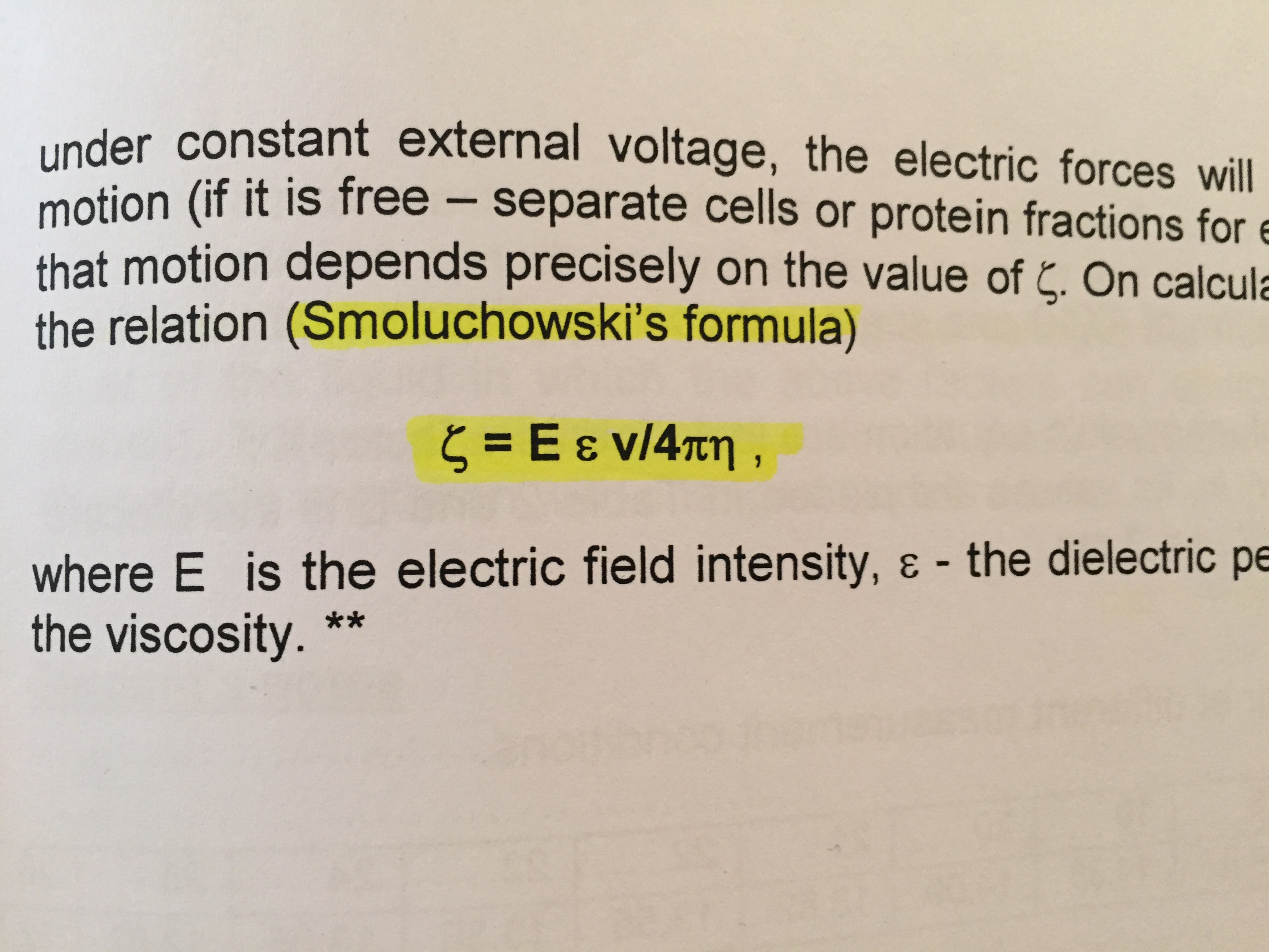
E= electric field intensity
ε= dielectric permeability of medium
η= viscosity
v= velocity
4π= 4 x 3.14 (pi to 2dp)
*Colloid= the dispersion of cells/insoluble matter in a fluid
---------------------------------------------------
Zeta potential [mV] | Stability behaviour of the colloid |
from 0 to ±5, | Rapid coagulation or flocculation |
from ±10 to ±30 | Incipient instability |
from ±30 to ±40 | Moderate stability |
from ±40 to ±60 | Good stability |
more than ±61 | Excellent stability |