knowt logo

Wk 2B - Male and Female Reproductive System

Reproductive Development

Intrauterine Development

  • The sex of an individual is determined at the moment of conception by the chromosomes information supplied by the ovum and sperm

  • Week 5

    • formation of Mesonephric and Paramesonephric ducts

  • Week 7or 8

    • Males – Mesonephric ducts develops and give rise to epididymis, vas deferens, seminal vesicles

    • begins formation of testosterone and develops into male reproductive organs

  • Week 10

    • If testosterone is not present

    • Females – Paramesonephric ducts becomes dominant and develop into female reproductive organs

      • ovaries form

      • cells that will develop into eggs are already present (Oocytes)

    • Week 12

      • External genitals begins to develop

      • Males – penile tissue elongated

      • Females – uterus, labia minora and labia majora form

Pubertal Development

  • is the stage of life at which a person becomes capable of sexual reproduction

  • Secondary sex characteristics develop during puberty
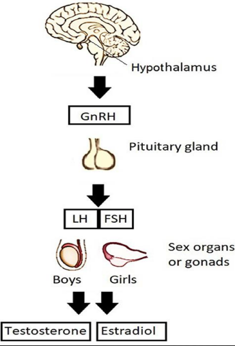
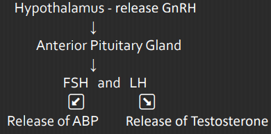
FSH and LH Function:

  1. Initiate the production of androgen and estrogen

  2. Initiate secondary sex characteristics

  3. Production of eggs and influence menstrual cycles

  • Girls – age 9 to 12 years Hormones

    • Estrogen (Estrone E1, Estradiol E2, Estriol E3)

  • Boys – age 12 to 14 years Hormones:

    • Androgen (Testosterone)

Estrogen Function:

  1. Development of the uterus, fallopian tubes, vagina

  2. Female fat distribution

  3. Hair patterns

  4. Breast development (Thelarche) – starts 1-2 years before menstruation.

  • Secondary sex characteristics – are physical characteristics developing at puberty which distinguishes between sexes

Secondary Sex Characteristics

Girls
  1. Growth spurt

  2. Increase in transverse diameter of pelvis

  3. Breast development (Thelarche)

  4. Growth of pubic hair and axillary hair (Adrenarche)

  5. Vaginal secretion

  6. Onset of menstruation (Menarche 12.4 y/o)

  • Menarche - first menstrual period

  • Average Age – 12.4 years of age

  • Average Range – 9-17 years old

Delayed Menstruation Causes:
- Stress
- Extreme exercises
- Low body weight
- Polycystic ovary syndrome (PCOS)

Boys
  1. Increase in weight

  2. Growth of testes

  3. Growth of face, axillary, and pubic hair

  4. Voice changes

  5. Penile growth

  6. Increase in height

  7. Spermatogenesis (production of sperm)

  • Spermatozoa produced in a cyclic pattern, produced in a continuous process.

  • Testosterone – primary androgenic hormone

Androgen Hormones are responsible for
1. Muscular Development
2. Physical Growth
3. Increase in Sebaceous gland secretions

Female Reproductive System

  • Gynecology - is the study of female reproductive organ

External Structures

  1. Mons Veneris

    • a pad of adipose tissue, located over the symphysis pubis

    • Covered by a triangle of coarse curly hair that protects the pubic bone from trauma

  2. Labia Mjora

    • are two folds of tissue, position lateral to the labia minora

    • Cover and protects the external genitalia, urethra, and vagina

  3. Labia Minora

    • posterior to mons veneris spread two hairless folds of connective tissue.

    • Protect the clitoris, urinary and vaginal orifice

  4. Clitoris

    • is a small rounded organ of erectile tissue (approximately 1-2 cm.)

    • serve as center of sexual arousal and orgasm

  5. Vestibule

    • the flattened, smooth surface inside the labia minora that encloses the vaginal and urethral opening

  6. Bartholin’s Gland

    • (Vulvovaginal Glands) located just lateral to the vaginal opening

    • Secretes mucus to lubricate the vagina during coitus.

  7. Fourchette

    • is the ridge of tissue joining the labia minora and majora,

    • Serve as site of Episiotomy during childbirth

  8. Hymen

    • a tough elastic semicircle tissue that covers the opening of the vagina

    • Hematocolpometra – imperforate hymen

  9. Skene’s Gland

    • (paraurethral glands) lateral to the urinary meatus

    • secretes mucous which serve as lubricants during sexual intercourse

  10. Perineum

    • muscular structure in between the vagina and anus. Support the genitourinary system

Internal Structures

  1. Ovaries

    • approximately 4 cm. long by 2 cm. in diameter, 1.5 cm. thick and shape of almonds

    • secretes hormones

    • Functions:

      • Produce estrogen and progesterone

      • Produce mature and discharge egg cells

      • Initiate and regulates menstrual cycles

    • 3 Principal Division of Ovaries

      1. Protective layer of surface epithelium

      2. Cortex – where the immature oocytes (Primordial) mature into ova

      3. Central medulla – contains nerves, blood vessels, lymphatic tissue and smooth muscle tissue

  2. Fallopian Tubes

    • provides a place for fertilization of the ovum by sperm. (approximately 10 cm. long)

    • Parts

      1. Interstitial

        • is the part of the tube that lies within the uterine wall and site for ectopic pregnancy

      2. Isthmus

        • is the distal portion of the tube that is cut or sealed in a tubal ligation or tubal sterilization procedure

      3. Ampulla

        • is the third and the longest portion of the tube.

        • It is the portion that fertilization of ovum occurs.

      4. Infundibular

        • distal segment of the tube. (2 cm. long and funnel shaped)

        • covered by fimbria (small hairs) that help to guide ovum into the fallopian tube

  3. Uterus

    • Weight

      • Non-pregnant - 60 grams

      • Pregnant - 1000 grams

    • Size

      • 5-7 cm. long

      • 5 cm. wide

      • 2.5 cm. thick (non-pregnant)

    • Function

      1. Receive the ovum from the fallopian tube.

      2. Provide a place for implantation and nourishment.

      3. Furnish protection to a growing fetus.

      4. Provide a place for menstruation, pregnancy, and labor.

3 Divisions of Uterus

  1. Fundus

    • is the body of the uterus, uppermost part and forms the bulk of the organ.

    • During pregnancy, it is the portion of the structure that expands to contain the growing fetus.

  2. Isthmus of the Uterus

    • is a short segment between the body and the cervix. In the nonpregnant uterus, it is only 1 to 2 mm in length.

    • It is the portion of the uterus that is cut when a fetus is born by a cesarean birth

  3. Breast

    • are located anterior to the pectoral muscle and breast tissue extend into the axilla.

    • remain in halted stage of development until a rise in estrogen at puberty causes them to increase in size

    Parts of Breast

    1. Milk Glands (Lobules)

      • with approximately 20 lobes that produced milk by acinar cells and deliver it to the nipple via a lactiferous duct

    2. Nipple

      • composed of smooth muscle capable of erection on manual or sucking stimulation

      • has 20 small openings through which milk is secrete

    3. Areola

      • a darkly pigmented out to about 4 cm., the area appears rough on the surface and contains many sebaceous glands (Montgomery tubercles)

    4. Ampulla

      • located just posterior to the nipple serves as reservoir for milk before breastfeeding

  4. Cervix

    • is the lowest portion of the uterus.

    • Internal os – opening of the canal at the junction of the cervix into the uterus

    • External os – distal opening to the vagina (Estimating the level of the fetus in the birth canal)

Layers of Uterus

  1. Endometrium

    • inner layers and important for menstrual function.

    • It becomes thick each month under the influence of estrogen and progesterone capable of supporting a pregnancy

    • If pregnancy does not occur, this is the layer that is shed as the menstrual flow

  2. Myometrium

    • composed of smooth muscle that offers extreme strength to the organ

    • Function:

      1. It prevent regurgitation of menstrual blood into the tubes

      2. It holds the internal cervical as closed during pregnancy to prevent a preterm birth

  3. Perimetrium

    • outermost layer, add further strength and support to the organ

Male Reproductive System

  • Andrology - is the study of the male reproductive system

External Structures

  1. Scrotum

    • is a rugated, skin-covered, muscular pouch hanging below the penis containing the testes.

    • Support the testes and help regulate the temperature of sperm.

  2. Testes

    • are two ovoid glands, that lie in the scrotum (2-3 cm. wide)

    • Produced and store sperm Produced hormones

    • composed of lobules containing interstitial cells

    • Lobules

      1. Seminiferous tubules – produce spermatozoa

      2. Leydig’s cells – responsible for the production of testosterone

  3. Penis

    • is composed of three cylindrical masses of erectile tissue in the penis shaft

      1. Corpus Cavernosa

        • two lateral columns of erectile tissue

      2. Corpus Spongiosum

        • encases the urethra, located on the underside of the penis

      3. Ischiocavernosus muscle

        • under stimulation, venous and arterial blood in erectile tissue leads to distention and erection of the penis

Internal Structures

  1. Epididymis

    • seminiferous tubules of each testes

    • 20 ft. long, takes 12-20 days for the sperm to travel the length of epididymis, 64 days to reach maturity

    • Serve as reservoir for sperm storage and maturation.

    • Conducting sperm from the tubule to the vas deferens.

    • A total of 65 - 75 days for the sperm to reach maturity

    • Aspermia – absence of sperm

    • Oligospermia – fewer than 20 million sperm per millimeter

  2. Vas deferens (Ductus Deferens)

    • Hallow tube surrounded by arteries and veins

    • carries sperm from epididymis through the inguinal canal into the abdominal cavity.

  3. Seminal vesicles

    • are pouches that lie along the bladder

    • Secretes viscous alkaline liquid portion of the semen.

    • Increase sperm motility

  4. Prostate gland

    • a chest-nut size gland that lies below the bladder

    • Secretes a thin, alkaline fluid that protects sperm from being immobilized.

    • Reduces the acidity of the vagina

    • Benign Prostatic Hypertrophy

      • enlargement of the prostate gland

      • swelling interferes with both urination and fertility

  5. Bulbourethral Glands

    • glands lies beside the prostate gland

    • Secretes an alkaline fluid to help ensure the safe passage of spermatozoa

  6. Urethra

    • It is 8 inches (18-20 cm.) long

    • vessel of transport of urine and semen

  7. Ejaculatory Ducts

    • two ejaculatory ducts pass through the prostate gland and join the seminal vesicles to the urethra.

Spermatozoa
  • ARE MALE SEX CELLS THAT CARRY A MAN’S GENETIC MATERIAL.

  • Sperm Count

    • 33 - 46 million sperm per ml. of seminal fluid

    • 50 million per ejaculation

    • 50% of sperm are motile

    • 30% are normal in shape and form

  • During ejaculation, semen receives contributions of fluid from

    • Prostate gland – 60%

    • Seminal vesicle - 30%

    • Epididymis - 5%

    • Bulbourethral glands – 5%

  • Spermatogenesis – the formation and development of sperm.

  • Testosterone - responsible for the development of the secondary male characteristics
EA

Wk 2B - Male and Female Reproductive System

Reproductive Development

Intrauterine Development

  • The sex of an individual is determined at the moment of conception by the chromosomes information supplied by the ovum and sperm

  • Week 5

    • formation of Mesonephric and Paramesonephric ducts

  • Week 7or 8

    • Males – Mesonephric ducts develops and give rise to epididymis, vas deferens, seminal vesicles

    • begins formation of testosterone and develops into male reproductive organs

  • Week 10

    • If testosterone is not present

    • Females – Paramesonephric ducts becomes dominant and develop into female reproductive organs

      • ovaries form

      • cells that will develop into eggs are already present (Oocytes)

    • Week 12

      • External genitals begins to develop

      • Males – penile tissue elongated

      • Females – uterus, labia minora and labia majora form

Pubertal Development

  • is the stage of life at which a person becomes capable of sexual reproduction

  • Secondary sex characteristics develop during puberty

FSH and LH Function:

  1. Initiate the production of androgen and estrogen

  2. Initiate secondary sex characteristics

  3. Production of eggs and influence menstrual cycles

  • Girls – age 9 to 12 years Hormones

    • Estrogen (Estrone E1, Estradiol E2, Estriol E3)

  • Boys – age 12 to 14 years Hormones:

    • Androgen (Testosterone)

Estrogen Function:

  1. Development of the uterus, fallopian tubes, vagina

  2. Female fat distribution

  3. Hair patterns

  4. Breast development (Thelarche) – starts 1-2 years before menstruation.

  • Secondary sex characteristics – are physical characteristics developing at puberty which distinguishes between sexes

Secondary Sex Characteristics

Girls
  1. Growth spurt

  2. Increase in transverse diameter of pelvis

  3. Breast development (Thelarche)

  4. Growth of pubic hair and axillary hair (Adrenarche)

  5. Vaginal secretion

  6. Onset of menstruation (Menarche 12.4 y/o)

  • Menarche - first menstrual period

  • Average Age – 12.4 years of age

  • Average Range – 9-17 years old

Delayed Menstruation Causes:
- Stress
- Extreme exercises
- Low body weight
- Polycystic ovary syndrome (PCOS)

Boys
  1. Increase in weight

  2. Growth of testes

  3. Growth of face, axillary, and pubic hair

  4. Voice changes

  5. Penile growth

  6. Increase in height

  7. Spermatogenesis (production of sperm)

  • Spermatozoa produced in a cyclic pattern, produced in a continuous process.

  • Testosterone – primary androgenic hormone

Androgen Hormones are responsible for
1. Muscular Development
2. Physical Growth
3. Increase in Sebaceous gland secretions

Female Reproductive System

  • Gynecology - is the study of female reproductive organ

External Structures

  1. Mons Veneris

    • a pad of adipose tissue, located over the symphysis pubis

    • Covered by a triangle of coarse curly hair that protects the pubic bone from trauma

  2. Labia Mjora

    • are two folds of tissue, position lateral to the labia minora

    • Cover and protects the external genitalia, urethra, and vagina

  3. Labia Minora

    • posterior to mons veneris spread two hairless folds of connective tissue.

    • Protect the clitoris, urinary and vaginal orifice

  4. Clitoris

    • is a small rounded organ of erectile tissue (approximately 1-2 cm.)

    • serve as center of sexual arousal and orgasm

  5. Vestibule

    • the flattened, smooth surface inside the labia minora that encloses the vaginal and urethral opening

  6. Bartholin’s Gland

    • (Vulvovaginal Glands) located just lateral to the vaginal opening

    • Secretes mucus to lubricate the vagina during coitus.

  7. Fourchette

    • is the ridge of tissue joining the labia minora and majora,

    • Serve as site of Episiotomy during childbirth

  8. Hymen

    • a tough elastic semicircle tissue that covers the opening of the vagina

    • Hematocolpometra – imperforate hymen

  9. Skene’s Gland

    • (paraurethral glands) lateral to the urinary meatus

    • secretes mucous which serve as lubricants during sexual intercourse

  10. Perineum

    • muscular structure in between the vagina and anus. Support the genitourinary system

Internal Structures

  1. Ovaries

    • approximately 4 cm. long by 2 cm. in diameter, 1.5 cm. thick and shape of almonds

    • secretes hormones

    • Functions:

      • Produce estrogen and progesterone

      • Produce mature and discharge egg cells

      • Initiate and regulates menstrual cycles

    • 3 Principal Division of Ovaries

      1. Protective layer of surface epithelium

      2. Cortex – where the immature oocytes (Primordial) mature into ova

      3. Central medulla – contains nerves, blood vessels, lymphatic tissue and smooth muscle tissue

  2. Fallopian Tubes

    • provides a place for fertilization of the ovum by sperm. (approximately 10 cm. long)

    • Parts

      1. Interstitial

        • is the part of the tube that lies within the uterine wall and site for ectopic pregnancy

      2. Isthmus

        • is the distal portion of the tube that is cut or sealed in a tubal ligation or tubal sterilization procedure

      3. Ampulla

        • is the third and the longest portion of the tube.

        • It is the portion that fertilization of ovum occurs.

      4. Infundibular

        • distal segment of the tube. (2 cm. long and funnel shaped)

        • covered by fimbria (small hairs) that help to guide ovum into the fallopian tube

  3. Uterus

    • Weight

      • Non-pregnant - 60 grams

      • Pregnant - 1000 grams

    • Size

      • 5-7 cm. long

      • 5 cm. wide

      • 2.5 cm. thick (non-pregnant)

    • Function

      1. Receive the ovum from the fallopian tube.

      2. Provide a place for implantation and nourishment.

      3. Furnish protection to a growing fetus.

      4. Provide a place for menstruation, pregnancy, and labor.

3 Divisions of Uterus

  1. Fundus

    • is the body of the uterus, uppermost part and forms the bulk of the organ.

    • During pregnancy, it is the portion of the structure that expands to contain the growing fetus.

  2. Isthmus of the Uterus

    • is a short segment between the body and the cervix. In the nonpregnant uterus, it is only 1 to 2 mm in length.

    • It is the portion of the uterus that is cut when a fetus is born by a cesarean birth

  3. Breast

    • are located anterior to the pectoral muscle and breast tissue extend into the axilla.

    • remain in halted stage of development until a rise in estrogen at puberty causes them to increase in size

    Parts of Breast

    1. Milk Glands (Lobules)

      • with approximately 20 lobes that produced milk by acinar cells and deliver it to the nipple via a lactiferous duct

    2. Nipple

      • composed of smooth muscle capable of erection on manual or sucking stimulation

      • has 20 small openings through which milk is secrete

    3. Areola

      • a darkly pigmented out to about 4 cm., the area appears rough on the surface and contains many sebaceous glands (Montgomery tubercles)

    4. Ampulla

      • located just posterior to the nipple serves as reservoir for milk before breastfeeding

  4. Cervix

    • is the lowest portion of the uterus.

    • Internal os – opening of the canal at the junction of the cervix into the uterus

    • External os – distal opening to the vagina (Estimating the level of the fetus in the birth canal)

Layers of Uterus

  1. Endometrium

    • inner layers and important for menstrual function.

    • It becomes thick each month under the influence of estrogen and progesterone capable of supporting a pregnancy

    • If pregnancy does not occur, this is the layer that is shed as the menstrual flow

  2. Myometrium

    • composed of smooth muscle that offers extreme strength to the organ

    • Function:

      1. It prevent regurgitation of menstrual blood into the tubes

      2. It holds the internal cervical as closed during pregnancy to prevent a preterm birth

  3. Perimetrium

    • outermost layer, add further strength and support to the organ

Male Reproductive System

  • Andrology - is the study of the male reproductive system

External Structures

  1. Scrotum

    • is a rugated, skin-covered, muscular pouch hanging below the penis containing the testes.

    • Support the testes and help regulate the temperature of sperm.

  2. Testes

    • are two ovoid glands, that lie in the scrotum (2-3 cm. wide)

    • Produced and store sperm Produced hormones

    • composed of lobules containing interstitial cells

    • Lobules

      1. Seminiferous tubules – produce spermatozoa

      2. Leydig’s cells – responsible for the production of testosterone

  3. Penis

    • is composed of three cylindrical masses of erectile tissue in the penis shaft

      1. Corpus Cavernosa

        • two lateral columns of erectile tissue

      2. Corpus Spongiosum

        • encases the urethra, located on the underside of the penis

      3. Ischiocavernosus muscle

        • under stimulation, venous and arterial blood in erectile tissue leads to distention and erection of the penis

Internal Structures

  1. Epididymis

    • seminiferous tubules of each testes

    • 20 ft. long, takes 12-20 days for the sperm to travel the length of epididymis, 64 days to reach maturity

    • Serve as reservoir for sperm storage and maturation.

    • Conducting sperm from the tubule to the vas deferens.

    • A total of 65 - 75 days for the sperm to reach maturity

    • Aspermia – absence of sperm

    • Oligospermia – fewer than 20 million sperm per millimeter

  2. Vas deferens (Ductus Deferens)

    • Hallow tube surrounded by arteries and veins

    • carries sperm from epididymis through the inguinal canal into the abdominal cavity.

  3. Seminal vesicles

    • are pouches that lie along the bladder

    • Secretes viscous alkaline liquid portion of the semen.

    • Increase sperm motility

  4. Prostate gland

    • a chest-nut size gland that lies below the bladder

    • Secretes a thin, alkaline fluid that protects sperm from being immobilized.

    • Reduces the acidity of the vagina

    • Benign Prostatic Hypertrophy

      • enlargement of the prostate gland

      • swelling interferes with both urination and fertility

  5. Bulbourethral Glands

    • glands lies beside the prostate gland

    • Secretes an alkaline fluid to help ensure the safe passage of spermatozoa

  6. Urethra

    • It is 8 inches (18-20 cm.) long

    • vessel of transport of urine and semen

  7. Ejaculatory Ducts

    • two ejaculatory ducts pass through the prostate gland and join the seminal vesicles to the urethra.

Spermatozoa
  • ARE MALE SEX CELLS THAT CARRY A MAN’S GENETIC MATERIAL.

  • Sperm Count

    • 33 - 46 million sperm per ml. of seminal fluid

    • 50 million per ejaculation

    • 50% of sperm are motile

    • 30% are normal in shape and form

  • During ejaculation, semen receives contributions of fluid from

    • Prostate gland – 60%

    • Seminal vesicle - 30%

    • Epididymis - 5%

    • Bulbourethral glands – 5%

  • Spermatogenesis – the formation and development of sperm.

  • Testosterone - responsible for the development of the secondary male characteristics

robot