Four Stages of Muscle Contraction, Sliding Filament Theory
The four stages of muscle contraction are excitation, contraction, recharge and relaxation. Prior to these stages, the muscle is resting.
Muscle contraction is initiated by a neural action potential (from the brain). The neural action potential travels via a motor neurone to the motor end-plate, which creates an action potential over the muscle fibre’s sarcoplasm, inward along the ‘T’ tubules (see below). This is excitation.
This triggers the release of calcium ions (Ca++) from the ‘T’ vesicles in the sarcoplasmic reticulum into the sarcoplasm.
The calcium ions bind to troponin molecules on actin filaments. Troponin has a high affinity for calcium ions. This causes tropomyosin molecules on the thin actin filaments move, exposing the active binding sites to myosin.
==Use this model **to help you remember:==
==Lock key: Calcium ions==
==Lock: Troponin==
==Chain: Tropomyosin==
==Bicycle: Actin==
==Athlete: Myosin==
==Energy bar: ATP==
Energy released from ATP is used to pull the actin filaments towards the centre of the sarcomere, resulting in the shortening of the muscle. This is called the power stroke. The greater the overlap of the filaments, the stronger the contraction.
The myosin detaches from the actin and the cross-bridge is broken when an ATP molecule binds to the myosin head. Ca++ is removed and the process is repeated. When the ATP is then broken down the myosin head can again attach to an actin binding site further along the actin filament and repeat the power stroke.
This attach, detach, reattach of cross-bridges is called the ratchet mechanism, which repeats, ‘recharges’, as long as ATP and Ca++ is available.
Once the impulse stops, the Ca++ is pumped back to the sarcoplasmic reticulum and the actin returns to its resting position, causing the muscle to lengthen and relax.
This is Huxley’s sliding theory of muscle contraction.
Key word definitions:
Actin: Thin protein filament found in the myofibril
Adenosine triphosphate (ATP): The energy currency of the body. Found in all cells; when broken down it releases stored energy.
Motor neurones: Nerves that carry information from the central nervous system to the skeletal muscles.
Myofibril: Part of a muscle fibre. Contains sarcomeres and the contractile proteins actin and myosin.
Myosin: Thick protein filament found in the myofibril.
Tropomyosin: Thread-like protein that winds around the surface of the actin.
Troponin: Globular protein on actin filament.
Extra information:
T tubules are invaginations of the sarcolemma (cell membrane) that penetrate deep into the muscle fibre, allowing for the rapid transmission of action potentials throughout the muscle fibre. This allows for synchronous contraction of all sarcomeres. T tubules also play a role in regulating calcium release from the SR during muscle contraction.
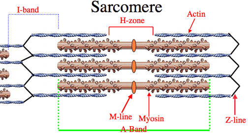
Contracted muscle: H band is smaller. I band is smaller. A band remains the same length. Z lines get closer together.
 The M-line is the middle of the sarcomere.
The Z-line is the end of the sarcomere.
The A-band contains both actin and myosin.
The H-zone contains myosin only.
The I-band contains actin only.