Sensory Systems
Hearing
Sensation and Perception
Sensation: the acquisition of sensory information
Perception: the interpretation of sensory information
In this unit, we will begin to explore how the brain processes information from the outside world through the senses. When we talk about the senses, we talk about sensation and perception. Sensation is the acquisition of sensory information, and perception is the interpretation of that information. To understand the world around us, our brain must do both.
Sensory Receptors
Respond to a particular form of energy, such as sound or light
Converts that energy into a neural response or action potential
Signal transduction: the conversion of sensory information into neural signalling by sensory receptors
For sensation, our sensory systems need a specialized type of cell, called a sensory receptor. This is not the same thing as the protein we learned about that the neurotransmitters bind to. In this case, a sensory receptor is a cell that responds to a particular form of energy, such as sound. The function of the receptor is to convert that energy into a neural response or action potential. The idea that our sensory receptors convert information, such as sound, light, or taste, into neuronal signalling is called signal transduction. In this unit, we will learn several forms of signal transduction, starting with sounds.
Anatomy of the Ear
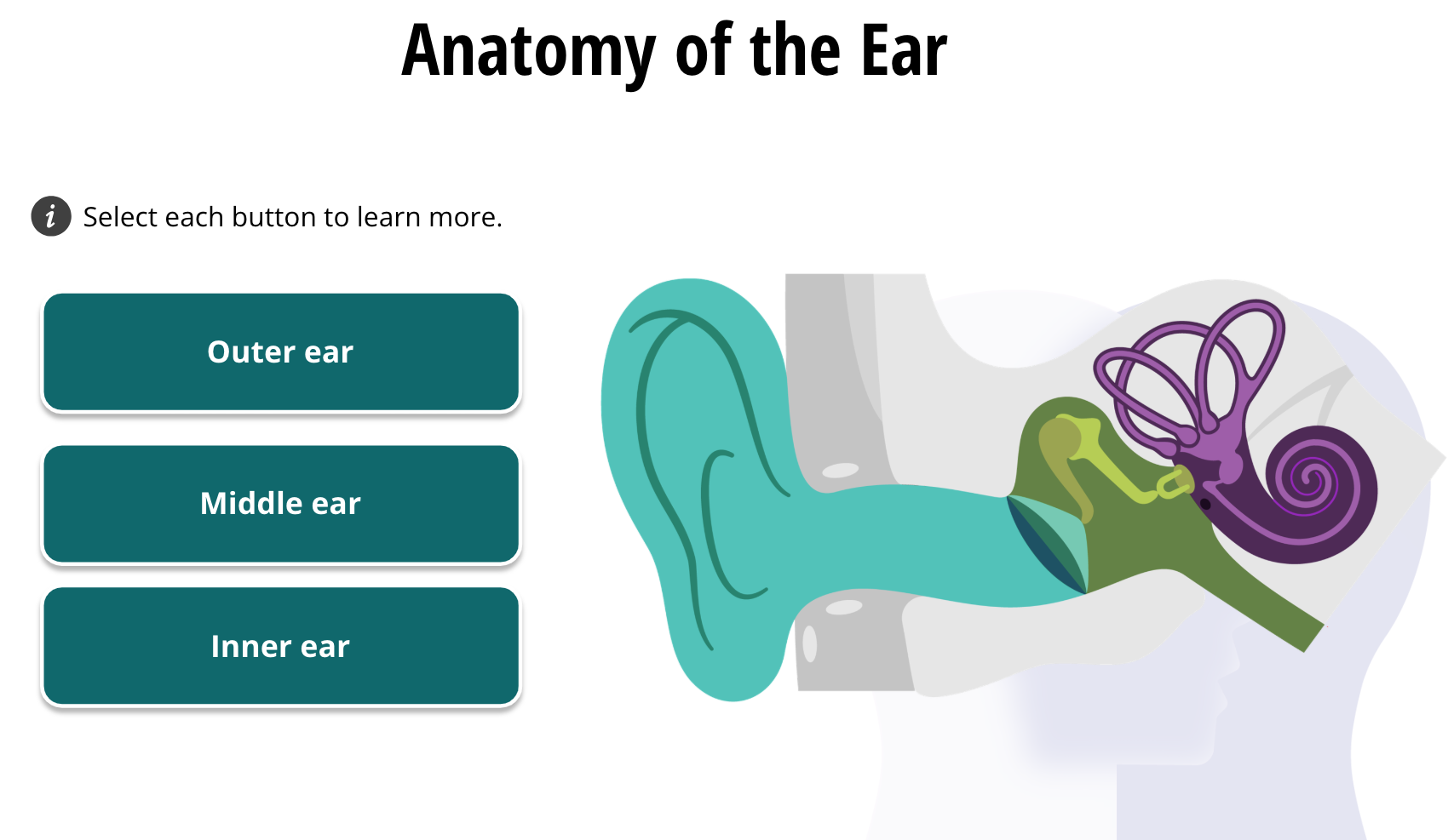
We will begin with our sense of hearing. To hear, we use our ears, of course. When neuroscientists say “ear,” we actually refer to all of the structures shown in this figure. They are categorized as the outer ear, middle ear, and inner ear.
Outer Ear
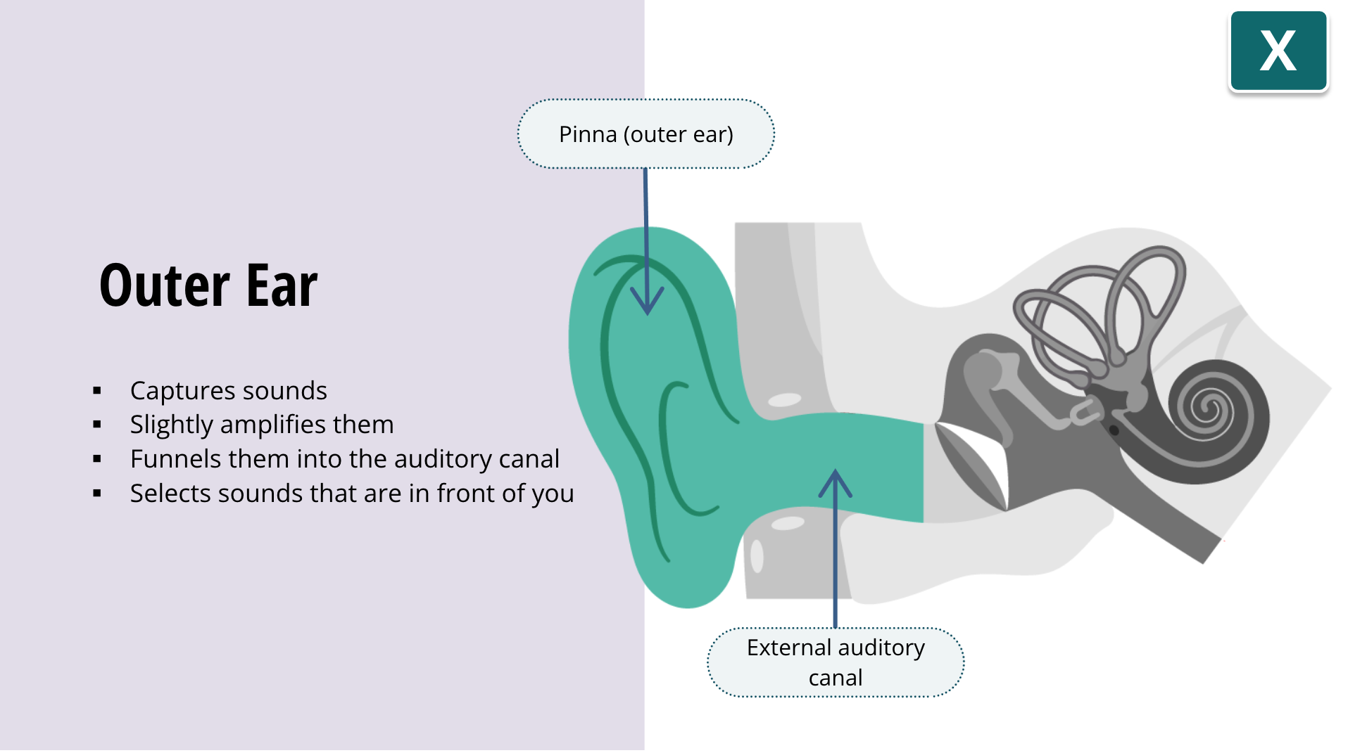
Captures sounds
Slightly amplifies sounds
Funnels sounds into the auditory canal
Selects sounds that are where you are facing
The ear on the side of your head is called the outer ear or pinna. It captures sounds and slightly amplifies them and funnels them into the auditory canal. By its shape, it also selects for sounds that are in front of you, helping you focus on listening to what you are facing, and somewhat excluding irrelevant sounds behind you. You may have noticed that dogs and cats can turn their ears toward a sound that is not directly in front of them. Unfortunately, we cannot do that.
Middle Ear
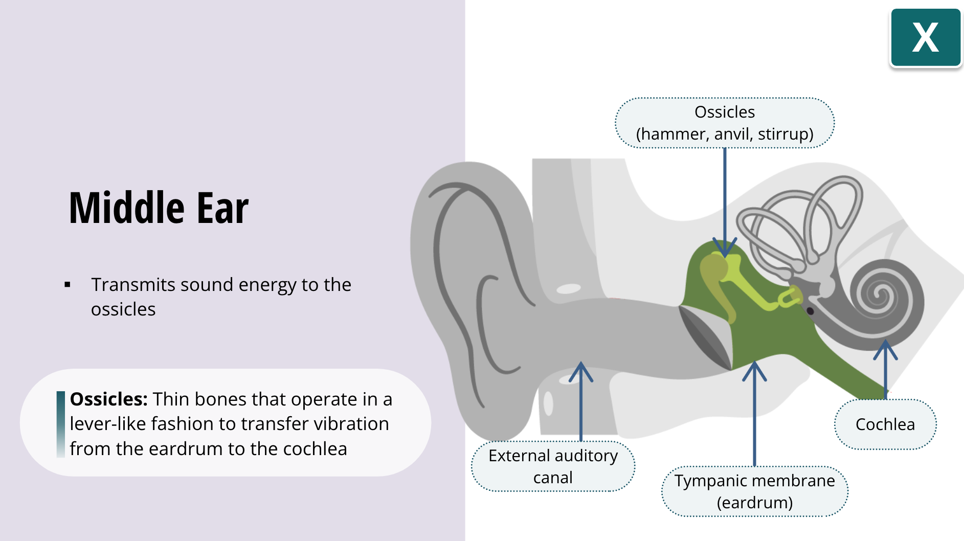
Transmits sound energy to the ossicles
The first part of the middle ear is the eardrum, or tympanic membrane. This is a thin membrane that stretches across the end of the auditory canal, and transmits sound energy to another area of the ear, called the ossicles. The ossicles are tiny bones that operate in a lever-like fashion to transfer vibration from the eardrum to the cochlea. You may be familiar with the common names for the ossicles: namely the hammer, anvil, and stirrup
Inner Ear
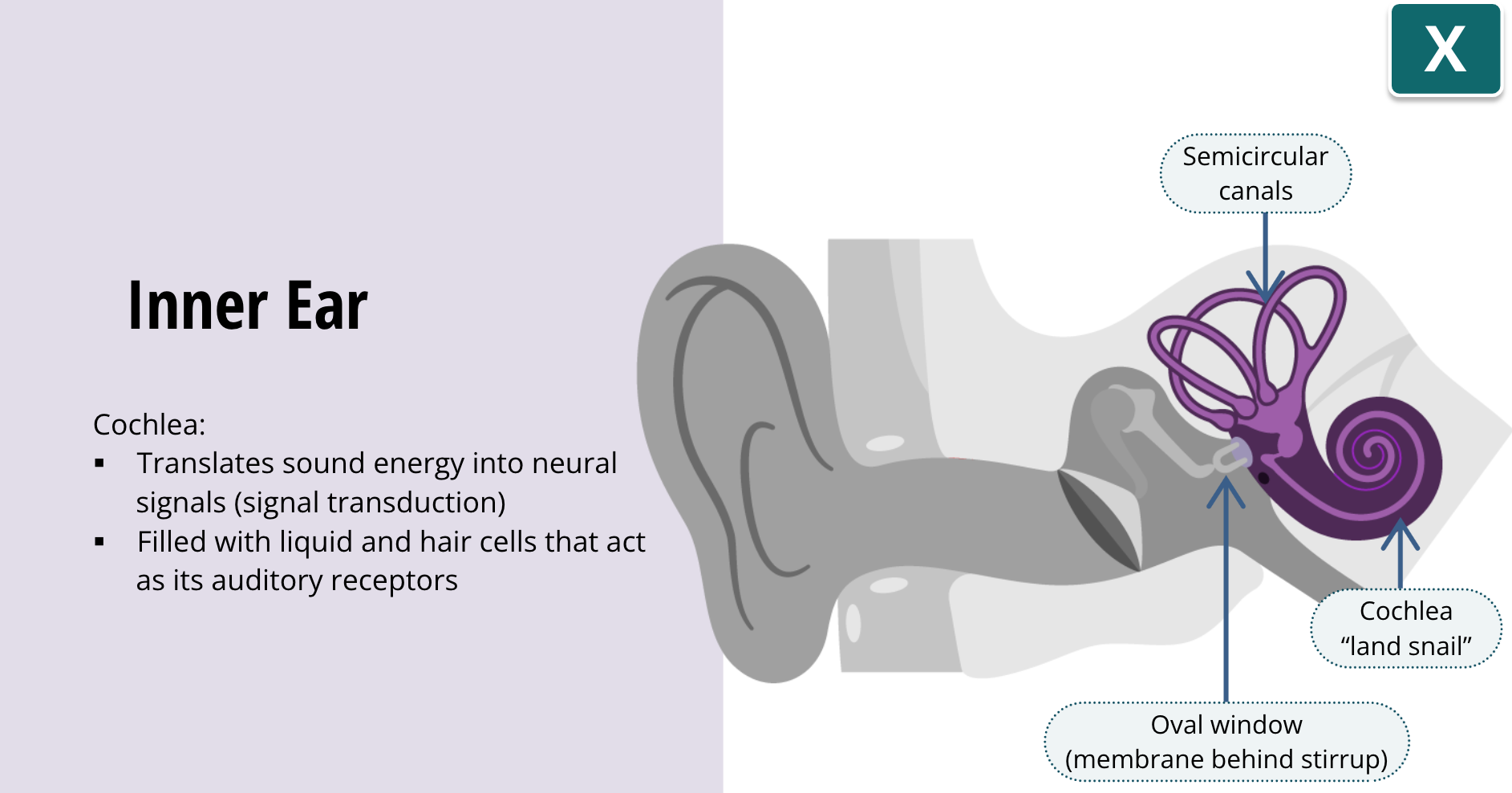
Translates sound energy into neural signals (signal transduction)
Filled with liquid and hair cells that act as its auditory receptors
Within the inner ear, we have the snail-shaped cochlea and the semicircular canals. You can ignore the semicircular canals, because they are important for balance and do not participate in hearing. Cochlea is Latin for “land snail” because of its shape, and it is where sound energy gets translated into neural signals. In other words, it is where signal transduction occurs. It is estimated that the cochlea has over a million moving parts. It is filled with liquid and hair cells that act as its auditory receptors. When the stirrup vibrates, it pushes against another membrane, called the oval window. The oval window in turn pushes against the cochlear fluid to transfer these vibrations into the cochlea.
Hair Cells
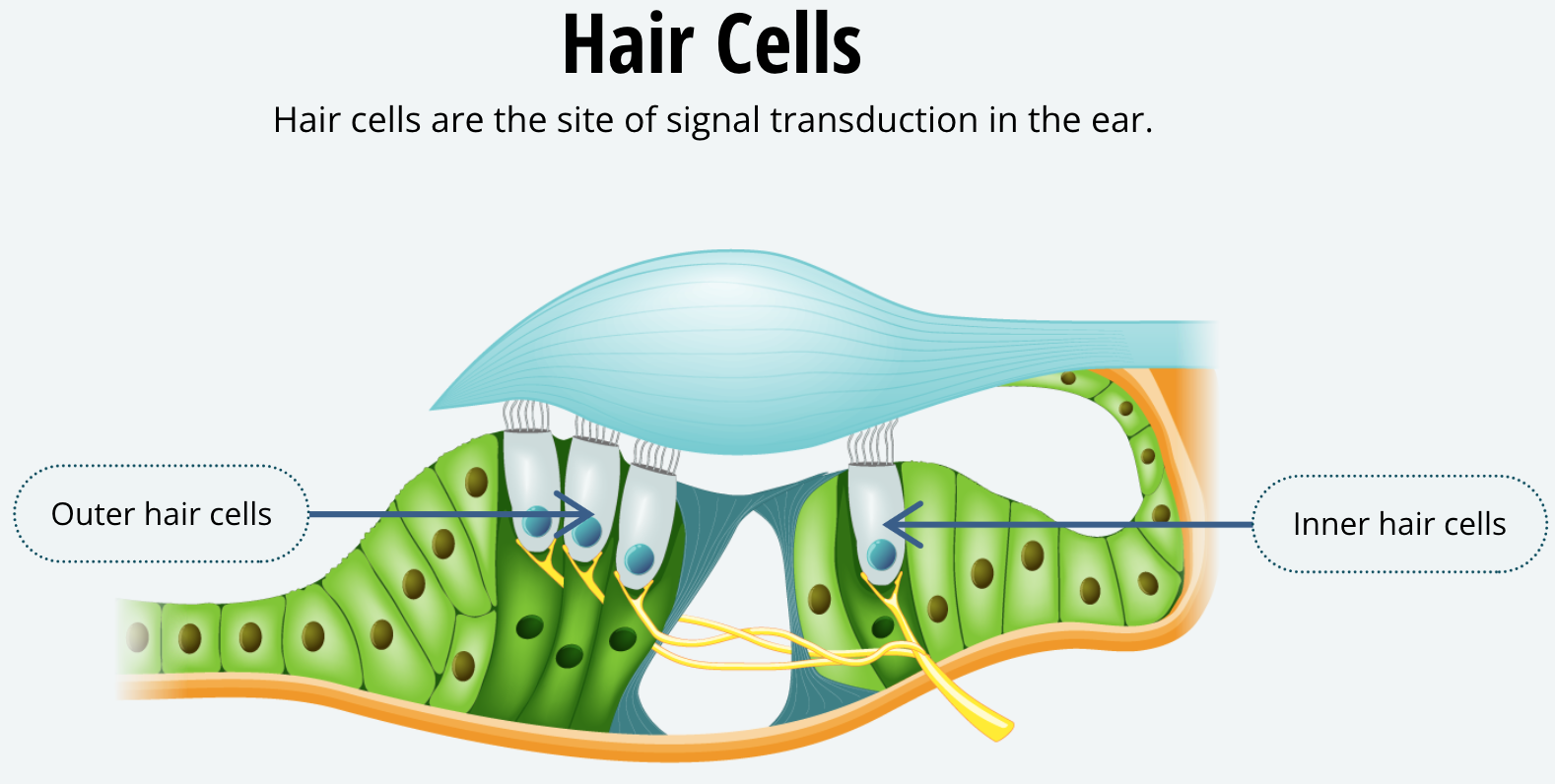
These vibrations within the cochlear fluid wash over the hair cells, and they convert those vibrations into neuronal signals. Basically, vibration of the cochlear fluid bends the hair cells. When the cells bend, potassium and calcium ion channels mechanically open. Thus, when they bend, they depolarize the auditory nerve that is attached to the auditory receptors. In this way, movement of the hair cells transduces sounds into action potentials. These cells are very sensitive; the amount of hair cell movement required to produce a sensation is equivalent to the Eiffel tour leaning just the width of your thumb.
Auditory Pathways to the Brain
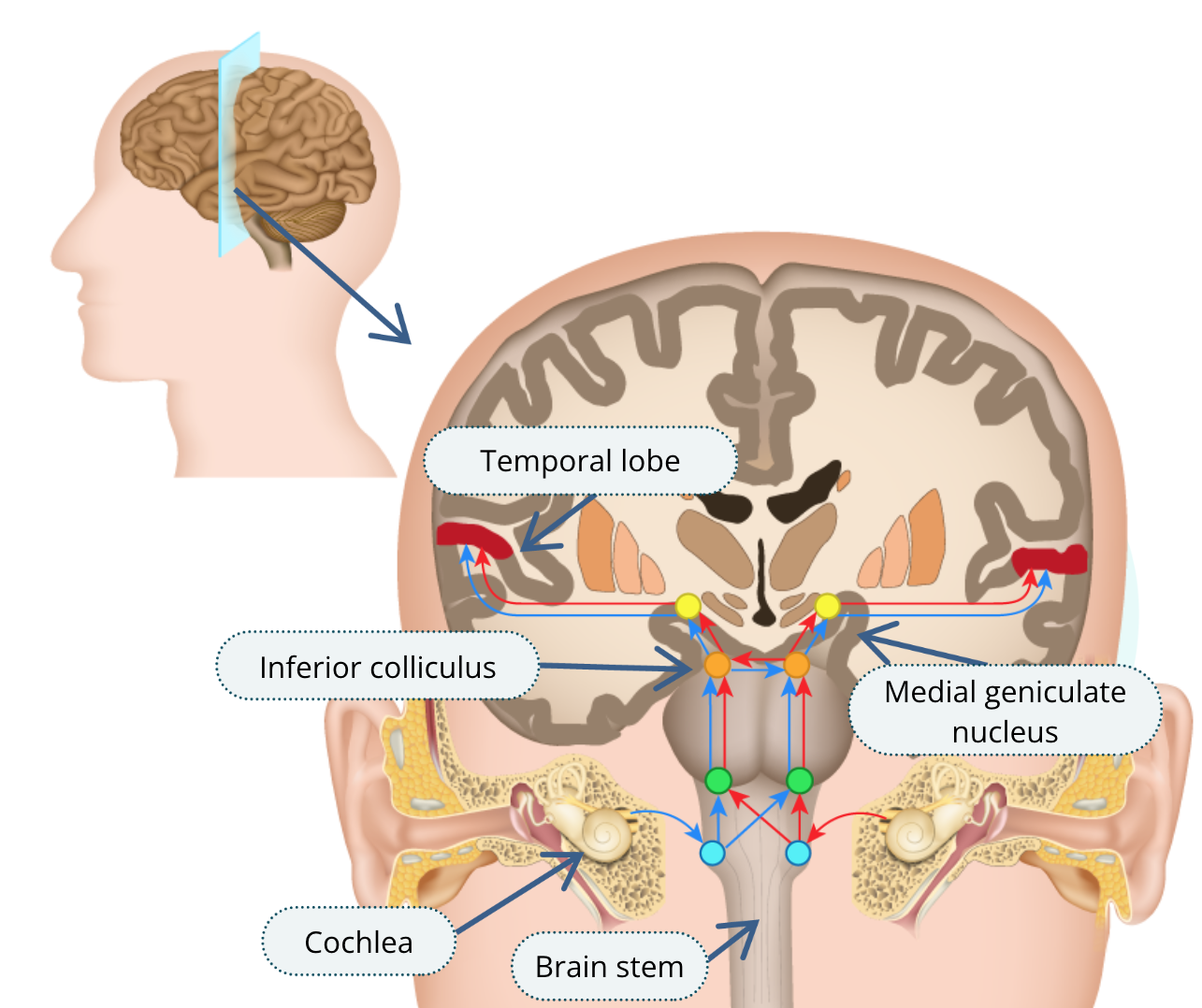
Now that the brain has sensed a sound and converted it to a neuronal signal, it is time to get on with the job of perception or making sense of that sound. The auditory nerves travel from both cochleae and they enter the brain through both sides of the brain stem. They synapse with other neurons in the inferior colliculus, and these neurons then connect to the medial geniculate nucleus of the thalamus, which in turn relays this information to each temporal lobe.
How the Brain Processes Sound
You may recall that the temporal lobe is the primary auditory processing area of the cortex. Neurons from each ear send information to both temporal lobes, but there are more connections with the lobe on the opposite side. This means that sounds heard in your left ear are predominantly, but not exclusively, processed in the right hemisphere. We have discovered that the left hemisphere is dominant for language in most people, and the right hemisphere is often better at other tasks, such as identifying melodies. The work of the auditory system is hardly finished once we have heard a sound. Beyond the primary auditory cortex, there are several secondary auditory areas involved in processing complex sounds and understanding their meaning.
In humans, the auditory information also travels well beyond the auditory areas. For example, the brain combines information from other senses to locate where a sound is coming from. Another area processes specific information about the content of the sounds. For example, monkeys have a very specific area of the cortex where neurons will fire only when the sound is a call of their own species. We call these the “where” and the “what” streams of auditory processing.
Vision
The Complexities of Vision
There are about 126 million light receptors in the human eye
The optic nerve contains 1 million neurons
The eye information transmission rate is about 100 million bits per second
Vision is a slightly more complex system in the brain, compared to hearing. For example, there are about 126 million light receptors in the human eye, and the optic nerve contains a million neurons. The eye transmits information to the brain at the same rate as an Ethernet connection, about 100 million bits per second. What the brain does with this information is equally impressive.
Anatomy of the Eye
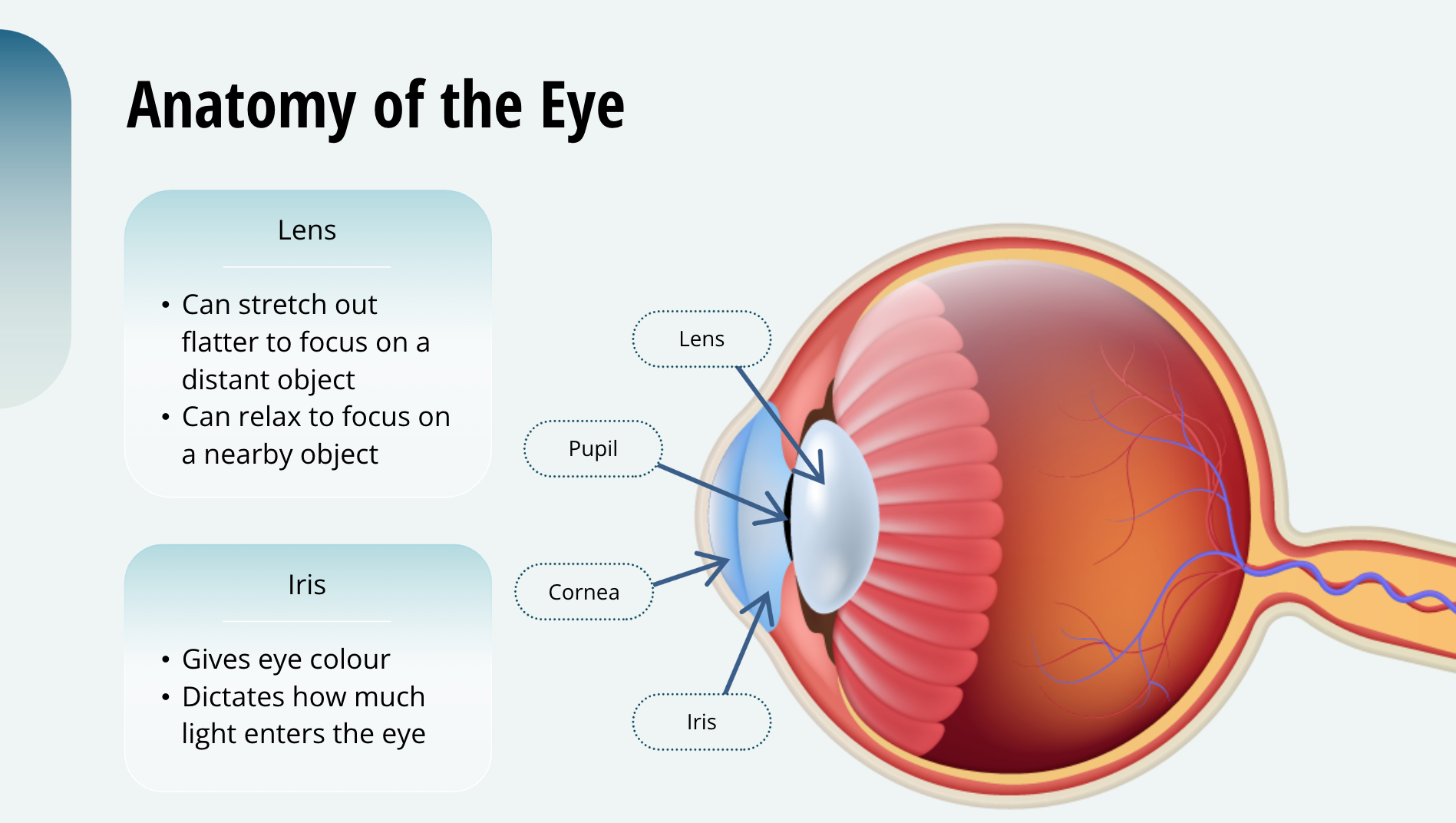
You may be already familiar with the anatomy of the eye. The transparent part of the outer covering is called the cornea, and behind the cornea is the lens. The lens is flexible, and the muscles that attach to it can stretch it out flatter to focus on the image of a distant object; or the muscles can relax to make the lens thicker to focus on a nearby object. The lens is partly covered by the iris, which is what gives your eye colour. The iris is, in fact, a circular muscle whose opening forms the pupil. The iris dictates how much light enters your eye by contracting in bright light to make the pupil smaller, and relaxing in dim light, letting the pupil dilate to allow more light in.
Signal Transduction in the Eye
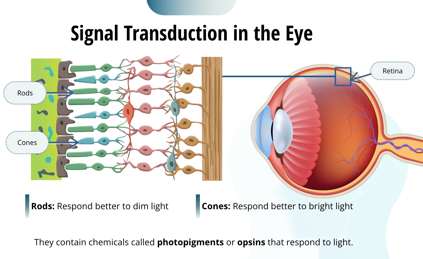
The retina is the light-sensitive structure at the back of the eye. It contains two types of light sensitive photoreceptors, called rods and cones, and the neurons that are connected to them. They are responsible for signal transduction, converting light into neural impulses. Rods respond better to dim light, and the cones respond better to bright light. In fact, when you are outside on a sunny day, most of your rods are overstimulated and unable to function. When you are in dim light, such as a darkened theatre, your rods are mostly active, and your cones are under-stimulated. The reason why it takes time for your eyes to adjust to the dark, when entering a dim room from the bright outside, is because your overstimulated rods need time to recover and become operational again. How the rods and cones convert light into neural impulses is too advanced for this course. However, you should know that the rods and cones contain chemicals called photopigments or opsins that respond to light. When exposed to light, they set off a number of reactions that lead to the depolarization or hyperpolarization of the neurons connected to the rods and cones.
Colour Blindness
Complete colour blindness: Very few people have an inherited lack of cones, relying on rods for vision
Partial colour blindness: Most people who experience colour blindness have a defect in one cone system
Colour blindness is an interesting condition that has played an important role in our understanding of colour perception. There are very few people with complete colour blindness (about one in every 100,000). They usually have inherited a lack of cones, so they only rely on rods for vision. However, most who experience colour blindness have partial colour blindness. In those cases, they are more likely have a defect in one of their cone systems rather than a complete lack of cones. In this figure, do you see the number 21 or 74? If you see 21, you may have a colour deficiency. If you see 74, you, like most people, have normal colour vision.
Anatomy of the Blind Spot
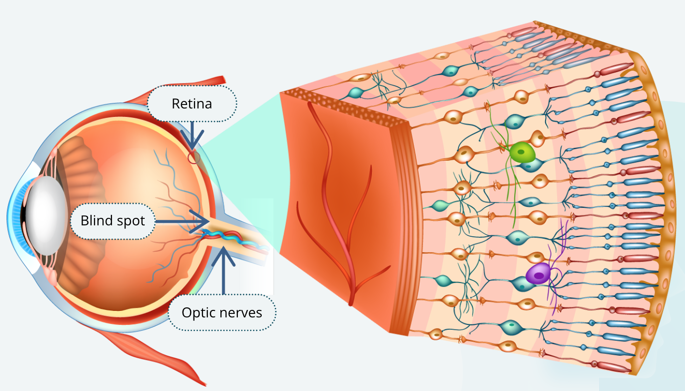
The ganglion cells of the retina join together and exit each eye to form two optic nerves. The portal at which they exit is full of axons and has no room for photoreceptors. Thus, this part of the retina cannot process visual information. The point of exit of the nerve is your blind spot. You normally do not notice your blind spot, because your brain fills in the missing visual information for you.
Getting Visual Information to the Brain
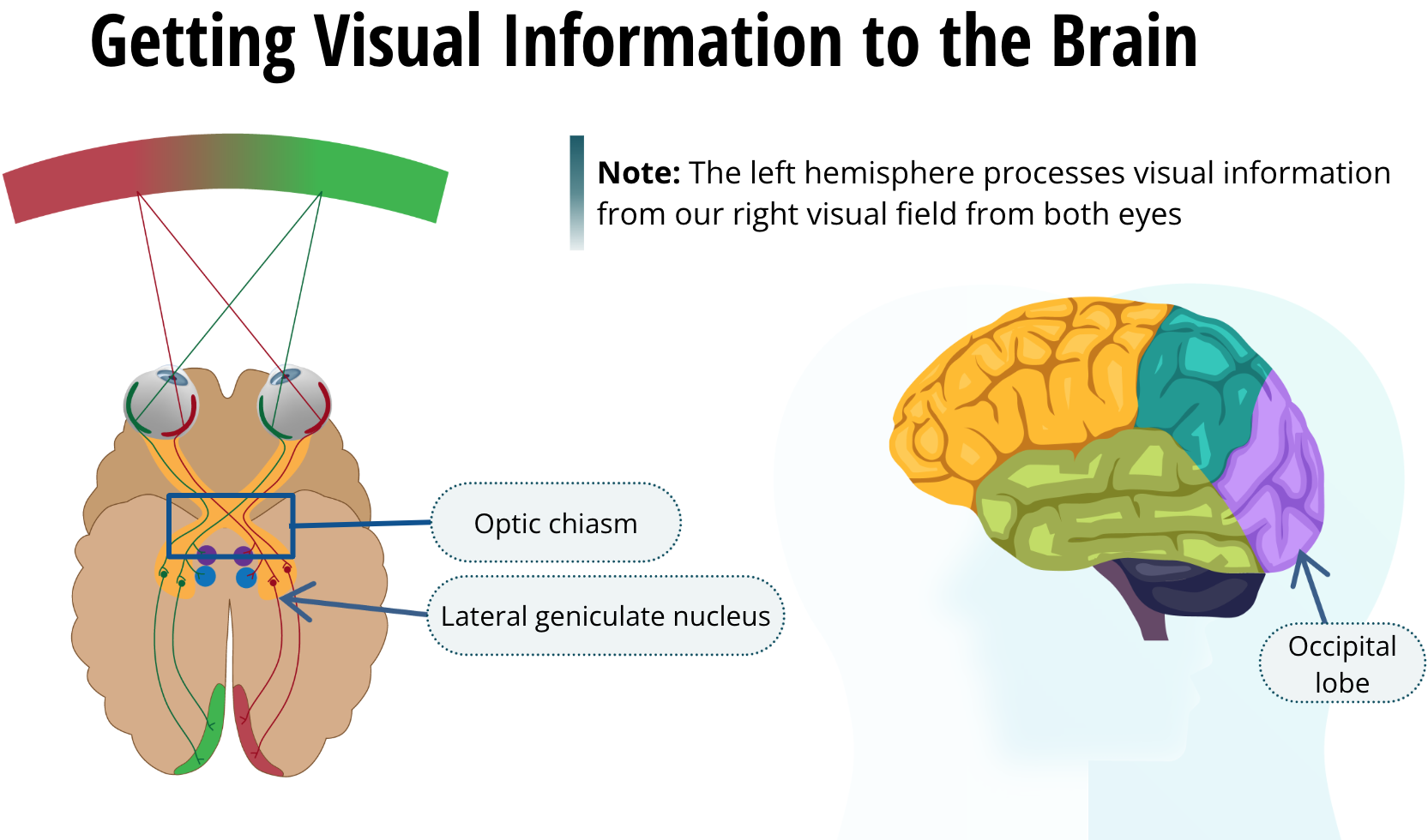
The two optic nerves converge at a point called the optic chiasm before separating again and travelling to their first synapse in the lateral geniculate nucleus of the thalamus. At the optic chiasm, axons from the nasal or medial side of the retina cross over to the occipital lobe in the opposite hemisphere. Axons from the outer or lateral side of the retina do not cross over, but project to the occipital lobe within the same hemisphere. Because of this arrangement, everything we see in our right visual field is processed by the left hemisphere and vice versa. It is important to note that it would be incorrect to think that our left hemisphere processes visual information from our right eye. It processes visual information from our right visual field from both eyes.
How Visual Information is Sent to the Occipital Lobe on the Opposite Side
Theories of Vision
During the past century, neuroscientists have made many strides in understanding how our brain processes visual information. Indeed, there are many advanced courses on this topic alone. For example, we have made strides in understanding colour vision. And there have been many competing theories on how the brain processes complex visual information.
How the Brain Processes Visual Information
Parvocellular system → small ganglion neurons → discrimination of fine details and colour |
Magnocellular system → big ganglion neurons → brightness, contrast, and movement |
The visual system has two pathways for visual analysis. Most visual information follows two routes from the retina through the brain. These are called the parvocellular system and the magnocellular system. These words come from the Latin words parvus and magnus, meaning small and large respectively. Thus, it should not be surprising that the parvocellular ganglion cells are smaller than magnocellular neurons. The parvocellular neurons are specialized for the discrimination of fine detail and colour. On the other hand, the magnocellular ganglion neurons are specialized in brightness, contrast, and movement.
Parvocellular vs. Magnocellular Processing
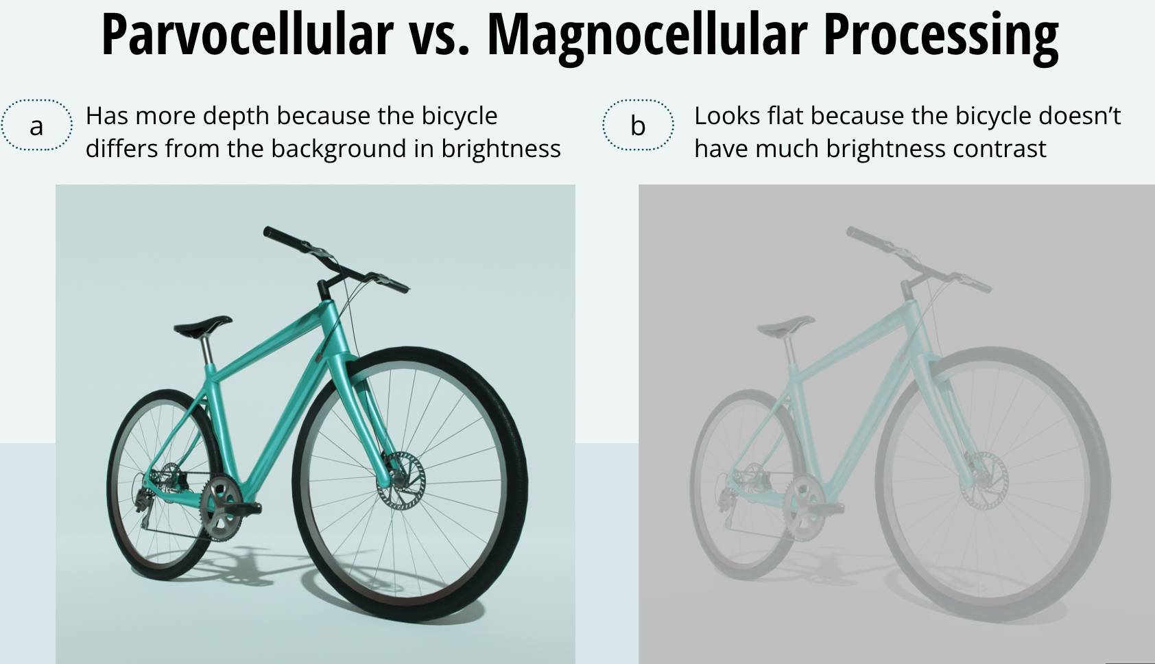
We see evidence of these differences every day. Notice how the bicycle in frame (a) has considerably more depth than the bicycle in (b). This is because the bicycle differs from the background in brightness, so the image stimulates your magnocellular system maximally. The bicycle in frame (b) looks flat. It has colour contrast, but not much brightness contrast. Thus, it provides little stimulation to the magnocellular system.
The What vs. Where Pathways
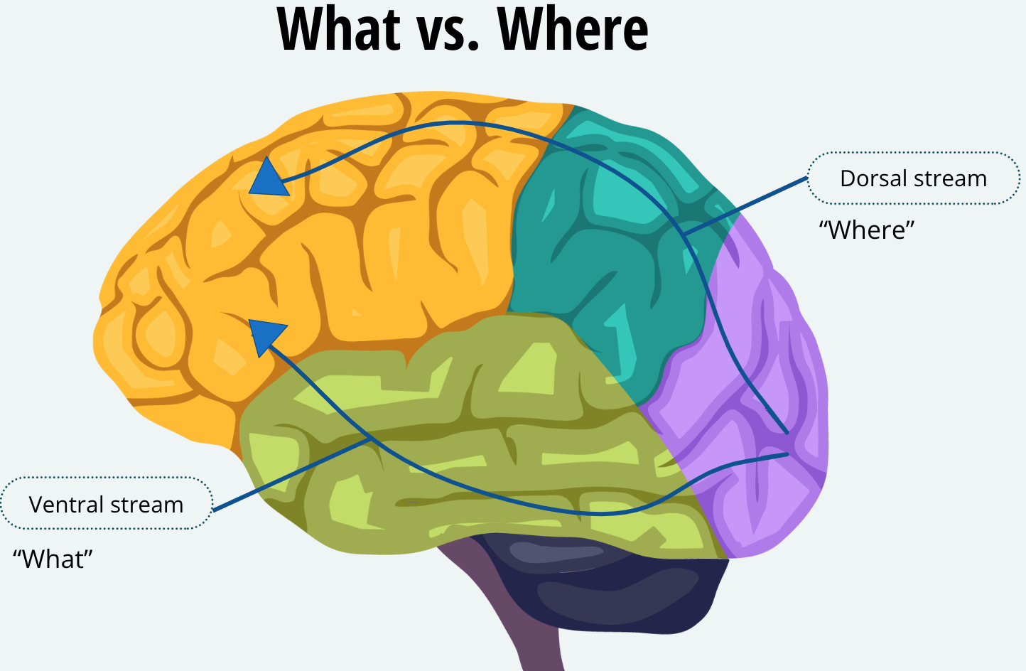
Both pathways travel to the lateral geniculate nucleus, and then to the primary visual cortex in the occipital lobes. Although these two systems are highly interconnected, the parvocellular system dominates the ventral stream, which connects the visual cortex to the temporal lobe. The magnocellular system dominates the dorsal stream from the visual cortex to the parietal lobes. Like the two auditory pathways we learned about in the previous lesson, the ventral stream is often referred to as the “what” of visual processing and the dorsal stream is referred to as the “where.”
Taste
The Chemical Senses
Olfaction - Our sense of smell
Gustation - Our sense of taste
The senses of taste and smell are also referred to as our chemical senses. They are able to detect some of the many chemicals that bombard us daily. They are older senses in that they connect differently and involve different parts of the brain involved in emotion. When we talk about smell, neuroscientists refer to this sense as olfaction, and the sense of taste is called gustation.
Detecting the Chemicals in our World
Early in evolution, organisms lived in the sea and had to detect chemical substances that signalled food, poison, or even sex. Considering it in this manner, things have not changed much in three billion years. Animals, including humans, have to depend on their chemical senses to identify nourishment, such as the tanginess of a hot and sour soup or the smell of mom’s fresh apple pie. We also have to identify noxious substances, such as the bitterness of a poisonous plant.
Basic Tastes
Salty
Sweet
Sour
Bitter
Umami
Although the number of different chemicals we can detect is countless, and the number of flavours we can sense is also unimaginable, research so far suggests that we can detect just five basic tastes. And we use these in combination to sense all the other flavours that we do. The basic tastes include the four widely known ones, which are saltiness, sourness, sweetness, and bitterness. Perhaps less familiar is the fifth taste quality known as umami. Umami is a Japanese word meaning “delicious” and is defined by the savoury taste of glutamate, found in the food additive MSG, or monosodium glutamate. You may not be able to easily think of umami as one of your favourite flavours, but its taste is sensed from certain amino acids, which make up proteins. We need proteins for nutrition, so once again Mother Nature wanted to make sure we enjoyed the taste. So, if you are a lover of a juicy BBQ steak, you can thank your basic umami taste.
How do we Sense Different Flavours?
The correspondence between taste and the chemistry of the molecule can be obvious. For example, most salts taste salty. But many of the substances we are able to taste can vary widely in their chemistry, and yet taste similar. For example, while sugars, such as sucrose or fructose, taste sweet, so do artificial sweeteners like aspartame, which is very different chemically. This leaves us with the question: how do we sense the immeasurable number of flavours that we do?
Flavour Discrimination
Basic Tastes — Each food acts on a combination of these tastes to produce its unique flavour
Olfaction — Important for informing our sense of taste
Other Sensory Modalities — Temperature, texture, and pain contribute to the unique flavour of food
The answer to this question is complex and there are many reasons, including how the brain perceives taste. In a nutshell, there are three principles that contribute to a food’s flavour. First, each food acts on a combination of the five basic tastes in a unique way to produce its unique flavour. Second, the sense of smell, or olfaction, is critically important to how we taste food. An experiment you can try is have a friend close his eyes and plug their nose, and you put a drop of different flavours on his tongue with a straw (such as vanilla extract or almond extract) and ask them to tell you what the flavour they’re tasting. In fact, they won’t be able to do that if his nose is plugged, but they will if they unplug their nose. The third reason we can taste all the flavours we do is that other sensory modalities, such as temperature, texture, and even pain, contribute to the unique flavour of food. For example, beer only tastes good when it is cold, apples only taste good when they are crisp, and hot and sour soup is yummy because the key ingredient in hot peppers, capsaicin, activates our pain receptors.
Anatomy of the Tongue
Papillae (“bumps”):
Each papilla has from 1 to several thousand taste buds
Each taste bud has 50 to 150 taste receptor cells
People typically have 2000 to 5000 taste buds on their tongue
Most papillae are sensitive to more than one taste and distributed and distributed throughout the tongue
If you look in the mirror and shine a flashlight on your tongue, you will see small, rounded bumps on the front and sides, and larger ones in the back. These are called papillae, which is Latin for “bumps.” Each papilla has from one to several thousand taste buds. And, in turn, each taste bud has anywhere from 50 to 150 taste receptor cells. People typically have about 2,000 to 5,000 taste buds on their tongue. Previously, people mistakenly thought that certain areas of the tongue have specific taste buds for each basic taste. For example, bitterness in the back and sweetness at the tip of the tongue. This is incorrect. Most papillae are sensitive to more than one taste, and those are distributed throughout the tongue.
Signal Transduction
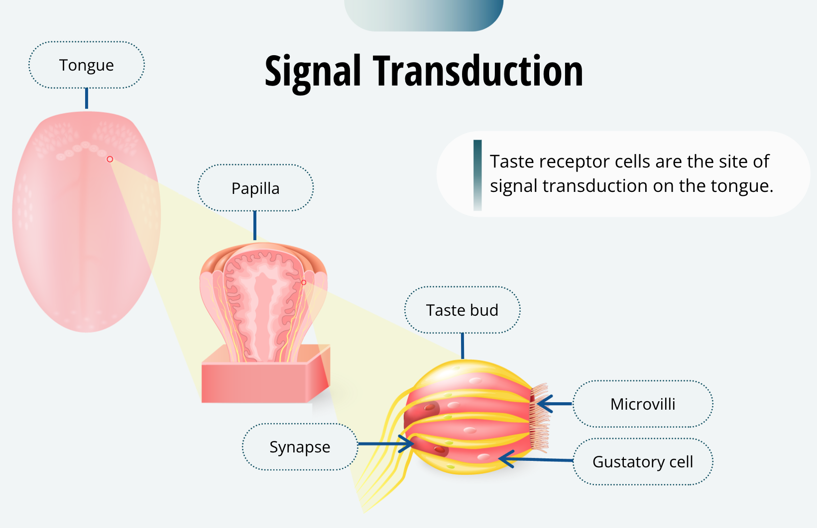
Taste receptor cells are the site of signal transduction on the tongue. These, like other sensory receptors, are not neurons per se, but they can generate an action potential in the neurons that synapse with them. They have microvilli that are exposed to the contents of the mouth, and form synapses with the gustatory neurons near the bottom of the taste bud. When the taste receptor cell is activated by the appropriate chemical, its membrane potential will change, causing it to produce an action potential in the gustatory neuron.
Taste Buds Regenerate
Before we move on from the tongue, there is one other thing worthy of note. The cells of the taste bud are constantly in a cycle of growth, death, and regeneration. Each taste bud on your tongue lasts for only about a week or two. That is why, if you burn your tongue on hot coffee for example, it takes a few days to get your sense of taste fully back. That is because there are new taste buds being generated to replace the ones you damaged with hot liquid. So, if taste receptors are not that specific to any one kind of taste, how are we able to detect so many different flavours? Another answer to this question lies within the brain.
How the Brain Processes Taste
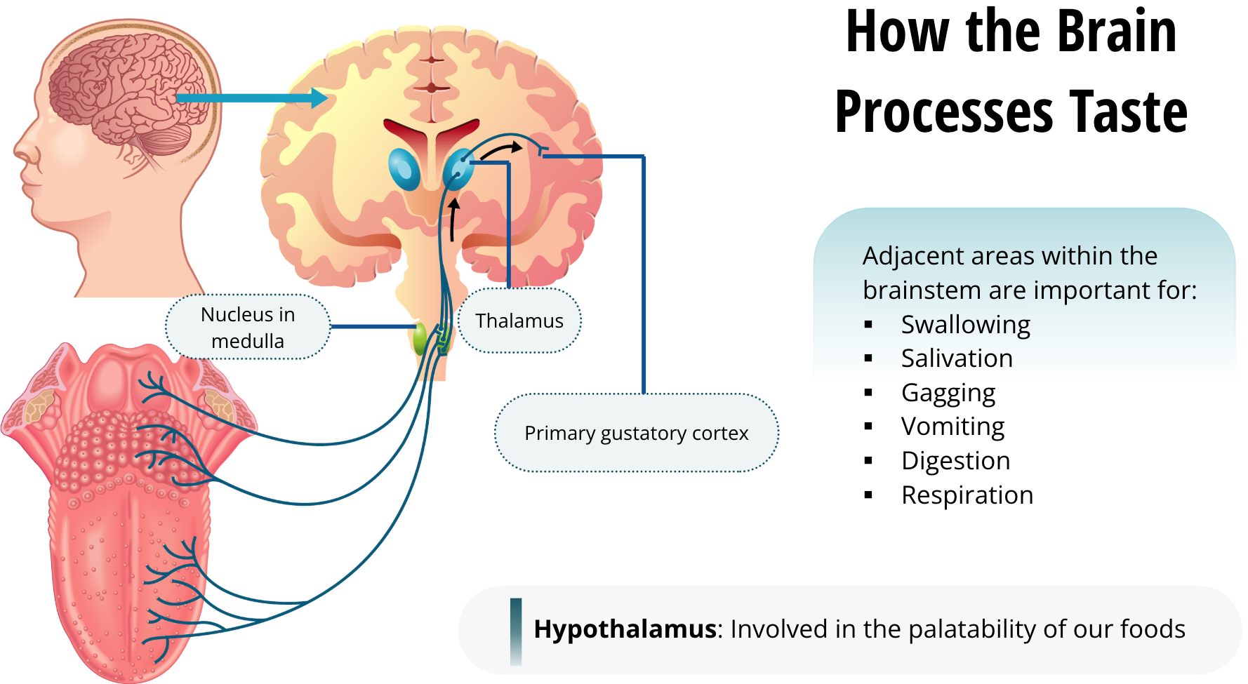
The tongue sends information to the brain via the gustatory neurons. They first synapse onto neurons in the medulla within the brain stem in an area called the gustatory nucleus. Like all other sensory information, this pathway then projects to the thalamus and ultimately to the cortex, specifically to the primary gustatory cortex, which is located in the parietal lobe just above the lateral fissure. This is presumed to be where the conscious experience of taste is processed. The gustatory nucleus also sends projections to other adjacent areas within the brainstem. These include areas important for swallowing, salivation, gagging, vomiting, and even digestion and respiration.
Gustatory information is also sent to the hypothalamus and this pathway is thought to be involved in the palatability of our foods and the motivation to eat. So here is something to think about: the taste of your food is conveying a lot of information to your brain that you are not even usually consciously aware of.
Smell
The Good and The Bad
As we learned in the previous lesson, the sense of smell, or olfaction, is one of the chemical senses. Olfaction brings us both good and bad. It combines with gustation to help us identify food flavours, but it also alerts us to potentially harmful substances, such as rotten food. With regard to our sense of smell, the bad news outweighs the good. It is estimated that our olfactory system can detect several thousand substances, but only about 20% of those would be considered pleasant smelling. Mother Nature designed us to be more perceptive of things that smell bad and that are potentially harmful to eat or otherwise encounter.
Anatomy of the Sinuses
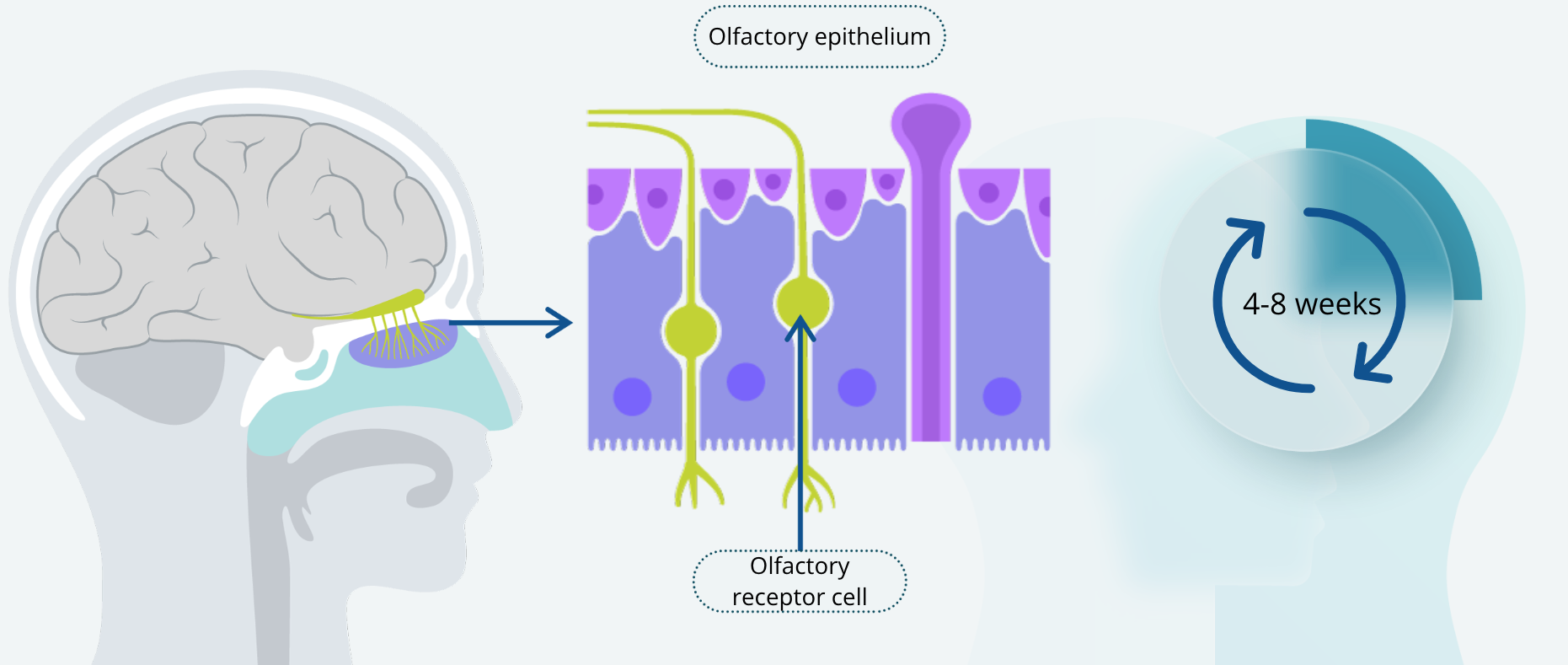
Our nose is actually just a vent through which we take air into our sinuses; it does not smell per se. We detect scents using a thin layer of cells in our sinus cavities called the olfactory epithelium. Signal transduction is carried out at this location by olfactory receptor cells. Unlike taste receptor cells, olfactory receptor cells are actually neurons that send axons into the central nervous system. Like taste receptor cells, olfactory receptor cells are constantly being regenerated, with a life cycle of about four to eight weeks. This makes them unique among all other types of neurons. Most neurons are never replaced — once we lose them, they are gone — yet olfactory receptor cells are constantly being replaced.
The Size of the Olfactory Epithelium
Broadly speaking, the size of the olfactory epithelium corresponds to how well we can detect different smells or odorants — which is a term used to describe chemical stimuli in the air. For example, we are relatively poor at detecting odorants whereas dogs are excellent sniffers. The size of the human olfactory epithelium is about 10 cm2, where the dog’s is about 170 cm2. In addition, dogs have about 100 times more receptors per cm2 than humans
Signal Transduction of Smell
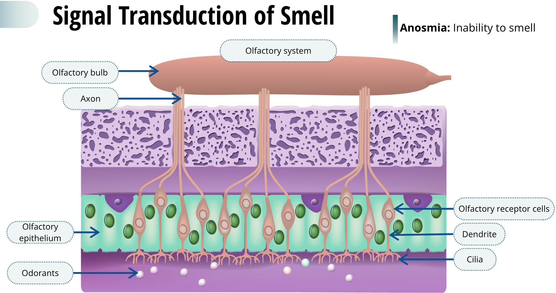
When we sniff, we bring air into the nasal passage, but only a very small percentage of that air passes over the olfactory epithelium. Odorants in the air dissolve in a layer of mucus on the olfactory epithelium, prior to activating the olfactory receptor cells. This mucus also contains many other substances, including antibodies, enzymes, and odorant-binding proteins. The binding proteins are thought to help concentrate the odorants in the mucus, so they can be better detected. The antibodies are important to help defend us from viruses and bacteria that may be air-born and infect us via the olfactory cells. Olfactory receptor cells have a single thin dendrite with a knob at the end that extends out several long cilia — which are tiny, hair-like projections from a cell. Odorants within the mucus bind to the cilia and cause an action potential in the olfactory receptor cell. These cells send axons to a pair of olfactory bulbs, which are located inside the skull just above the sinuses. The olfactory axons that cross through the skull to the olfactory bulbs are very fragile. Sometimes, a blow to the head can permanently sever these axons. The resulting condition, called anosmia, leaves the patient without the ability to smell.
Odorant Receptor Proteins
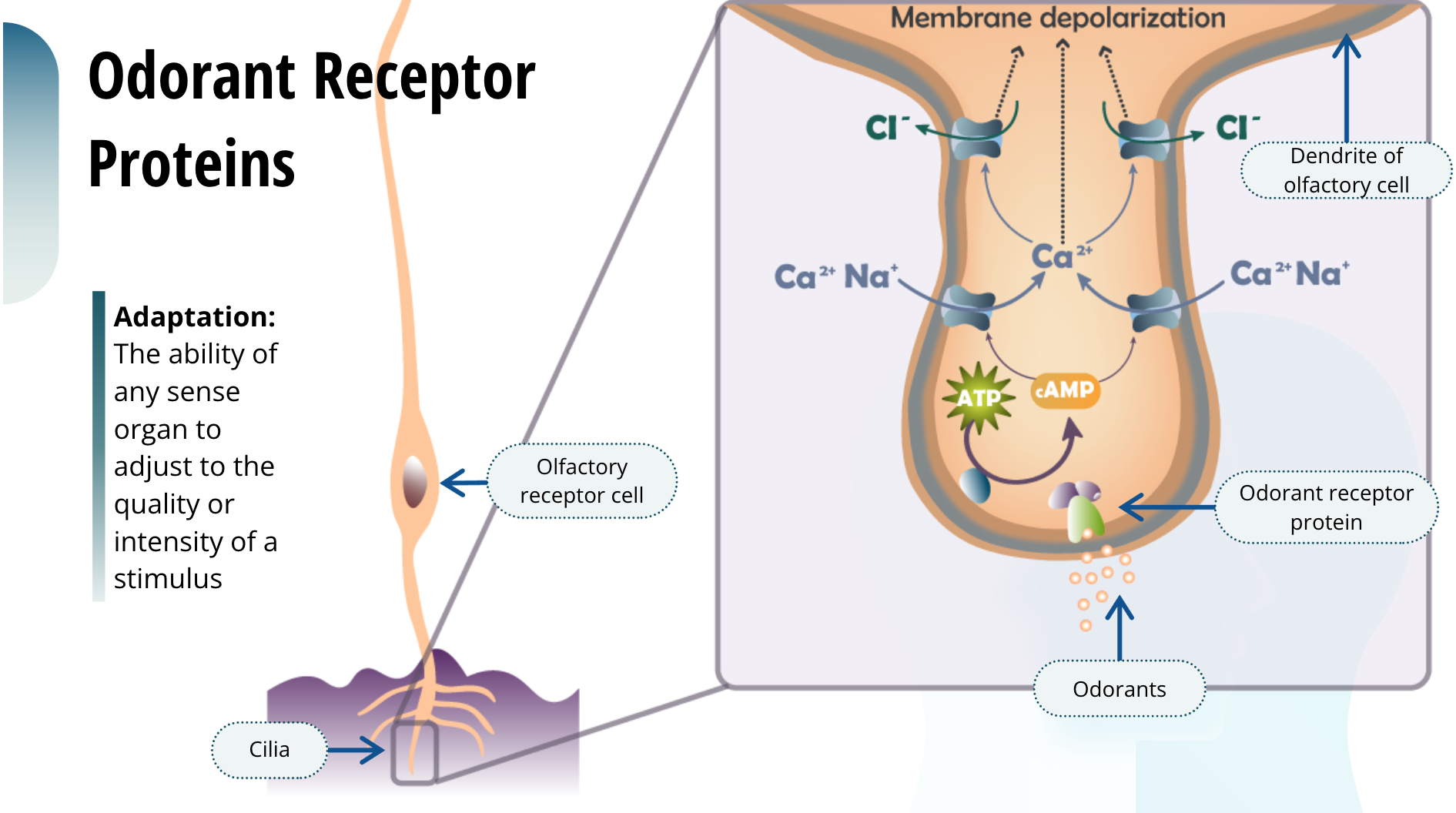
The basic mechanism of signal transduction for olfaction lies with the odorant receptor proteins that are embedded in the surface of the cilia. These proteins selectively bind with odorants and cause a depolarization of the olfactory receptor cell. As you might think, we have many different types of odorant receptor proteins that are specific to each type of odorant. In fact, over 1,000 different genes have been discovered so far in the rodent that encode for different odorant receptor proteins. Have you ever noticed that when you visit that one aunt who has four cats, you immediately smell the litter box when you enter her home, but once you have been there a while you stop smelling it? “Aunt Cat Lady” claims to never be able to smell it. All of our senses are capable of undergoing some form of adaptation, which is the ability of any sense organ to adjust to the quality or intensity of a stimulus. But the olfactory system does this much more quickly than our other senses. The reason behind this is that even in the continued presence of an odorant, the olfactory receptor cell itself stops being responsive after about a minute. This is also the reason why when you keep sniffing a pleasant smell, after a short period you can no longer smell it.
Getting Scent Information to the Brain
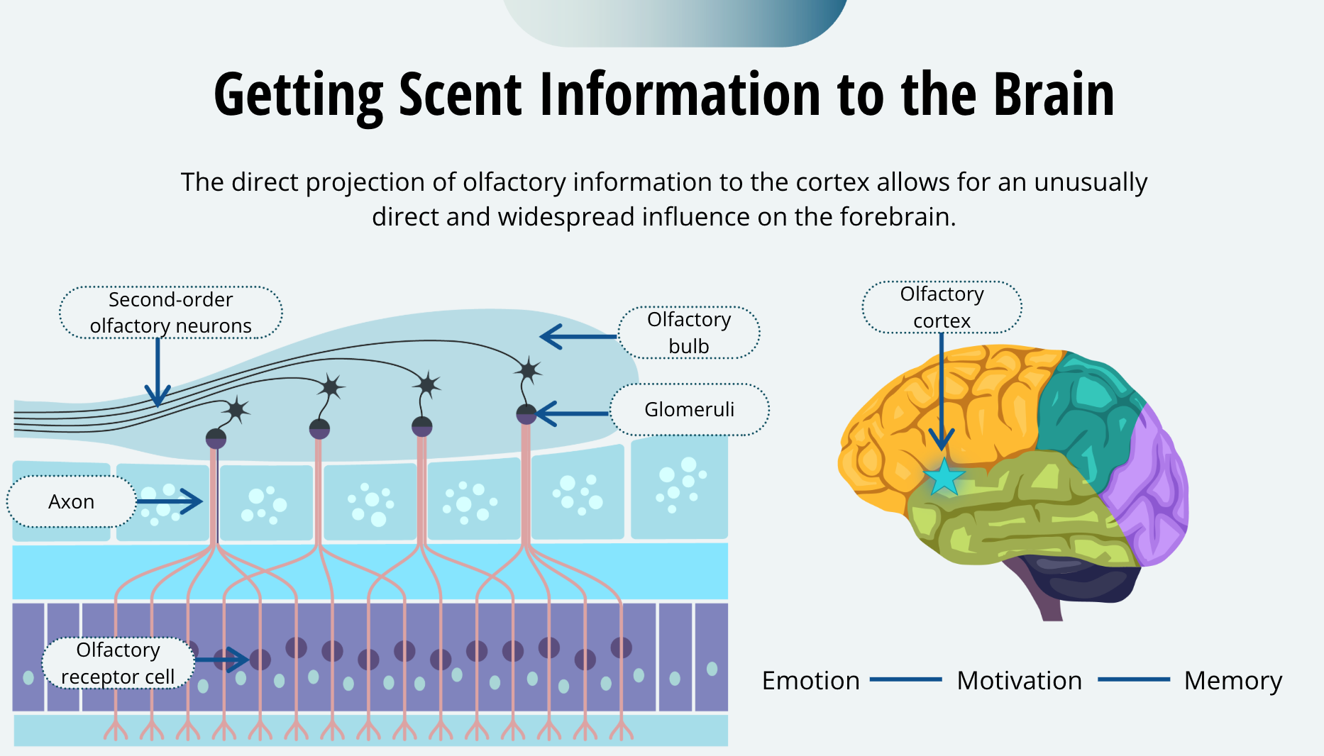
The olfactory bulbs contain spherical structures called glomeruli. Within the glomeruli, the axon terminals of the olfactory receptor cells synapse with the dendrites of second-order olfactory neurons. The second-order olfactory neurons, in turn, project to several areas in the brain. One of the more important targets of olfactory information is the olfactory cortex, which is located in the temporal lobe, tucked inside, just below the lateral fissure. This anatomical organization makes olfaction unique among the senses. All other senses first relay information through the thalamus prior to projecting to the cortex. This direct projection of olfactory information to the cortex allows for an unusually direct and widespread influence on the forebrain. This is probably why smells are so important in other domains of behaviour, such as emotion, motivation, and even memory.
Smell and Memory
Have you ever noticed how sometimes you encounter a very specific smell, and it evokes a very strong memory? For example, the smell of new pencils may remind you of your first day of school, or that distinct smell of mom’s apple pie takes you back to happy family celebrations. Olfaction is important for a widespread number of brain functions, and smells are an integral part of how our brain perceives our environment.
Pheremones
Vomeronasal organ: An auxiliary olfactory sense organ in the sinus cavity that detects pheremones
Pheremones: Chemicals released by the body which are important signals of reproductive behaviours, marking territory and indicating aggression or submission, and play a critical role in animal communication
Within the sinus cavity, at least for many animals, is an auxiliary olfactory sense organ called the vomeronasal organ. It is able to detect a different class of chemical signals called pheromones. Pheromones are chemicals that are released by the body and are important signals for reproductive behaviours, marking territory, and indicating aggression or submission. We have no doubt that pheromones play a critical role in communication between some animals. You may have often been awakened in the night by the screeching and howling of neighbourhood cats. When a female cat is about to become fertile, or is in heat, it releases pheromones to notify the male cats that it will soon be receptive to mating. The ensuing cacophony of every male cat in the area shows us that this is an effective means for cat reproduction.
Pheremones in Humans
Humans only have a vestigial vomeronasal organ in childhood
Female roommates may co-cycle with their menstrual cycle
The story of humans and pheromones is another matter and is quite controversial. On one hand, it is thought that humans only have a vestigial vomeronasal organ, which disappears after childhood. On the other hand, there is some evidence that female roommates may, over time, co-cycle with their menstrual cycles because of pheromones. Perhaps there are other ways our brain can sense the chemical world around us that we just have not discover yet.
Touch
Senses of the Skin
Touch
Cold
Warm
Pain
Itch
Position and movement
The body has several skin senses, including touch. Traditionally, the skin senses were thought to be touch, cold, warm, and pain. However, there is recent evidence that there may be a specific sense for itch, it was thought to be a variant of pain. Regardless of how many there actually are, it is important to know that each has distinctive receptors and a separate pathway to the brain. These, along with systems that inform us about the position and movement of our limbs, are collectively called the somatosensory system.
Skin Receptors
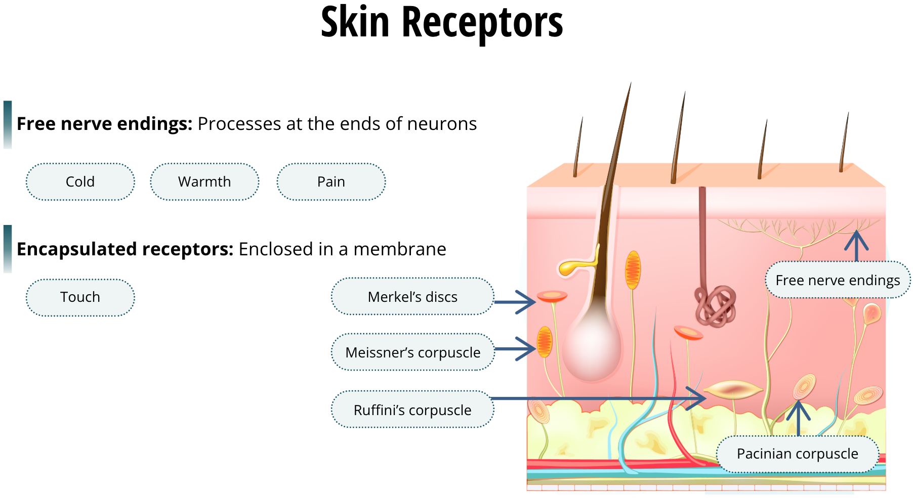
There are two general types of receptors in your skin. Free nerve endings are simply processes at the ends of neurons. Free nerve endings detect cold, warm, and pain. All the other receptors are encapsulated receptors, which are more complex structures. Encapsulated receptors are enclosed in a membrane, and they are involved in the signal transduction of detecting touch. When we use lidocaine as a local anesthetic, it blocks the voltage-gated sodium channels in the pain-sensing neurons. You should recall that if the sodium channel cannot open, the neuron will not be able to depolarize. This is pretty much how local anesthetics work. Because touch is a complex sense that conveys several types of information, it requires many different types of encapsulated receptors. In the superficial layers of the skin, we have the Meissner’s corpuscles, which exhibit a brief burst of impulses, while Merkel’s discs exhibit a more sustained neuronal response. Because they are both located at the surface of the skin, they can detect the texture and fine detail of objects. In fact, it is these two types of receptors that are important in the reading of Braille. Alternatively, Pacinian corpuscles and Ruffini endings are located in the deeper layer of the skin. They detect stretching of the skin and contribute to our sensation of the shape of a grasped object. Because the density of skin receptors varies throughout the body, so does sensitivity. The skin receptors are densest in the fingertips and lips, and those areas are the most sensitive to touch.
The Dermatomes
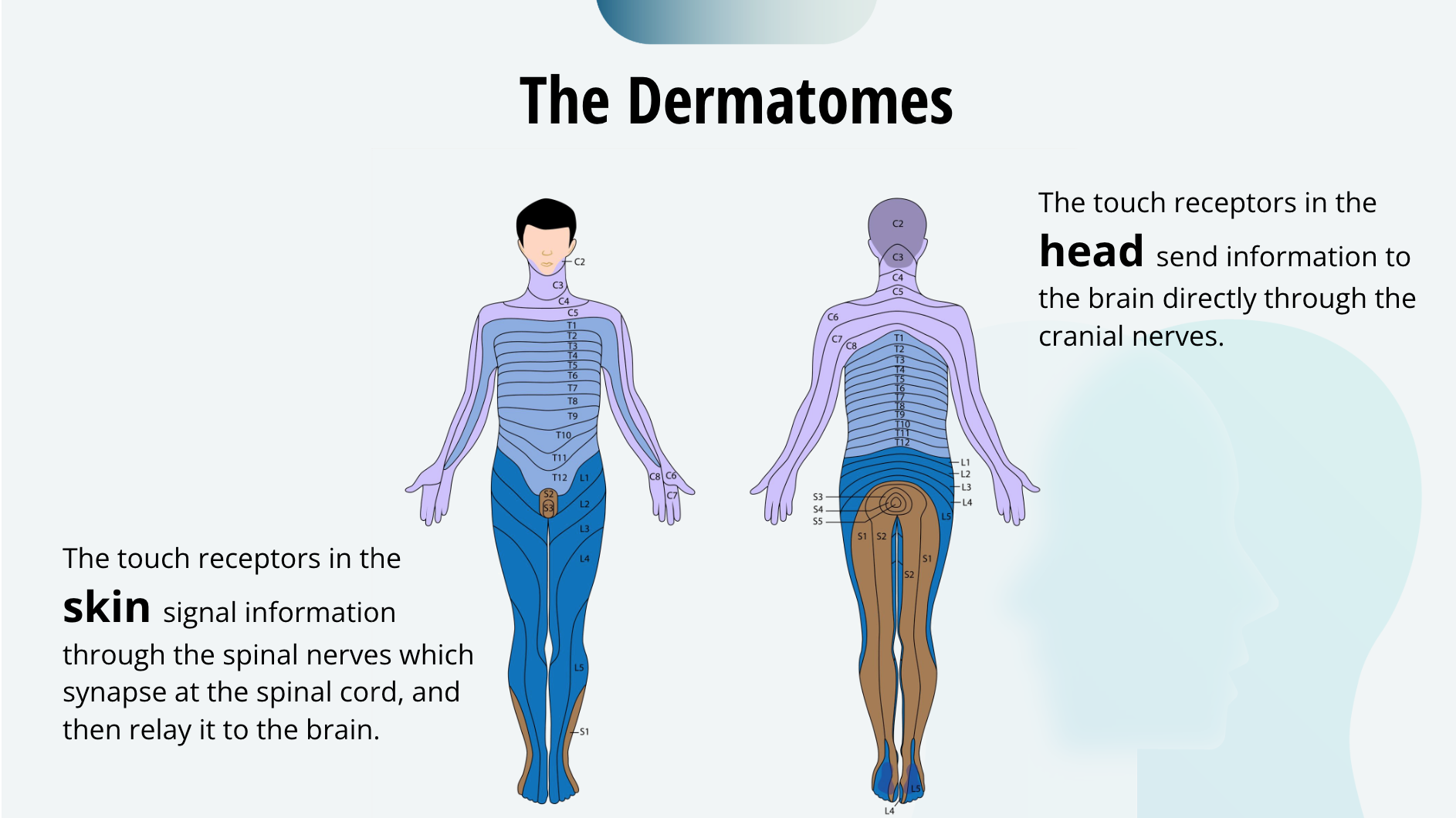
The body is divided into segments called dermatomes, each of which has its own spinal nerve. This figure makes them look more distinct than they are, as there is considerable overlap between them. The touch receptors in the skin signal information through the spinal nerves which synapse at the spinal cord, and then relay it to the brain. Or in the case of your head, they send information to the brain directly through the cranial nerves. Touch senses in your head do not pass through the spinal cord.
The Somatosensory Cortex
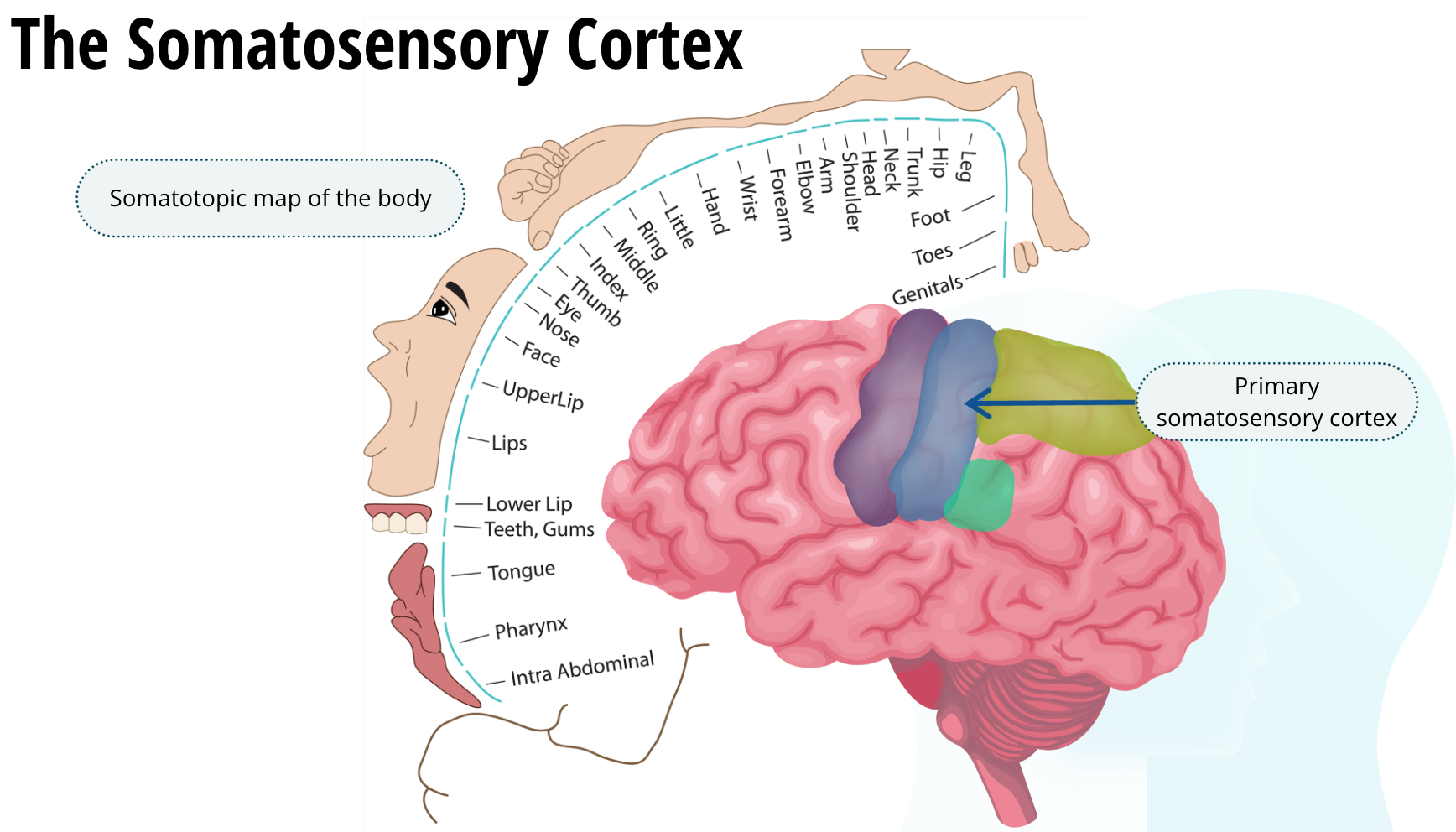
From there, the information about touch goes to the somatosensory cortex. Like we saw with the auditory and visual systems, most of the information crosses over to the other hemisphere. So, the touch of an object in your right hand is processed by the left side of your brain. As we discussed, Dr. Penfield first mapped out the somatosensory cortex. Each adjacent part of the body is represented in the adjacent parts of the somatosensory cortex. We call this a somatotopic map of the body.
How the Brain Processes Touch Information
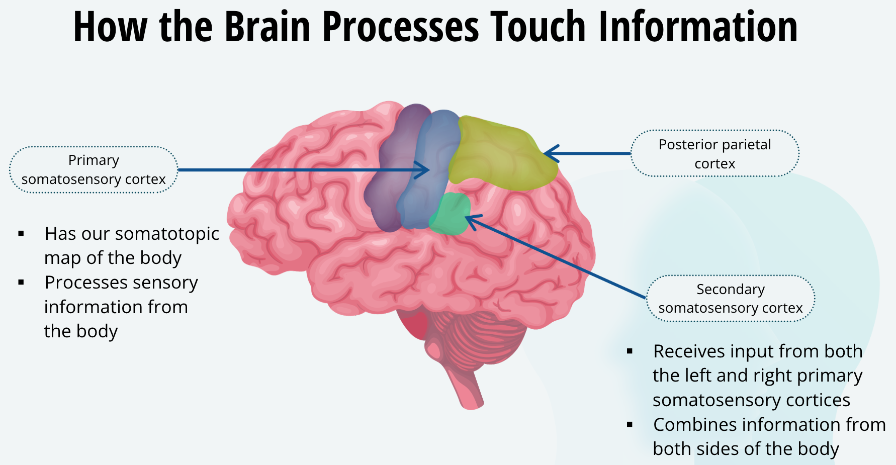
As you can see, the somatosensory cortex can be divided into the primary somatosensory cortex, the smaller secondary somatosensory cortex, and another association area called the posterior parietal cortex. Touch information is first sent to the primary somatosensory cortex, where we have our somatotopic map of the body, and this area processes sensory information from the body. The secondary somatosensory cortex receives input from both the left and right primary somatosensory cortices; thus, it combines information from both sides of the body. Finally, the posterior parietal cortex is an association area, and it brings together the body’s senses, vision, and audition to coordinate them all, so that you can move through the world, while at the same time integrating all of your senses to help you do that with a unified body image.
Having a Unified Body Image
A fascinating yet tragic condition called body integrity identity disorder demonstrates our need to have a unified body image. People with this disorder have no apparent brain damage or emotional or mental disorder. Yet, they are absolutely convinced that one of their limbs does not belong to them. They often request that the offending limb be amputated. Dangerously, some have tried to perform the amputation themselves at home, when no surgeon would agree to amputate a perfectly healthy limb.