Unit 2 Cram Sheet - MedInt, PLTW
2.1
Genes, Chromosomes, and DNA
A chromosome is tightly coiled DNA. The human body contains 23 pairs of chromosomes: 22 pairs of autosomes & one pair of sex chromosomes. These chromosomes are inherited from your parents, and from the moment of conception (fertilization) they are your genetic code - your DNA.
The chromosomes are typically only visible during cell division - the rest of the time, DNA is a jumbled mess that is invisible with a light microscope. This DNA, which forms chromosomes, holds genes.
Genes are the coding sections of DNA, and their job is to provide the instructions for building proteins. Your body is composed of proteins. They are the workers of your body & are essentially responsible for every trait you have: eye color, blood type, skin color, and diseases you have, Chromosomes themselves can be the cause of disease, as can defective genes.
In short, too many chromosomes: bad 😔. Not enough chromosomes: bad 😔. Inheriting a copy of defective DNA (bad genes): also bad 😡.
Genetic Testing Overview
Genetic testing is often performed by a genetic counselor. A genetic counselor is a trained professional who helps individuals & families understand and adjust to a genetic diagnosis or the possibility of having a hereditary disorder. Genetic counselors interpret family history information and educate patients & professionals about genetic diseases. In addition, genetic counselors address psychological and ethical issues associated with a genetic disorder and/or a genetic test result.
Genetic counseling can help a family understand the risks of having a child with a genetic disorder, the medical facts about an already-diagnosed medical condition, and other information necessary for a person or couple to make decisions suitable to their cultural, religious, and moral beliefs. To keep things simple, they help with the testing & provide information people need to make informed choices.
Types of Genetic Disorders
Genetic testing reveals whether or not a DNA-based problem is present. These genetic disorders are caused by abnormalities in an individual’s genetic material. There are four different types of genetic disorders: single-gene, multifactorial, chromosomal, & mitochondrial.
Single-gene disorders
A single-gene disorder is a change or a mutation in one gene. Sickle cell anemia and cystic fibrosis are good examples of these. Single-gene disorders may be classifies as autosomal dominant, autosomal recessive, or sex-linked.
Multifactorial disorders
These are caused by multiple bad genes AND the environment in combination. Breast cancer is an example of this. People are more prone to breast cancer if they have certain forms of certain genes, but they are not guaranteed to inherit that disease. Their chances go up a lot if they make certain lifestyle choices, such as the use of alcohol or smoking.
Mitochondrial disorders
Mitochondrial disorders are fairly rare, and are caused by mutations in the DNA of the mitochondria. If the mitochondria are defective, the body has a difficult time making ATP, which is needed to fuel all cell processes. These are ONLY passed from mother to child. Leber’s hereditary optic neuropathy is an example of this.
Chromosomal disorders
Chromosomal disorders involve inheriting either not enough chromosomes or extras. This happens when either a sperm or egg are made with the wrong number of chromosomes. Diseases where you inherit extra chromosomes include Down’s syndrome.
Down’s syndrome is also known as Trisomy-21. This is because a person with Down’s syndrome has inherited an extra cope of chromosome 21. Tri- means three, and these people have three copies of a chromosome when they are only supposed to have two.
These disorders are easily revealed with a karyotype, a picture of the chromosomes where they have been paired based on size, banding pattern, a centromere position, then arranged from biggest to smallest.
Types of Genetic Screening
Carrier Screening
Carrier screening is a test that is typically done on adult copies who are considering having children, & want to determine if those children could inherit any diseases. Most of the time, there is a family history of something like cystic fibrosis or Tay Sachs disease in the family that the couple wants to ensure that they won’t pass to their child. This process is simple: a blood sample is drawn, the DNA is extracted and amplified using PCR, and the DNA undergoes testing for the disease(s) they are concerned about. This may involve DNA sequencing or gel electrophoresis - sometimes both.
Preimplantation Genetic Diagnosis (PGD)
Preimplantation Genetic Diagnosis (PGD) is a bit different. This procedure is often used by people with known autosomal dominant or sex-linked conditions that they do not want to pass to their children. Here, eggs and sperm are harvested from prospective parents. The eggs are fertilized by the sperm in vitro (in a petri dish) and the embryos are allowed to develop to the 8-cell stage. After the embryos are that big, one single cell from each embryo is removed. The DNA is extracted from that one cell, amplified, & tested for the presence of the trait parents do not want. Healthy embryos are selected and implanted in the mother for development. This technology has several ethical dilemmas surrounding it.
Fetal Screening/Prenatal Diagnosis
Fetal screening/prenatal diagnosis is performed on fetuses while they are still in utero. Amniocentesis or chronic villus sampling are used to extract cells from the fetus for testing. Amniocentesis involves inserting a large needle through the abdomen and into the uterus, where amniotic fluid (the fluid surrounding and protecting the child) is removed. This fluid contains cells shed from the baby: skin cells, cells from the lining of the small intestine, or cells from the bladder. The cells in this fluid provide the DNA needed to perfoem genetic testing. Typically, this procedure requires the use of ultrasound to locate the baby. It is normally performed after the baby is 14 weeks old.
Chronic villus sampling, on the other hand, can be done earlier. Here, chorionic villus cells are removed from the placenta. This is done by inserting a needle vaginally and directing that needle to the placenta. A sall sample of those cells - which are identical to the cells inside the baby - and removed & used for testing. Just like with amniocentesis, ultrasound is used to locate the baby as well as the placenta so the procedure can be done safely. Both procedures carry some risk of miscarriage.
PCR
PCR stands for polymerase chain reaction. This is a laboratory procedure that produces multiple copies of a specific DNA sequence. This can be a copy of a single gene, a large segment of DNA, or the entire genome of an individual. PCR is a three step process that usually takes place in a thermal cycle. Three “ingredients” are added to a sample on DNA so that copies can be made: Taq polymerase, DNA primers, & DNA nucleotides. These are included in a little pellet called a PCR bead.
Denaturation
The first step of PCR is known as denaturation. The temperature in the thermal cycler cranks up to 95 degrees C (nearly boiling). The high temperatures break up the hydrogen bonds that hold the double-stranded DNA together. Think of a zipper being completely unzipped, with the two halves falling away from each other. Denaturation is required so new DNA can be “grown”.
Annealing
The second step of PCR is known as annealing. The thermal cycler cools to 55 degrees C, and the DNA primers (which were added to the DNA mixture early on in the bead) are ready to do their job. Think of annealing as gluing. In this stage, the DNA primers (short sequences of DNA that target the beginning of the section of DNA being copied) bind to the section of DNA that scientists wish to cope. The primer is there so that the DNA is “primed” (readied) for copying. It tells Taq polymerase what section of DNA it should copy.
Extension
Finally, extension occurs. The temperature here is 72 degrees C, and requires both Taq polymerase and DNA nucleotides. In bacteria, Taq polymerase is used to copy bacterial DNA before bacterial cells divide. Scientists use this polymerase to copy DNA during PCR. Taq polymerase attaches to the DNA at the site of the primer. After attaching, it flows down the DNA strand, adding complementary nucleotides to the DNA so that it becomes double-stranded. When Taq polymerase is done doing its job, there are two double-stranded pieces of DNA made from the original one.
Gel Electrophoresis
Three steps of DNA processing are taken first. After we have made copies and cut the copies (see isolation, amplification, & restriction), Gel electrophoresis is performed. Here, an agarose gel is prepared and placed into a buffer solution. The gel contains wells at one end, to which the DNA is added. Markers (standard fragments of known lengths) are added first. Following this are the samples for the patient or patients being tested.
After the DNA is placed into the wells in the agarose gel, it is charged with an electrical current that seperates the fragments. This occurs because DNA has a slightly negative charge. Due to the placement of the agarose gel in the buffer, DNA is pulled based on attractive forces to the positive end of the electrophoresis chamber. The pulling separates DNA fragments, which are stained after the process is completed, and can then be read. Part od this involves determining the size of the DNA fragments. To do this, the “known” bands of the markers are used as a comparison for the sizes of the other fragments. The sizes are used to figure out which gene versions (alleles) a person has inherited. The gel results can be used to determine which version of a gene a patient has.

Isolation
This process begins with the isolation of DNA. Cells are taken from somewhere (blood, saliva, cheek swabbing, etc.) and the cells are lysed (blown up). The blown ip cells and their contents are all mixed together, so a new procedure is used to separate the DNA from the cell waste. This is centrifugation. Centrifugation (fast spinning) separates the heavy cell components from the other waste products (plasma, spit, etc.). At the end of this process, a small pellet of cell parts - including DNA - can be found at the bottom of the spun tube, while the supernatant (fluid on the top of the pellet) is merely waste that can be discarded. To that tiny tube, a small amount of Chelex is added. Chelex forces the DNA to precipitate out and separate itself from the remainder of the cell wasye in the tube. After this happens, the supernatant (which in this case contains the desired DNA) is moved to a new tube, while the pellet full of garbage is discarded.
So.
get cells. blow up cells. spin cells. dump waste. add Chelex. move DNA-holding supernatant to new tube.
Amplification (PCR)
im not writing allat again go look up at the previously written PCR steps
Restriction
After the section of DNA we want has been copied, we next need a way to find out if people have the “bad” version of it or not. It’s important to do something so that the different versions of the gene can be distinguished. This is done with restriction enzymes. Restriction enzymes are molecular scissors that recognize specific DNA sequences and cut the nucleotide strands. This allows identification of the single nucleotide polymorphisms, or SNPs.
Single nucleotide polymorphisms are tiny differences in the DNA of individuals that make them unique. They are 1-nucleotide differences in the DNA. SNPs can be the reason for disease, so being able to identify them is incredibly useful. This is one method that is used to identify “bad” genes.
2.2
Gene Therapy
Gene therapy is a type of disease treatment in which faulty genes are replaced with functional copies. It involves replacing “bad” genes with “good” ones, or flipping genetic switches that are making things go wrong. Gene therapy has the potential to eradicate certain inherited disease by providing people affected with diseases with normal genes that will produce what the body needs.
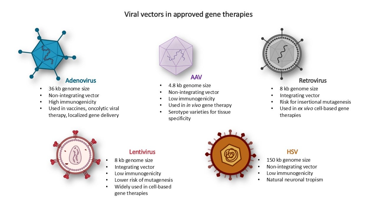
Vectors
Vectors are methods of transportation do deliver healthy genes to affected cells. This method is highly experimental in most cases, although it has been used to successfully treat certain diseases in humans. Vectors are almost always viruses, as they are able to transport DNA to the specific cell that needs it.
Remember that vectors can only carry up to a certain size of gene.
Common Vector types
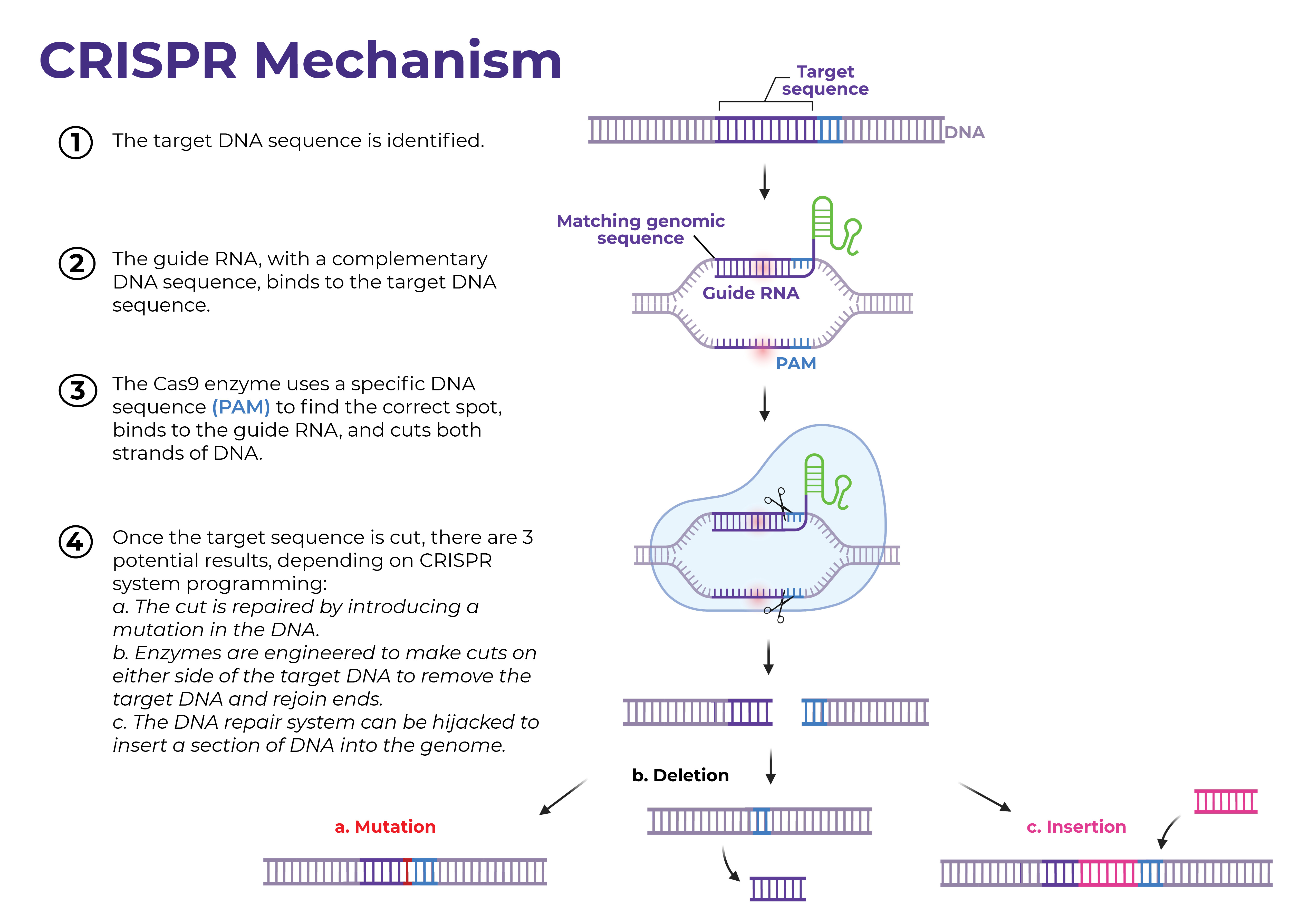
CRISPR
RISPR stands for “clustered interspaced short palindromic repeats.” Biologists use the term to describe the “genetic appearance” of a system that was discovered in microbes – including bacteria and archaea – as early as 1987. For a long time, no one really understood what it did, but around 2005, researchers discovered CRISPR is an immune system. It’s used by microbes to help protect themselves from invading viruses. To stop the invaders, the microbes use CRISPR to recognize and eliminate specific trespassers.
When a virus or other invader enters a bacterial cell, the bacterium incorporates some of the trespasser’s DNA into its own genome so it can find and eliminate the virus during future infections.
cas9