knowt logo

Introduction to the Skeletal System

Introduction

SKELETON

Skeletal Framework of Vertebrates

Shape

Support

Protection

Muscle attachment (locomotion)

Exoskeleton (Dermal Skeleton)

Primitive trait

Teeth, membrane bones of skull (higher chordates = dermal)

Endoskeleton

Distinguishing characteristic (chordates)

Appears earlier in ontogeny

GENERAL STRUCTURE

Mineralized connective tissue deposited in collagen (Matrix)

Bone

Cartilage

Enameloids

Dentin

Ligaments vs Tendons

Ligaments—Bones to bones

Tendons—Muscles to bones

FORMATION OF MINERALIZED STRUCTURES

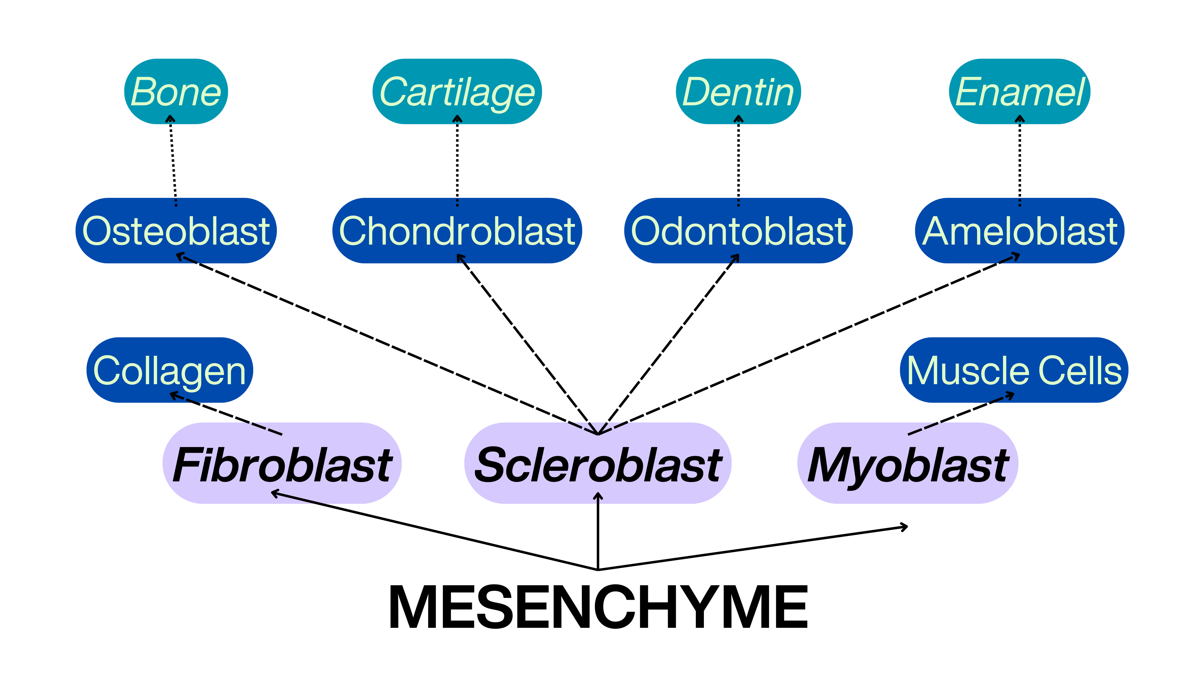
FORMATION OF SKELETAL TISSUES

To memorize the formation of skeletal tissues, you can use the mnemonic "Fabulous Frogs Create Colorful Bundles, Dancing Delightfully". Here’s how it breaks down:

  • Fabulous - Fibroblasts

  • Frogs - Fibrils

  • Create - Collagen Fibers

  • Colorful Bundles- Collagen Bundles

  • Dancing - Dense Connective Tissue

  • Delightfully- Deposition of Minerals

CARTILAGE

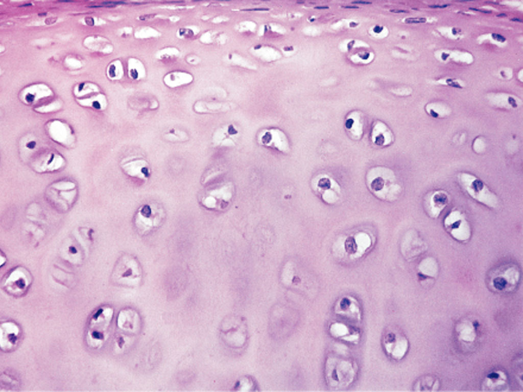
Avascular (no blood) — Specialized connective tissue of chondrocytes in lacunae in a matrix (chondromucin)

Perichondrium — Dense connective tissue

Mesodermal — Origin of cartilage; head and gill region (originate from neural crest cells)

*no canaliculi, no blood vessels

FORMATION OF CARTILAGE

To help you memorize the key components in the formation of cartilage, you can use the mnemonic "Charming Mice Form Amazing Fuzzy Chins". Here’s how it breaks down:

  • Charming - Chondroblast

  • Mice - Mucopolysaccharides in matrix

  • Form - Formation of perichondrium

  • Amazing - Addition of cartilage from perichondrium, fibroblasts, and chondroblasts

  • Tunnels - Transforming to become Chondrocytes

TYPES OF CARTILAGE

Hyaline Cartilage

Most abundant, least differentiated

Precursor (replacement bone)

Articular surfaces of bone

Fibrous Cartilage

Intervertebral discs

Attachment of tendons and ligaments

Elastic Cartilage

External ear and epiglottis

Calcified Cartilage

Calcium salts are deposited

Within interstitial substance of hyaline cartilage/fibrocartilage

BONE

Vascular (with blood) — Connective tissue (calcified bone matrix)

Bone matrix — Osteoblasts and osteocytes (water & mucopolysaccharides; cementing substance)

  • Inorganic component — Calcium hydroxyapatite (Calcium, Phosphate, & Hydroxyl ions)

  • Organic component — Type I Collagen (amorphous ground substance)

TYPES OF BONE CELLS

Osteogenic Cells/ Scleroblasts

Stem cells (mesenchyme)

Foundation of connective tissues

Osteoblasts

Bone formation

Growth regulation

Secretes organic and mineral substances (ossification)

Osteocytes

Osteoblasts —> own intercellular deposits

Maintains cellular activities (bone tissue)

Osteoclasts

Contains enzymes for bone resorption (release of ion stores)

Contains hormone receptors for regulation

CLASSIFICATION OF BONES

SHAPE

Long bone

Short bone

Sesamoid bone

Flat bone

Irregular bone

STRUCTURE

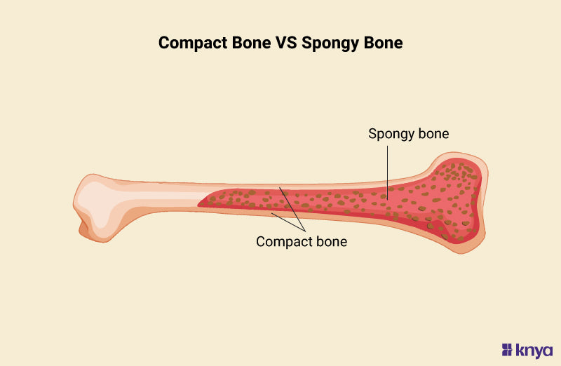
Compact bone

  • Lamellae around haversian canal

  • Haversian system — Canal + Surrounding lamellae (absent in amphibians)

Spongy bone

  • Bone trabeculae + bone marrow

ORIGIN

Intramembranous Ossification

  • Membrane bones from connective tissue

  • Bones of lower jaw, skull, pectoral girdle, dermal bones, & vertebrae (teleosts, urodeles, apodans)

  • Either: Compact or Spongy, Lamellar or Non-Lamellar

Endochondral Ossification

  • Bone from hyaline cartilage

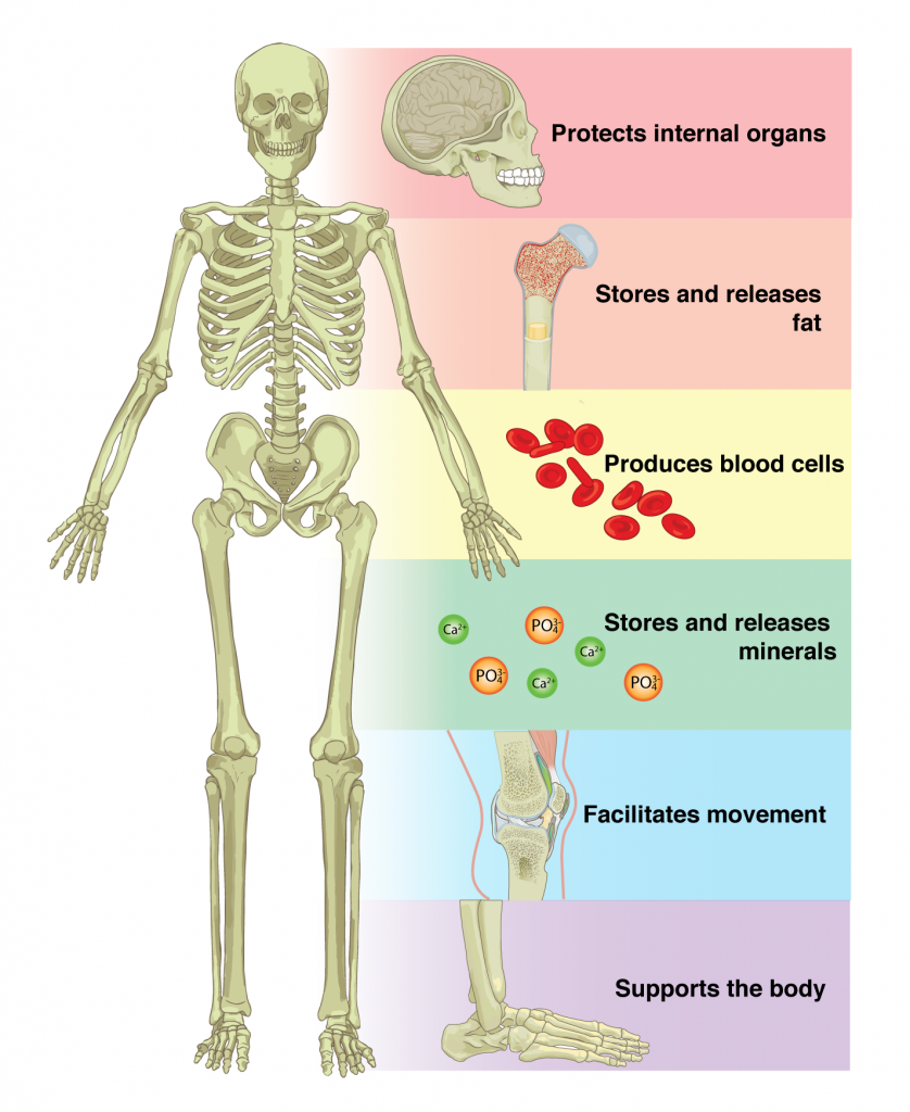
FUNCTION

Support (soft tissues, muscles)

Locomotion

Protect vital organs (skull, ribs, vertebrae)

Hematopoiesis (RBC production in bone marrow)

Reservoir (Calcium, Phosphate)

POSITION

Axial Skeleton

  • Notochord, vertebral Column

  • Ribs + Sternum; Skull & visceral skeleton

Appendicular skeleton

  • Pectoral + Pelvic girdles

  • Skeleton — paired fins, paired limbs; median fins of fishes

Heterotopic skeleton

HETEROTOPIC BONES

Formation of marrow-containing bone outside of normal skeleton

OS CORDIS

Deer, Bovines

OS PENIS

Dogs, Basal Primates, Other Mammals

OS CLITORIDIS

Female Mammals

GIZZARD BONE

Doves

TONGUE

Bats

GULAR POUCH

South American Lizard

DIAPHRAGM

Camel

UPPER EYELID

Crocodilians

OS ROSTRI

Swine

CLOACAL BONES

Lizards

DENTIN

Dentinal Tubules — Canaliculi, coated in enamel

Found in scales of basal ray-finned & elasmobranch fishes + teeth

Skeletal Remodeling

CALCIUM STORAGE + WITHDRAWAL

Calcium — Deposited/withdrawn from storage (amount of calcium in serum)

Withdrawal — Regulation of parathyroid gland & calcitonin

SKELETAL REMODELING

Purpose — Accommodates organs it protects (possibly from stress; THICKER BONES)

Characteristics — Roughened surface areas, bony ridges, prominence in muscle attachments from sustained muscle use

Tendons, Ligaments, and Joints

TENDONS VERSUS LIGAMENTS

TENDONS

MUSCLE-TO-BONES

Maximal resistance to tension when muscle contracts

Continuous with epimysium of muscles + perichondrium (periosteum of cartilages/bones)

LIGAMENTS

BONE-TO-BONE

Less regular than tendon; directly continuous with periosteum

Largest ligament in mammals (nuchal ligament)

TYPES OF JOINTS

Joint — Where two bones/cartilages meet

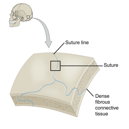
FIBROUS (synarthroses)

Minimal to no movement

Function — Hold bones together

  • Sutures — Flat bones of cranium

  • Syndesmoses — Between two bones

  • Gomphoses — Specialized joint; holds teeth in plate

CARTILAGINOUS (amphiarthroses)

Limited movement

Function — stretching or compression

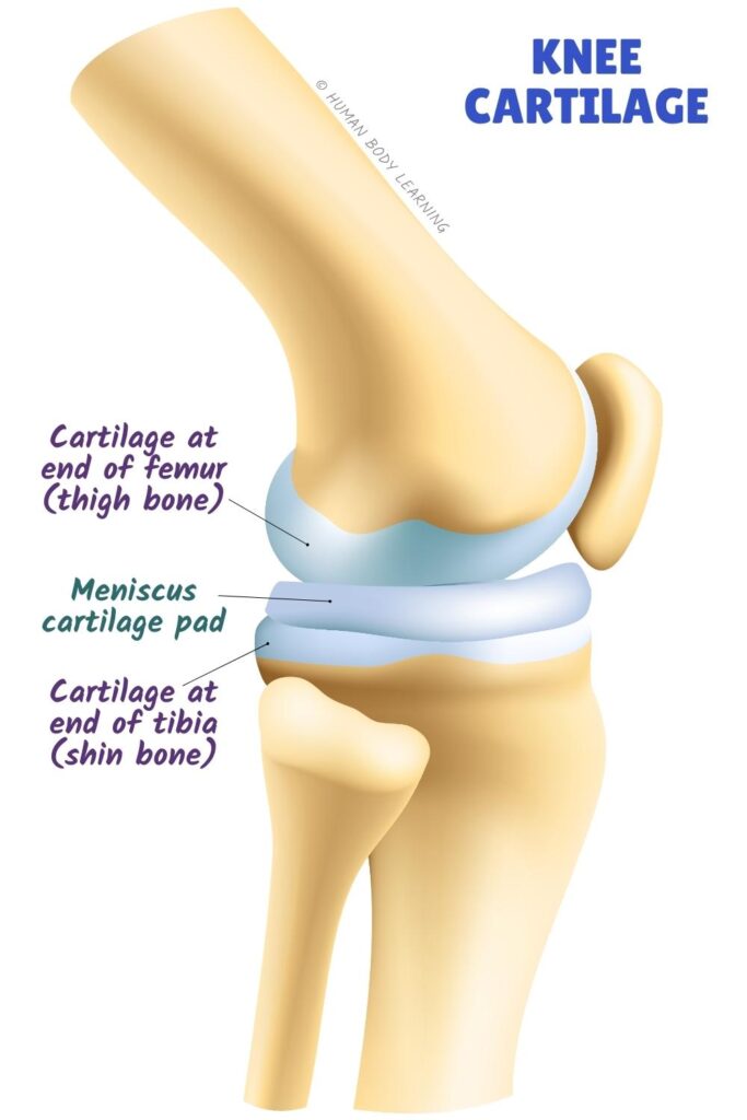
  • Hyaline cartilage joint (synchondroses) — Growing bone; lost in mature animals

  • Fibrocartilaginous joint (symphyses) — Mature skeleton; ossifies with age

SYNOVIAL (diarthroses/true joints)

Wide range of movement

Single plane or multiple planes

Number (articulating surfaces)Simple, complex, compound

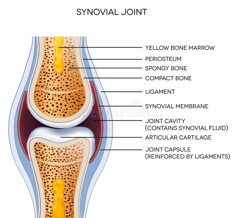
Components

  • Two bones — Covered by hyaline cartilage

  • Joint cavity — Lies between two bones

  • Joint capsuleOuter, fibrous layer (protection, strengthening), & inner, synovial layer (viscous, slick synovial fluid)

FUNCTION AND MOVEMENTS OF SYNOVIAL JOINTS

FLEXION & EXTENSION

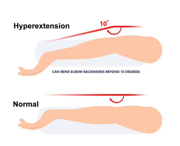
HYPEREXTENSION

ABDUCTION & ADDUCTION

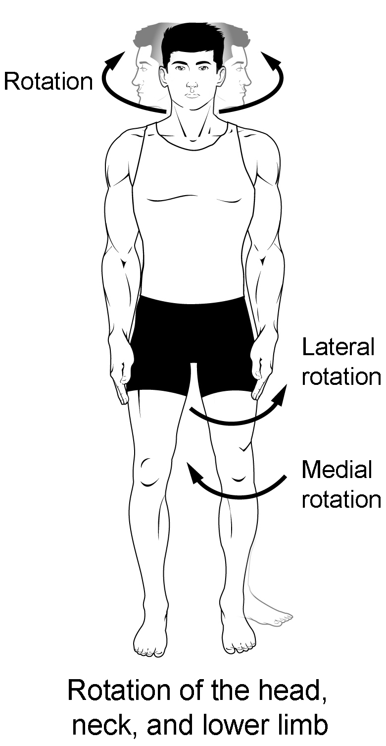
ROTATION

CIRCUMDUCTION

Elbow Hyperextension Injury - Symptoms, Causes & Treatment.

SHAPE/FORM OF SYNOVIAL JOINTS

PLANE

Gliding/sliding movements

Multi-axial

Example: Carpals of wrist

HINGE

Flexion & extension in one plane

Example: Elbow

PIVOT (TROCHOID)

One bone rotates to another

Example: Atlanto-axial joint, proximal radioulnar joint, distal radioulnar joint

ELLIPSOIDAL (CONDYLAR)

Two bones fit together (concave, convex)

Flexion, extension, abduction, & adduction

Example: Wrist joint

SADDLE

Similar but better movement than ellipsoidal

Example: Carpo-metacarpal joint, sternoclavicular joint

BALL-&-SOCKET

Allows all movements except gliding

Example: Shoulder, hop joints

RM

Introduction to the Skeletal System

Introduction

SKELETON

Skeletal Framework of Vertebrates

Shape

Support

Protection

Muscle attachment (locomotion)

Exoskeleton (Dermal Skeleton)

Primitive trait

Teeth, membrane bones of skull (higher chordates = dermal)

Endoskeleton

Distinguishing characteristic (chordates)

Appears earlier in ontogeny

GENERAL STRUCTURE

Mineralized connective tissue deposited in collagen (Matrix)

Bone

Cartilage

Enameloids

Dentin

Ligaments vs Tendons

Ligaments—Bones to bones

Tendons—Muscles to bones

FORMATION OF MINERALIZED STRUCTURES

FORMATION OF SKELETAL TISSUES

To memorize the formation of skeletal tissues, you can use the mnemonic "Fabulous Frogs Create Colorful Bundles, Dancing Delightfully". Here’s how it breaks down:

  • Fabulous - Fibroblasts

  • Frogs - Fibrils

  • Create - Collagen Fibers

  • Colorful Bundles- Collagen Bundles

  • Dancing - Dense Connective Tissue

  • Delightfully- Deposition of Minerals

CARTILAGE

Avascular (no blood) — Specialized connective tissue of chondrocytes in lacunae in a matrix (chondromucin)

Perichondrium — Dense connective tissue

Mesodermal — Origin of cartilage; head and gill region (originate from neural crest cells)

*no canaliculi, no blood vessels

FORMATION OF CARTILAGE

To help you memorize the key components in the formation of cartilage, you can use the mnemonic "Charming Mice Form Amazing Fuzzy Chins". Here’s how it breaks down:

  • Charming - Chondroblast

  • Mice - Mucopolysaccharides in matrix

  • Form - Formation of perichondrium

  • Amazing - Addition of cartilage from perichondrium, fibroblasts, and chondroblasts

  • Tunnels - Transforming to become Chondrocytes

TYPES OF CARTILAGE

Hyaline Cartilage

Most abundant, least differentiated

Precursor (replacement bone)

Articular surfaces of bone

Fibrous Cartilage

Intervertebral discs

Attachment of tendons and ligaments

Elastic Cartilage

External ear and epiglottis

Calcified Cartilage

Calcium salts are deposited

Within interstitial substance of hyaline cartilage/fibrocartilage

BONE

Vascular (with blood) — Connective tissue (calcified bone matrix)

Bone matrix — Osteoblasts and osteocytes (water & mucopolysaccharides; cementing substance)

  • Inorganic component — Calcium hydroxyapatite (Calcium, Phosphate, & Hydroxyl ions)

  • Organic component — Type I Collagen (amorphous ground substance)

TYPES OF BONE CELLS

Osteogenic Cells/ Scleroblasts

Stem cells (mesenchyme)

Foundation of connective tissues

Osteoblasts

Bone formation

Growth regulation

Secretes organic and mineral substances (ossification)

Osteocytes

Osteoblasts —> own intercellular deposits

Maintains cellular activities (bone tissue)

Osteoclasts

Contains enzymes for bone resorption (release of ion stores)

Contains hormone receptors for regulation

CLASSIFICATION OF BONES

SHAPE

Long bone

Short bone

Sesamoid bone

Flat bone

Irregular bone

STRUCTURE

Compact bone

  • Lamellae around haversian canal

  • Haversian system — Canal + Surrounding lamellae (absent in amphibians)

Spongy bone

  • Bone trabeculae + bone marrow

ORIGIN

Intramembranous Ossification

  • Membrane bones from connective tissue

  • Bones of lower jaw, skull, pectoral girdle, dermal bones, & vertebrae (teleosts, urodeles, apodans)

  • Either: Compact or Spongy, Lamellar or Non-Lamellar

Endochondral Ossification

  • Bone from hyaline cartilage

FUNCTION

Support (soft tissues, muscles)

Locomotion

Protect vital organs (skull, ribs, vertebrae)

Hematopoiesis (RBC production in bone marrow)

Reservoir (Calcium, Phosphate)

POSITION

Axial Skeleton

  • Notochord, vertebral Column

  • Ribs + Sternum; Skull & visceral skeleton

Appendicular skeleton

  • Pectoral + Pelvic girdles

  • Skeleton — paired fins, paired limbs; median fins of fishes

Heterotopic skeleton

HETEROTOPIC BONES

Formation of marrow-containing bone outside of normal skeleton

OS CORDIS

Deer, Bovines

OS PENIS

Dogs, Basal Primates, Other Mammals

OS CLITORIDIS

Female Mammals

GIZZARD BONE

Doves

TONGUE

Bats

GULAR POUCH

South American Lizard

DIAPHRAGM

Camel

UPPER EYELID

Crocodilians

OS ROSTRI

Swine

CLOACAL BONES

Lizards

DENTIN

Dentinal Tubules — Canaliculi, coated in enamel

Found in scales of basal ray-finned & elasmobranch fishes + teeth

Skeletal Remodeling

CALCIUM STORAGE + WITHDRAWAL

Calcium — Deposited/withdrawn from storage (amount of calcium in serum)

Withdrawal — Regulation of parathyroid gland & calcitonin

SKELETAL REMODELING

Purpose — Accommodates organs it protects (possibly from stress; THICKER BONES)

Characteristics — Roughened surface areas, bony ridges, prominence in muscle attachments from sustained muscle use

Tendons, Ligaments, and Joints

TENDONS VERSUS LIGAMENTS

TENDONS

MUSCLE-TO-BONES

Maximal resistance to tension when muscle contracts

Continuous with epimysium of muscles + perichondrium (periosteum of cartilages/bones)

LIGAMENTS

BONE-TO-BONE

Less regular than tendon; directly continuous with periosteum

Largest ligament in mammals (nuchal ligament)

TYPES OF JOINTS

Joint — Where two bones/cartilages meet

FIBROUS (synarthroses)

Minimal to no movement

Function — Hold bones together

  • Sutures — Flat bones of cranium

  • Syndesmoses — Between two bones

  • Gomphoses — Specialized joint; holds teeth in plate

CARTILAGINOUS (amphiarthroses)

Limited movement

Function — stretching or compression

  • Hyaline cartilage joint (synchondroses) — Growing bone; lost in mature animals

  • Fibrocartilaginous joint (symphyses) — Mature skeleton; ossifies with age

SYNOVIAL (diarthroses/true joints)

Wide range of movement

Single plane or multiple planes

Number (articulating surfaces)Simple, complex, compound

Components

  • Two bones — Covered by hyaline cartilage

  • Joint cavity — Lies between two bones

  • Joint capsuleOuter, fibrous layer (protection, strengthening), & inner, synovial layer (viscous, slick synovial fluid)

FUNCTION AND MOVEMENTS OF SYNOVIAL JOINTS

FLEXION & EXTENSION

HYPEREXTENSION

ABDUCTION & ADDUCTION

ROTATION

CIRCUMDUCTION

Elbow Hyperextension Injury - Symptoms, Causes & Treatment.

SHAPE/FORM OF SYNOVIAL JOINTS

PLANE

Gliding/sliding movements

Multi-axial

Example: Carpals of wrist

HINGE

Flexion & extension in one plane

Example: Elbow

PIVOT (TROCHOID)

One bone rotates to another

Example: Atlanto-axial joint, proximal radioulnar joint, distal radioulnar joint

ELLIPSOIDAL (CONDYLAR)

Two bones fit together (concave, convex)

Flexion, extension, abduction, & adduction

Example: Wrist joint

SADDLE

Similar but better movement than ellipsoidal

Example: Carpo-metacarpal joint, sternoclavicular joint

BALL-&-SOCKET

Allows all movements except gliding

Example: Shoulder, hop joints

robot