knowt logo

3 Major Structures of the Brain

Major Structures of the Brain

Definition of Terms

  1. Nucleus – cluster of neurons in the central nervous system (plural is nuclei)

  2. Tracts – bundle of nerve fibers (axons) - CNS

  3. Peduncle – Stem-like connector

  4. Gyrus – Kulot kulot (Plural is Gyri)

Structure of the Vertebrate Nervous System

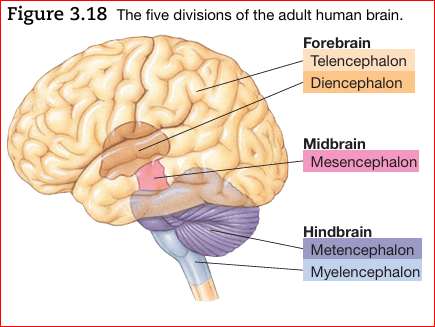
Three major divisions of the brain include:

  1. Hindbrain.

    • Myelencephalon (“marrow-brain”)

    • Rhombencephalon (“parallelogram-brain”)

    • Metencephalon (“afterbrain”)

  2. Midbrain.

    • Mesencephalon (“middle-brain”)

      • Major Structures: Tectum, tegmentum, superior colliculus, inferior colliculus, substantia nigra

  3. Forebrain.

    • Prosencephalon(“forward-brain”)

    • Diencephalon(“between-brain”)

      • Major Structures: Thalamus, Hypothalamus

    • Telencephalon (“endbrain”)

      • Major Structures: Cerebral cortex, hippocampus, basal ganglia

Hindbrain

The Hindbrain consists of the:

  1. Medulla.

  2. Pons.

  3. Cerebellum.

Myelencephalon (marrow-brain)

  • Located at the posterior portion of the brain

  • Hindbrain structures, the midbrain and other central structures of the brain combine and make up the brain stem (diencephalon, mesencephalon, metencephalon, and myelencephalon)

  • Sometimes called the Medulla or Medulla Oblangata

  • Basic Life Support

  • Situated between the Pons and the Brainstem

  • Composed largely of tracts carrying signals between the rest of the brain and the body

  • Contains the Reticular Formation (plays a role in arousal, attention, movement, maintenance of muscle tone, various cardiac, respiratory, and circulatory reflexes)


    *Encephalon means “within the head”

Reticular Formation

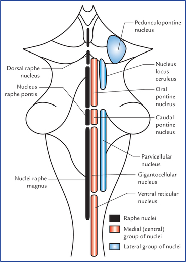
  • a complex network of about 100 tiny nuclei that occupies the central core of the brain stem from the posterior boundary of the myelencephalon to the anterior boundary of the midbrain.

  • named because of its netlike appearance (reticulum means “little net”).

  • referred to as the reticular activating system because parts of it seem to play a role in arousal.

  • Division of the Reticular Formation:

    1. Parvocellular Reticular Nuclei (Lateral Zone) – Regulate exhalation

    2. Gigantocellular reticular Nuclei (Medial Zone) – Motor Coordination

    3. Raphe Nuclei (Median) – Where Serotonin (5-HT) is synthesized; Mood regulation

    4. Somatic Motor Control (tone, balance, and posture – during body movements), also relays eye and ear signals to the cerebellum so that the cerebellum can integrate visual, auditory and motor coordination
Medullary Pyramids

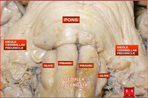
  • Bundle of fibers (Pyramid Tracts) that decussate and continue down the spinal cord on the contralateral side.

    Pyramidal Tracts:

    • Corticospinal Tract: Movement of Muscles of the body

    • Corticobulbar Tract: Facial Expression, Movement of the head, swallowing, phonation, and movements of the tongue.

Olivary Bodies

  • Termed as Medullary Olives or simply Olives

  • Pair of prominent oval structures that contain the olivary nuclei.

  • Two parts:

    1. Inferior Olivary Nucleus - part of the olivo-cerebellar system and is mainly involved in cerebellar (Learned, muscle memory) motor-learning and function

    2. Superior Olivary Nucleus - considered part of the pons and part of the auditory system, aiding in the perception of sound

      *Cerebral - still learning, consciously in the mind

Metencephalon

  • Houses many ascending and descending tracts and parts of the reticular formation

  • Pons – Regulation of Breathing (pneumotaxic center) and involved in the transmission of to and from other areas of the brain (cerebrum to the cerebellum)

  • Cerebellum – coordinate muscle movements, posture, and integrate sensory formation from the inner ear and proprioception (sense of one’s position and strength of effort) in the muscles and joint
Pons

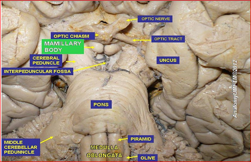
  • In Latin, pons means “bridge”

  • lies on each side of the medulla (ventral and anterior).

  • along with the medulla, contains the reticular formation and raphe system.

  • works in conjunction to increase arousal and readiness of other parts of the brain.

Reticular formation

  • descending portion is one of several brain areas that control the motor areas of the spinal cord.

  • ascending portion sends output to much of the cerebral cortex, selectively increasing arousal and attention.

*The raphe system also sends axons to much of the forebrain, modifying the brain’s readiness to respond to stimuli.

The Cerebellum

  • Means little brain

  • a structure located in the hindbrain with many deep folds.

  • helps regulate motor movement, balance and coordination.

  • is also important for shifting attention between auditory and visual stimuli.

Midbrain - Mesencephalon

The midbrain is comprised of the following structures:

  1. Tectum (Latin: roof) - Dorsal to the midbrain.

  2. Superior colliculus - visual-motor function

    & Inferior colliculus - auditory function (or colliculi)

    • *located on each side of the tectum and processes sensory information

      *colliculi means little hills

      *In lower vertebrates, the function of the tectum is entirely visual-motor, and it is sometimes referred to as the optic tectum.

  3. Tagmentum (Latin: Floor)- the intermediate level of the midbrain containing nuclei for cranial nerves and part of the reticular formation. Ventral to the tectum.

  4. Substantia nigra - gives rise to the dopamine-containing pathway facilitating readiness for movement

Two divisions: Tectum and Tegmentum

Tectum: Auditory and Visual Reflexes

Two bumps = Superior (Visual) and Inferior Colliculus (Auditory)

Tegmentum = Three colorful structures

  1. Periaqueductal Gray = Gray matter situated around the cerebral aqueduct

    • Mediates the analgesic effect of opiate drugs

  2. Substantia Nigra (black substance)– Eye-movement, motor planning, reward-seeking, learning, and addiction (mediated by the striatum)

  3. Red Nucleus – Vestigial in primate brains, = Motor Coordination (crawling in babies and arm swinging in walking)

    *Both the Myelencephalon and the Metencephalon form the Hindbrain

Diencephalon

Composed of two structures: the thalamus and the hypothalamus

Thalamus

  • large, two-lobed structure that constitutes the top of the brain stem. Both sit on each side of the Third Ventricle. Sensory “way-station”.

  • Composed of many different pairs of nuclei which project to the cortex

  • Massa intermedia – Joins both lobes. On the surface, we can see the white Lamina (layers) - composed of myelinated axons.

  • Internal Capsule – Fibers (corticospinal tracts) that join the telencephalon to the diencephalon. They carry motor information from the motor cortex downward.

  • Most understood Thalamic nuclei: Sensory relay nuclei – nuclei that receive signals from sensory receptors, process them, and then transmit them to appropriate areas of sensory cortex.

  • lateral geniculate nuclei - relay stations in the visual

  • Byventral posterior nuclei - somatosensory systems

  • medial geniculate nuclei - auditory

Hypothalamus

  • Below the anterior thalamus (hypo means “below”).

  • Regulated behavior = Sleeping, eating, and sexual behavior.

  • Regulates hormones released by the pituitary gland

    • The literal meaning of pituitary gland is “snot gland”; it was discovered in a gelatinous state behind the nose of an unembalmed cadaver and was incorrectly assumed to be the main source of nasal mucus.

    • dangles from it on the ventral surface of the brain.

    • Associated with behaviors such as eating, drinking, sexual behavior and other motivated behaviors.

  • Optic Chiasm – the point at which the optic nerves from each eye come together.

    • X shape is created because some of the axons of the optic nerve decussate (cross over to the other side of the brain) via the optic chiasm.

    • The decussating fibers are said to be contralateral, and the nondecussating fibers are said to be ipsilateral.

  • Mamilliary bodiesSpherical nuclei which has a role in recollective memory. located on the inferior surface of the hypothalamus, just behind the pituitary.
Telencephalon

  • Largest division of the Human Brain and mediates the brains most complex functions. Initiates voluntary movements, interprets sensory input, and mediates complex cognitive processes such as learning, speaking, and problem solving.

The Cerebral Cortex

  • cerebral cortex (cerebral bark) - a layer of tissue that covers the cerebral hemispheres. Deeply convoluted (furrowed).

    • Convolutions have the effect of increasing the amount of cerebral cortex without increasing the overall volume of the brain.

    • most mammals are lissencephalic (smooth-brained).

    • the number and size of cortical convolutions appear to be related more to body size rather than intellectual capacities.

    • Because the cerebral cortex is mainly composed of small, unmyelinated neurons, it is gray and is often referred to as the gray matter. In contrast, the layer beneath the cortex is mainly composed of large myelinated axons, which are white and often referred to as the white matter.

  • divided into two halves

  • joined by two bundles of axons called the corpus callosum and the anterior commissure.

  • more highly developed in humans than other species.

  • Large furrows are called fissures whereas small ones are called sulci; ridges between fissures and sulci are called gyri.

  • Longitudinal fissure – Divides both hemispheres. Inside, cerebral commissures connect both hemispheres. The largest commissure is the corpus collosum.

  • Largest gyri = Precentral gyri (in the frontal lobe), postcentral gyri (parietal lobe), and the superior temporal gyri (temporal lobe).

Organization of the Cerebral Cortex:

  • Contains up to six distinct laminae (layers) that are parallel to the

  • surface of the cortex.

  • Cells of the cortex are also divided into columns that lie perpendicular to the laminae.

  • Divided into four lobes: occipital, parietal, temporal, and frontal.

Four Lobes:

  1. Frontal Lobe - has two distinct functional areas:

    • Precentral gyrus and adjacent frontal cortex have a motor function

    • Frontal cortex anterior to motor cortex performs complex cognitive functions, such as planning response sequences, evaluating the outcomes of potential patterns of behavior, and assessing the significance of the behavior of others.

  2. Parietal Lobe - two large functional areas in each parietal lobe:

    • Postcentral gyrus analyzes sensations from the body (e.g., touch)

    • the posterior parts of the parietal lobes play roles in perceiving the location of both objects and our own bodies and in directing our attention.

  3. Temporal Lobes - The cortex of each temporal lobe has three general functional areas:

    • Superior temporal gyrus is involved in hearing and language

    • Inferior temporal cortex identifies complex visual patterns

    • Medial portion of temporal cortex (which is not visible from the usual side view) is important for certain kinds of memory.

  4. Occipital Lobe - visual

Telencephalon Gyri

  1. Postcentral gyrus – analyzesensations of the body

  2. Anterior Paracentral Lobule –Continuation of the precentral gyrusand concerned with somatosensationof distal limbs

  3. Precentral Gyrus – Motor Function

  4. Posterior paracentral lobule precuneus – Innervation of the contralateral lower extremities, and control of defecation and urination.

  5. Gyrus Rectus/Orbital Gyri – Function is unclear and is viewed as primitive development but is linked with personality and hypersexuality with men

  6. Superior Parietal Lobule – Somatosensory

    Activity

  7. Inferior Frontal Gyrus – Speech production

  8. Inferior Parietal Lobule – Divided into

    Two Parts:

    • Supramarginal Gyrus – Integration of

    sensory information

    • Angular Gyrus – Receives visual

    information

  9. Broca’s Area – Speech Production(ONLY FOUND ON LEFT

  10. Wernicke’s Area – Language Comprehension(ONLY FOUND ON LEFT)

  11. Transverse Temporal Gyri of Heschl – Auditory Cortex

  12. Superior Temporal Gyrus – Auditory processing, and social cognition

  13. Middle Temporal Gyrus – Language processes

  14. Inferior Temporal Gyrus – Visual Perception

  15. Fusiform Gyrus – Object and Facial recognition

  16. Fusiform Facial Area – Recognition of Facial Expression\

  17. Superior and Inferior Occipital Gyri – Interpretation of visual stimuli

  18. Cuneate Gyrus – Letters and analysis of logical events

  19. Lingual Gyrus – Recognition of words

  20. Long and Short Gyrus of the Insula – Sensation of Taste and Olfaction

  21. Cingulate Gyrus – Emotion and Behavioral Regulation

  22. Parahippocampal Gyrus – Memory Encoding and Retrieval

  23. Dentate Gyrus – New episodic memory, spontaneous exploration of novel environments

Neocortex

  • New cortex

  • 90 percent of human cerebral cortex

  • six-layered cortex of relatively recent evolution

  • the layers of neocortex are numbered I through VI, starting at the surface.

  • the six layers of neocortex differ from one another in terms of the size and density of their cell bodies and the relative proportion of pyramidal and stellate cell bodies that they contain.

  • neocortex’s columnar organization: neurons in a given vertical column of neocortex often form a mini-circuit that performs a single function.

  • There are variations in the thickness of the respective layers from area to area

  • Cortical neurons has two categories:

    1. pyramidal (pyramid-shaped) cells - large multipolar neurons with pyramid-shaped cell bodies.

      • Apical dendrite - a large dendrite that extends from the apex of the pyramid straight toward the cortex surface and a very long axon.

    2. stellate (star-shaped) cells - small star shaped interneurons (neurons with a short axon or no axon).

Hippocampus

  • function: memory for spatial location

  • one important area of cortex that is not neocortex—it has only three major layers

  • located at the medial edge of the cerebral cortex as it folds back on itself in the medial temporal lobe

  • hippocampus means “sea horse”

Limbic system and of the basal ganglia

Limbic system is a circuit of midline structures that circle the thalamus (limbic means “ring”).

  • Four F’s of motivation: fleeing, feeding, fighting, and sexual behavior.

  • Major structures of the limbic system include the amygdala, the fornix, the cingulate cortex, the septum, the mammillary bodies and the hippocampus

  • Basal forebrain is comprised of several structures that lie on the dorsal surface of the forebrain.

    • Contains the nucleus basalis:

      • receives input from the hypothalamus and basal ganglia

      • sends axons that release acetylcholine to the cerebral cortex

      • Key part of the brains system for arousal, wakefulness, and attention

  • Pathway: Amygdala – Cingulate Cortex – Fornix – Septum Pellucidum

    1. Amygdala – Emotional Memory and fear

      • almond-shaped nucleus in the anterior temporal lobe

    2. Cingulate Cortex – Linking motivational outcomes to behavior. = Important for Schizophrenia and Depression

      • It is the large strip of cortex in the cingulate gyrus on the medial surface of the cerebral hemispheres, just superior to the corpus callosum; it encircles the dorsal thalamus (cingulate means “encircling”).

    3. Fornix – Function is not entirely sure but correlates well with Recall Memory

      • the major tract of the limbic system

      • encircles the dorsal thalamus

      • fornix means “arc

    4. Septum Pellucidum – Anatomical Barrier yet real function remains unclear.

      • a midline nucleus located at the anterior tip of the cingulate cortex

      • Includes the olfactory bulb, hypothalamus, hippocampus, amygdala, and cingulate gyrus of the cerebral cortex

      • associated with motivation, emotion, drives and aggression

    *Hippocampus is a large structure located between the thalamus and cerebral cortex.

    • Toward the posterior portion of the forebrain

    • critical for storing certain types of memory.

Basal Ganglia

  • Plays a role in the performance of voluntary motor responses.

    1. Amygdala - considered part of both limbic and the basal ganglia systems.

    2. caudate - (caudate means “tail-like”) in a posterior direction and in an anterior direction from amygdala. Long tail-like.

    3. Putamen - connected by a series of fiber bridges, located at the center.

      • Planning, learning and execution, motor preparation, specifying of amplitudes of movement, and movement sequences. Reinforcement Learning, implicit learning, category learning, “Hate Circuit” (with insula).

      • Recent studies have shown that for trans women, larger amounts of gray matter have been found BUT this does not imply much.

      • Together, the caudate and the putamen, which both have a striped appearance, are known as the striatum (striped structure)

    4. Globus pallidus (pale globe) - located medial to the putamen between the putamen and the thalamus.

      • Regulates movement at the subconscious level.

      • pallidotomy - damage is caused in order to reduce involuntary muscle tremor.

    *a pathway that projects to the striatum from the substantia nigra of the midbrain: Parkinson’s disease, a disorder characterized by rigidity, tremors, and poverty of voluntary movement, is associated with the deterioration of this pathway.

    *nucleus accumbens, which is in the medial portion of the ventral striatum. The nucleus accumbens is thought to play a role in the rewarding effects of addictive drugs and other reinforcers.

Canal system

  • The central canal is a fluid-filled channel in the center of the spinal cord.

  • The ventricles are four fluid-filled cavities within the brain containing cerebrospinal fluid.

DG

3 Major Structures of the Brain

Major Structures of the Brain

Definition of Terms

  1. Nucleus – cluster of neurons in the central nervous system (plural is nuclei)

  2. Tracts – bundle of nerve fibers (axons) - CNS

  3. Peduncle – Stem-like connector

  4. Gyrus – Kulot kulot (Plural is Gyri)

Structure of the Vertebrate Nervous System

Three major divisions of the brain include:

  1. Hindbrain.

    • Myelencephalon (“marrow-brain”)

    • Rhombencephalon (“parallelogram-brain”)

    • Metencephalon (“afterbrain”)

  2. Midbrain.

    • Mesencephalon (“middle-brain”)

      • Major Structures: Tectum, tegmentum, superior colliculus, inferior colliculus, substantia nigra

  3. Forebrain.

    • Prosencephalon(“forward-brain”)

    • Diencephalon(“between-brain”)

      • Major Structures: Thalamus, Hypothalamus

    • Telencephalon (“endbrain”)

      • Major Structures: Cerebral cortex, hippocampus, basal ganglia

Hindbrain

The Hindbrain consists of the:

  1. Medulla.

  2. Pons.

  3. Cerebellum.

Myelencephalon (marrow-brain)

  • Located at the posterior portion of the brain

  • Hindbrain structures, the midbrain and other central structures of the brain combine and make up the brain stem (diencephalon, mesencephalon, metencephalon, and myelencephalon)

  • Sometimes called the Medulla or Medulla Oblangata

  • Basic Life Support

  • Situated between the Pons and the Brainstem

  • Composed largely of tracts carrying signals between the rest of the brain and the body

  • Contains the Reticular Formation (plays a role in arousal, attention, movement, maintenance of muscle tone, various cardiac, respiratory, and circulatory reflexes)


    *Encephalon means “within the head”

Reticular Formation

  • a complex network of about 100 tiny nuclei that occupies the central core of the brain stem from the posterior boundary of the myelencephalon to the anterior boundary of the midbrain.

  • named because of its netlike appearance (reticulum means “little net”).

  • referred to as the reticular activating system because parts of it seem to play a role in arousal.

  • Division of the Reticular Formation:

    1. Parvocellular Reticular Nuclei (Lateral Zone) – Regulate exhalation

    2. Gigantocellular reticular Nuclei (Medial Zone) – Motor Coordination

    3. Raphe Nuclei (Median) – Where Serotonin (5-HT) is synthesized; Mood regulation

    4. Somatic Motor Control (tone, balance, and posture – during body movements), also relays eye and ear signals to the cerebellum so that the cerebellum can integrate visual, auditory and motor coordination

Medullary Pyramids

  • Bundle of fibers (Pyramid Tracts) that decussate and continue down the spinal cord on the contralateral side.

    Pyramidal Tracts:

    • Corticospinal Tract: Movement of Muscles of the body

    • Corticobulbar Tract: Facial Expression, Movement of the head, swallowing, phonation, and movements of the tongue.

Olivary Bodies

  • Termed as Medullary Olives or simply Olives

  • Pair of prominent oval structures that contain the olivary nuclei.

  • Two parts:

    1. Inferior Olivary Nucleus - part of the olivo-cerebellar system and is mainly involved in cerebellar (Learned, muscle memory) motor-learning and function

    2. Superior Olivary Nucleus - considered part of the pons and part of the auditory system, aiding in the perception of sound

      *Cerebral - still learning, consciously in the mind

Metencephalon

  • Houses many ascending and descending tracts and parts of the reticular formation

  • Pons – Regulation of Breathing (pneumotaxic center) and involved in the transmission of to and from other areas of the brain (cerebrum to the cerebellum)

  • Cerebellum – coordinate muscle movements, posture, and integrate sensory formation from the inner ear and proprioception (sense of one’s position and strength of effort) in the muscles and joint

Pons

  • In Latin, pons means “bridge”

  • lies on each side of the medulla (ventral and anterior).

  • along with the medulla, contains the reticular formation and raphe system.

  • works in conjunction to increase arousal and readiness of other parts of the brain.

Reticular formation

  • descending portion is one of several brain areas that control the motor areas of the spinal cord.

  • ascending portion sends output to much of the cerebral cortex, selectively increasing arousal and attention.

*The raphe system also sends axons to much of the forebrain, modifying the brain’s readiness to respond to stimuli.

The Cerebellum

  • Means little brain

  • a structure located in the hindbrain with many deep folds.

  • helps regulate motor movement, balance and coordination.

  • is also important for shifting attention between auditory and visual stimuli.

Midbrain - Mesencephalon

The midbrain is comprised of the following structures:

  1. Tectum (Latin: roof) - Dorsal to the midbrain.

  2. Superior colliculus - visual-motor function

    & Inferior colliculus - auditory function (or colliculi)

    • *located on each side of the tectum and processes sensory information

      *colliculi means little hills

      *In lower vertebrates, the function of the tectum is entirely visual-motor, and it is sometimes referred to as the optic tectum.

  3. Tagmentum (Latin: Floor)- the intermediate level of the midbrain containing nuclei for cranial nerves and part of the reticular formation. Ventral to the tectum.

  4. Substantia nigra - gives rise to the dopamine-containing pathway facilitating readiness for movement

Two divisions: Tectum and Tegmentum

Tectum: Auditory and Visual Reflexes

Two bumps = Superior (Visual) and Inferior Colliculus (Auditory)

Tegmentum = Three colorful structures

  1. Periaqueductal Gray = Gray matter situated around the cerebral aqueduct

    • Mediates the analgesic effect of opiate drugs

  2. Substantia Nigra (black substance)– Eye-movement, motor planning, reward-seeking, learning, and addiction (mediated by the striatum)

  3. Red Nucleus – Vestigial in primate brains, = Motor Coordination (crawling in babies and arm swinging in walking)

    *Both the Myelencephalon and the Metencephalon form the Hindbrain

Diencephalon

Composed of two structures: the thalamus and the hypothalamus

Thalamus

  • large, two-lobed structure that constitutes the top of the brain stem. Both sit on each side of the Third Ventricle. Sensory “way-station”.

  • Composed of many different pairs of nuclei which project to the cortex

  • Massa intermedia – Joins both lobes. On the surface, we can see the white Lamina (layers) - composed of myelinated axons.

  • Internal Capsule – Fibers (corticospinal tracts) that join the telencephalon to the diencephalon. They carry motor information from the motor cortex downward.

  • Most understood Thalamic nuclei: Sensory relay nuclei – nuclei that receive signals from sensory receptors, process them, and then transmit them to appropriate areas of sensory cortex.

  • lateral geniculate nuclei - relay stations in the visual

  • Byventral posterior nuclei - somatosensory systems

  • medial geniculate nuclei - auditory

Hypothalamus

  • Below the anterior thalamus (hypo means “below”).

  • Regulated behavior = Sleeping, eating, and sexual behavior.

  • Regulates hormones released by the pituitary gland

    • The literal meaning of pituitary gland is “snot gland”; it was discovered in a gelatinous state behind the nose of an unembalmed cadaver and was incorrectly assumed to be the main source of nasal mucus.

    • dangles from it on the ventral surface of the brain.

    • Associated with behaviors such as eating, drinking, sexual behavior and other motivated behaviors.

  • Optic Chiasm – the point at which the optic nerves from each eye come together.

    • X shape is created because some of the axons of the optic nerve decussate (cross over to the other side of the brain) via the optic chiasm.

    • The decussating fibers are said to be contralateral, and the nondecussating fibers are said to be ipsilateral.

  • Mamilliary bodiesSpherical nuclei which has a role in recollective memory. located on the inferior surface of the hypothalamus, just behind the pituitary.

Telencephalon

  • Largest division of the Human Brain and mediates the brains most complex functions. Initiates voluntary movements, interprets sensory input, and mediates complex cognitive processes such as learning, speaking, and problem solving.

The Cerebral Cortex

  • cerebral cortex (cerebral bark) - a layer of tissue that covers the cerebral hemispheres. Deeply convoluted (furrowed).

    • Convolutions have the effect of increasing the amount of cerebral cortex without increasing the overall volume of the brain.

    • most mammals are lissencephalic (smooth-brained).

    • the number and size of cortical convolutions appear to be related more to body size rather than intellectual capacities.

    • Because the cerebral cortex is mainly composed of small, unmyelinated neurons, it is gray and is often referred to as the gray matter. In contrast, the layer beneath the cortex is mainly composed of large myelinated axons, which are white and often referred to as the white matter.

  • divided into two halves

  • joined by two bundles of axons called the corpus callosum and the anterior commissure.

  • more highly developed in humans than other species.

  • Large furrows are called fissures whereas small ones are called sulci; ridges between fissures and sulci are called gyri.

  • Longitudinal fissure – Divides both hemispheres. Inside, cerebral commissures connect both hemispheres. The largest commissure is the corpus collosum.

  • Largest gyri = Precentral gyri (in the frontal lobe), postcentral gyri (parietal lobe), and the superior temporal gyri (temporal lobe).

Organization of the Cerebral Cortex:

  • Contains up to six distinct laminae (layers) that are parallel to the

  • surface of the cortex.

  • Cells of the cortex are also divided into columns that lie perpendicular to the laminae.

  • Divided into four lobes: occipital, parietal, temporal, and frontal.

Four Lobes:

  1. Frontal Lobe - has two distinct functional areas:

    • Precentral gyrus and adjacent frontal cortex have a motor function

    • Frontal cortex anterior to motor cortex performs complex cognitive functions, such as planning response sequences, evaluating the outcomes of potential patterns of behavior, and assessing the significance of the behavior of others.

  2. Parietal Lobe - two large functional areas in each parietal lobe:

    • Postcentral gyrus analyzes sensations from the body (e.g., touch)

    • the posterior parts of the parietal lobes play roles in perceiving the location of both objects and our own bodies and in directing our attention.

  3. Temporal Lobes - The cortex of each temporal lobe has three general functional areas:

    • Superior temporal gyrus is involved in hearing and language

    • Inferior temporal cortex identifies complex visual patterns

    • Medial portion of temporal cortex (which is not visible from the usual side view) is important for certain kinds of memory.

  4. Occipital Lobe - visual

Telencephalon Gyri

  1. Postcentral gyrus – analyzesensations of the body

  2. Anterior Paracentral Lobule –Continuation of the precentral gyrusand concerned with somatosensationof distal limbs

  3. Precentral Gyrus – Motor Function

  4. Posterior paracentral lobule precuneus – Innervation of the contralateral lower extremities, and control of defecation and urination.

  5. Gyrus Rectus/Orbital Gyri – Function is unclear and is viewed as primitive development but is linked with personality and hypersexuality with men

  6. Superior Parietal Lobule – Somatosensory

    Activity

  7. Inferior Frontal Gyrus – Speech production

  8. Inferior Parietal Lobule – Divided into

    Two Parts:

    • Supramarginal Gyrus – Integration of

    sensory information

    • Angular Gyrus – Receives visual

    information

  9. Broca’s Area – Speech Production(ONLY FOUND ON LEFT

  10. Wernicke’s Area – Language Comprehension(ONLY FOUND ON LEFT)

  11. Transverse Temporal Gyri of Heschl – Auditory Cortex

  12. Superior Temporal Gyrus – Auditory processing, and social cognition

  13. Middle Temporal Gyrus – Language processes

  14. Inferior Temporal Gyrus – Visual Perception

  15. Fusiform Gyrus – Object and Facial recognition

  16. Fusiform Facial Area – Recognition of Facial Expression\

  17. Superior and Inferior Occipital Gyri – Interpretation of visual stimuli

  18. Cuneate Gyrus – Letters and analysis of logical events

  19. Lingual Gyrus – Recognition of words

  20. Long and Short Gyrus of the Insula – Sensation of Taste and Olfaction

  21. Cingulate Gyrus – Emotion and Behavioral Regulation

  22. Parahippocampal Gyrus – Memory Encoding and Retrieval

  23. Dentate Gyrus – New episodic memory, spontaneous exploration of novel environments

Neocortex

  • New cortex

  • 90 percent of human cerebral cortex

  • six-layered cortex of relatively recent evolution

  • the layers of neocortex are numbered I through VI, starting at the surface.

  • the six layers of neocortex differ from one another in terms of the size and density of their cell bodies and the relative proportion of pyramidal and stellate cell bodies that they contain.

  • neocortex’s columnar organization: neurons in a given vertical column of neocortex often form a mini-circuit that performs a single function.

  • There are variations in the thickness of the respective layers from area to area

  • Cortical neurons has two categories:

    1. pyramidal (pyramid-shaped) cells - large multipolar neurons with pyramid-shaped cell bodies.

      • Apical dendrite - a large dendrite that extends from the apex of the pyramid straight toward the cortex surface and a very long axon.

    2. stellate (star-shaped) cells - small star shaped interneurons (neurons with a short axon or no axon).

Hippocampus

  • function: memory for spatial location

  • one important area of cortex that is not neocortex—it has only three major layers

  • located at the medial edge of the cerebral cortex as it folds back on itself in the medial temporal lobe

  • hippocampus means “sea horse”

Limbic system and of the basal ganglia

Limbic system is a circuit of midline structures that circle the thalamus (limbic means “ring”).

  • Four F’s of motivation: fleeing, feeding, fighting, and sexual behavior.

  • Major structures of the limbic system include the amygdala, the fornix, the cingulate cortex, the septum, the mammillary bodies and the hippocampus

  • Basal forebrain is comprised of several structures that lie on the dorsal surface of the forebrain.

    • Contains the nucleus basalis:

      • receives input from the hypothalamus and basal ganglia

      • sends axons that release acetylcholine to the cerebral cortex

      • Key part of the brains system for arousal, wakefulness, and attention

  • Pathway: Amygdala – Cingulate Cortex – Fornix – Septum Pellucidum

    1. Amygdala – Emotional Memory and fear

      • almond-shaped nucleus in the anterior temporal lobe

    2. Cingulate Cortex – Linking motivational outcomes to behavior. = Important for Schizophrenia and Depression

      • It is the large strip of cortex in the cingulate gyrus on the medial surface of the cerebral hemispheres, just superior to the corpus callosum; it encircles the dorsal thalamus (cingulate means “encircling”).

    3. Fornix – Function is not entirely sure but correlates well with Recall Memory

      • the major tract of the limbic system

      • encircles the dorsal thalamus

      • fornix means “arc

    4. Septum Pellucidum – Anatomical Barrier yet real function remains unclear.

      • a midline nucleus located at the anterior tip of the cingulate cortex

      • Includes the olfactory bulb, hypothalamus, hippocampus, amygdala, and cingulate gyrus of the cerebral cortex

      • associated with motivation, emotion, drives and aggression

    *Hippocampus is a large structure located between the thalamus and cerebral cortex.

    • Toward the posterior portion of the forebrain

    • critical for storing certain types of memory.

Basal Ganglia

  • Plays a role in the performance of voluntary motor responses.

    1. Amygdala - considered part of both limbic and the basal ganglia systems.

    2. caudate - (caudate means “tail-like”) in a posterior direction and in an anterior direction from amygdala. Long tail-like.

    3. Putamen - connected by a series of fiber bridges, located at the center.

      • Planning, learning and execution, motor preparation, specifying of amplitudes of movement, and movement sequences. Reinforcement Learning, implicit learning, category learning, “Hate Circuit” (with insula).

      • Recent studies have shown that for trans women, larger amounts of gray matter have been found BUT this does not imply much.

      • Together, the caudate and the putamen, which both have a striped appearance, are known as the striatum (striped structure)

    4. Globus pallidus (pale globe) - located medial to the putamen between the putamen and the thalamus.

      • Regulates movement at the subconscious level.

      • pallidotomy - damage is caused in order to reduce involuntary muscle tremor.

    *a pathway that projects to the striatum from the substantia nigra of the midbrain: Parkinson’s disease, a disorder characterized by rigidity, tremors, and poverty of voluntary movement, is associated with the deterioration of this pathway.

    *nucleus accumbens, which is in the medial portion of the ventral striatum. The nucleus accumbens is thought to play a role in the rewarding effects of addictive drugs and other reinforcers.

Canal system

  • The central canal is a fluid-filled channel in the center of the spinal cord.

  • The ventricles are four fluid-filled cavities within the brain containing cerebrospinal fluid.