MIDTERMS STUDY GUIDE: Bio
UNIT 1: Hierarchy and the Chemistry of Life
1A. Introduction to organic compounds (3.1 - 3.3)
Explain why carbon can form large, diverse molecules.
Carbon can make many different molecules because it can bond with four other atoms at once. This lets it form long chains and shapes, making a huge variety of molecules.
Define inorganic and organic compounds.
Inorganic compounds - chemicals that don’t primarily come from living things and usually don’t contain carbon-hydrogen (C-H) bonds. Ex: water, salts, and metals.
Organic compounds - chemicals that mostly contain carbon and hydrogen. They are usually found in living things. Ex: sugar, fats, and proteins.
List the four main classes of macromolecules important to life.
Carbohydrates (ex. sugars, starches), Proteins (ex. enzymes, muscles), Lipids (ex. fats, oils), and Nucleic Acids (ex. DNA, RNA)
Explain the relationship between monomers and polymers.
Monomers are small building blocks, and polymers are long chains made by linking many monomers together. For example, individual sugar molecules (monomers) can join to form a sugar chain (polymer).
Compare dehydration synthesis and hydrolysis.
Dehydration synthesis - when two molecules join together, and a water molecule is removed
Hydrolysis - when a water molecule is added to break a bond between two molecules.
So, dehydration makes bigger molecules, while hydrolysis breaks them down
1B. Carbohydrates (3.4 - 3.7)
Name the monomers of carbohydrates
monosaccharides (like glucose and fructose)
Describe the composition and structure of carbohydrates
Carbohydrates are made of sugar molecules, which are made of carbon, hydrogen, and oxygen. They can be simple sugars (like glucose) or long chains of sugars (like starch). The basic formula for most carbohydrates is CH₂O.
Identify examples of monosaccharides, disaccharides, and polysaccharides
Monosaccharides - glucose, fructose, galactose
Disaccharides - sucrose (table sugar), lactose (milk sugar), maltose
Polysaccharides - starch, glycogen, cellulose
Explain the 2 roles of carbohydrates and provide examples of each
Being an energy source - carbs provide quick energy. Ex: glucose is used by our cells for energy
Being a storage compartment - carbs can be stored for later use Ex: starch in plants and glycogen in animals store energy for when it’s needed
Classify the polysaccharides: starch, chintin, cellulose, and glycogen in terms of the type of organism that they are found in and their function
Starch: found in plants; stores energy. Chitin: found in animals and fungi; provides structure. Cellulose: found in plants; provides structure. Glycogen: found in animals; stores energy.
Describe the structures, function, properties, and types of carbohydrate molecules common in the human diet.
Carbohydrates in the human diet include sugars, starches, and fiber. Sugars (like glucose and sucrose) provide quick energy and are found in fruits and sweets. Starches (like in potatoes, bread, and rice) store energy and take longer to digest. Fiber (found in vegetables, fruits, and whole grains) helps with digestion and keeps the digestive system working well, but our body can’t digest it.
Be able to identify carbohydrates from pictures.

1C. Lipids (3.8 - 3.11)
Name the subunits of lipids
fatty acids and glycerol
Describe the composition and structure of triglycerides
Triglycerides are made up of one glycerol molecule and three fatty acids. The glycerol is a small molecule, and the fatty acids are long chains of carbon, hydrogen, and oxygen atoms. They are connected in a way that forms a "backbone" of glycerol with three fatty acid tails attached.
Explain the difference between saturated and unsaturated fatty acids
Saturated fatty acids - have no double bonds between carbon atoms, and all carbon atoms are "saturated" with hydrogen. They are usually solid at room temperature (e.g., butter).
Unsaturated fatty acids - have one or more double bonds between carbon atoms, which means they have fewer hydrogen atoms. They are usually liquid at room temperature (e.g., olive oil).
Explain why fats are hydrophobic
Fats are hydrophobic because they are made of long chains of carbon and hydrogen, which don’t mix well with water. Water is a polar molecule, while fats are nonpolar, so they repel each other. This is why fats don’t dissolve in water.
Describe the structures, functions, properties, and types of lipid molecules.
Lipids include fats, oils, phospholipids, and steroids. Fats and oils store energy and provide insulation. Phospholipids form cell membranes, while steroids, like cholesterol, act as hormones. Lipids are hydrophobic, energy-rich, and play key roles in protection, regulation, and structure in the body.
Describe the structural properties of phospholipids that make them essential for cell membranes.
Phospholipids have a unique structure with a hydrophilic (water-loving) head and two hydrophobic (water-hating) tails. This allows them to form a bilayer in cell membranes, where the heads face outward toward water, and the tails face inward, away from water. This structure creates a barrier that protects the cell while allowing certain substances to pass in and out.
Be able to identify lipids from pictures.

1D. Proteins (3.12 - 3.14)
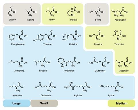
Name the monomers of proteins
amino acids
Describe the structure of an amino acid and explain the relevance of the R group
An amino acid consists of a central carbon atom attached to four groups: an amino group (-NH₂), a carboxyl group (-COOH), a hydrogen atom (H), and a variable R group (side chain). The R group is what makes each amino acid unique. It determines the amino acid's properties, such as whether it’s hydrophobic, hydrophilic, acidic, or basic. These properties affect how the amino acid behaves in a protein, influencing its shape, function, and how it interacts with other molecules.
Describe the structures, functions, properties, and types of proteins.
Proteins are made of amino acids and have different shapes. They do jobs like speeding up reactions (enzymes), building structures (collagen), and carrying molecules (hemoglobin). Types include enzymes, structural proteins, transport proteins, antibodies, and hormones. Their properties depend on their amino acids.
Describe the 4 levels of protein structure and identify the types of chemical bonds and interactions that play a role at each level
primary structure - The sequence of amino acids in a chain. Peptide bonds link the amino acids.
secondary structure - Local folds like alpha helices and beta sheets. Hydrogen bonds help stabilize these folds.
tertiary structure - The overall 3D shape of the protein. Hydrophobic interactions, hydrogen bonds, ionic bonds, and disulfide bonds (strong covalent bonds between cysteine amino acids) hold the shape together
quaternary structure - Multiple protein chains (subunits) coming together. Non-covalent interactions (like hydrogen bonds and hydrophobic interactions) or covalent bonds (like disulfide bonds) hold the subunits together
Explain why a change in the amino acid sequence of a protein can impact that proteins function
A change in the amino acid sequence can alter a protein's shape, making it unable to function properly, like failing to bind or catalyze reactions. Even small changes can disrupt its activity.
Explain how denaturation affects protein structure
Denaturation is when a protein’s structure unravels, losing its shape. This happens due to factors like heat or pH changes. Without its proper shape, the protein can't function correctly.
Be able to identify amino acids from pictures.
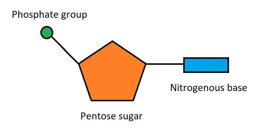
1E. Nucleic Acids (3.15 - 3.16)
Name the monomers of nucleic acids
nucleotides
Compare the structures and functions of DNA and RNA, noting similarities and differences.
DNA - double-stranded, has the sugar deoxyribose, and uses the bases A, T, C, G. It stores genetic information.
RNA - single-stranded, has the sugar ribose, and uses A, U, C, G (with uracil instead of thymine). It helps in protein synthesis and carries genetic information from DNA.
Be able to identify nucleotides from pictures.
Enzymes Quiz:
1F. Chemical reactions either release or store energy (5.11)
Vocab: endergonic reaction, energy coupling, exergonic reaction, metabolic pathway, metabolism
endergonic reaction - chemical reaction that requires energy to occur, absorbs energy from surroundings
energy coupling - process where energy from an exergonic (energy-releasing) reaction is used to drive an endergonic (energy-absorbing) reaction
exergonic reaction - chemical reaction that releases energy, the products have less energy than the reactants, so energy is given off, often as heat or light
metabolic pathway - series of chemical reactions in a cell, where the product of one reaction becomes the reactant for the next
metabolism - chemical reactions that happen in a cell or organism to maintain life; breaking down molecules for energy (catabolism) and building new molecules (anabolism)
Identify endergonic and exergonic reactions (should be able to identify from graph)
:max_bytes(150000):strip_icc()/endergonic-vs-exergonic-609258_final-2904b2c359574dfcb65a9fca2d54179a.png)
Describe the relationship between catabolic or anabolic reactions with endergonic or exergonic reactions
Catabolic reactions break down molecules and release energy, so they are exergonic.
Anabolic reactions build molecules and require energy, making them endergonic.
In short, catabolic = exergonic (energy-releasing) and anabolic = endergonic (energy-consuming).
1G. How Enzymes function (5.13)
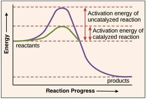
Vocab: activation energy, catalyst, energy barrier, enzyme
activation energy - energy needed to start a chemical reaction. It’s like a "push" that helps molecules react with each other
catalyst - substance that speeds up a chemical reaction without being used up in the process. It lowers the activation energy needed for the reaction to occur.
energy barrier - amount of energy needed to start a chemical reaction. It’s the "hurdle" that reactants must overcome for a reaction to happen.
enzyme - protein that speeds up chemical reactions in the body. It lowers the energy needed for a reaction to occur, making processes like digestion and metabolism happen faster.
Explain why cells need enzymes
Cells need enzymes because they speed up chemical reactions, allowing important processes to happen quickly and efficiently at the right temperature. Without enzymes, reactions would be too slow to support life.
Describe the relationship between enzymes and activation energy
Enzymes lower the activation energy needed for a reaction to start. This makes reactions happen faster and more easily.
Be able to read and label enzyme graphs using the terms: activation energy, exergonic or endergonic reaction, reactants, and products
1H. A specific enzyme catalyzes each cellular reaction (5.14)
Vocab: active site, allosteric site, cofactors, coenzymes, induced fit, substrate
active site: the region on an enzyme where the substrate binds and a chemical reaction is catalyzed
allosteric site: different part of an enzyme where molecules can bind, changing the enzyme's shape and affecting its activity. It’s like a “control” switch for the enzyme
cofactors: small molecules or ions that help enzymes work properly
coenzymes: type of cofactor, usually organic molecules, that help enzymes by carrying chemical groups or electrons during reactions
induced fit: model describing how an enzyme's active site changes shape when the substrate binds
substrate: the molecule that an enzyme acts on to help start a chemical reaction
Describe how protein structure relates to specificity of enzymes
Enzymes are proteins with a unique shape. This shape allows them to only bind to certain molecules (substrates), like a key fitting into a lock. This is why each enzyme works with specific substances.
Explain the steps of a catalyzed reaction (the catalytic cycle)
binding - The substrate (molecule) attaches to the enzyme’s active site.
reaction - The enzyme helps convert the substrate into a product by speeding up the reaction.
release - The product is released from the enzyme.
reset - The enzyme is ready to bind to a new substrate and repeat the process
Identify factors that influence the optimal conditions for enzyme function
Enzyme activity depends on temperature, pH, substrate concentration, enzyme concentration, and salt levels. Each enzyme has ideal conditions, and if these conditions are off, the enzyme may not work well.
Predict the effects that a change in one of these factors would have on enzyme activity
Changes in temperature, pH, substrate, enzyme concentration, and salt levels can slow down or stop enzyme activity. Each factor has an optimal range for the enzyme to work best.
UNIT 2: Cell Structure, Function, Specialization
2B. Introduction to the Cell (4.1 - 4.4)
Describe the structure of a plasma membrane and relate these parts to the functions of the plasma membrane. (also see 3.10, 5.1, 5.2)
The plasma membrane is a phospholipid bilayer with proteins, cholesterol, and carbohydrates. It controls what enters and exits the cell, supports the cell, and helps with communication and recognition.
Distinguish between the structures of prokaryotic and eukaryotic cells. (more details in 4.5-4.22)
Prokaryotic cells are simpler, smaller, and lack a nucleus. Their DNA is in a region called the nucleoid. They also don't have membrane-bound organelles, like mitochondria or the endoplasmic reticulum. Examples include bacteria and archaea.
Eukaryotic cells are larger and more complex, with a defined nucleus that holds their DNA. They have membrane-bound organelles, such as the mitochondria, Golgi apparatus, and endoplasmic reticulum. Examples include plant, animal, and fungal cells.
Explain why compartmentalization is important in eukaryotic cells.
Compartmentalization in eukaryotic cells helps separate different functions into specific areas, making processes more efficient and controlled. It allows the cell to perform complex tasks without interference between them.
Describe the structures and functions of the four basic functional compartments of eukaryotic cells. (also see 4.22)
Eukaryotic cells have four key parts: the nucleus (controls the cell), the cytoplasm (holds organelles), the endomembrane system (processes proteins and waste), and the mitochondria (produce energy). These parts help the cell function efficiently.
Compare the structures of plant and animal cells. Note the function of each cell part.
Plant and animal cells both have a nucleus, cytoplasm, and mitochondria. Plant cells also have a cell wall, chloroplasts for photosynthesis, and a large vacuole for storage. Animal cells have lysosomes for waste breakdown and centrioles for cell division. These structures help each cell type do its specific job.
Describe the parts of the cell theory.
The cell theory has three main ideas: First, all living things are made of cells. Second, the cell is the basic unit that carries out all life functions. Third, all cells come from existing cells through cell division. These ideas are essential for understanding how life works at the cellular level.
2C. The Endomembrane System (4.7 - 4.12)
Describe the structures and functions of the components of the endomembrane system, including smooth and rough endoplasmic reticulum, Golgi apparatus, lysosomes, vacuoles, vesicles, and peroxisomes.
smooth ER - network of tubules and sacs that lack ribosomes on their surface, giving it a smooth appearance. a part of lipid production, detoxification, and calcium storage
rough ER - network of flattened sacs covered with ribosomes on its surface, giving it a rough appearance. a part of protein synthesis, protein folding, and protein transport.
reticulum - network of membranes within the cell, which can be rough (with ribosomes) or smooth (without ribosomes). a part of transport, synthesis, and storage of proteins
golgi apparatus - made up of stacked, flattened sacs call cisternae. A part of protein modification, packaging, and transport
lysosomes - small, membrane-bound sacs containing digestive enzymes. A part of digestion and cell cleanup
vacuoles - membrane-bound sacs filled with fluid or stored substances. They are larger in plant cells. A part of storage and support.
vesicles - small, membrane-bound sacs. A part of transport and storage
peroxisomes - small, membrane-bound organelles containing enzymes. A part of detoxification and fat metabolism
2D. Energy-Converting Organelles (4.13 - 4.15)
Compare the structures and functions of chloroplasts and mitochondria.
both energy-producing organelles, but they have different functions and structures
Chloroplasts - The structure of chloroplasts is highly specialized to support their function in photosynthesis:
Outer membrane: Semi-permeable barrier.
Inner membrane: Selective permeability and contains transport proteins.
Intermembrane space: Space between the two membranes.
Stroma: Fluid-filled space containing enzymes, DNA, and ribosomes; site of the Calvin cycle.
Thylakoids: Membrane-bound sacs containing chlorophyll; site of light-dependent reactions.
Grana: Stacks of thylakoids, increasing surface area for light absorption.
Lamellae: Membranes that connect thylakoid stacks.
Chlorophyll: Pigment in thylakoids that captures light energy for photosynthesis.
Chloroplast DNA: Genetic material within the chloroplast, encoding some of its proteins.
Ribosomes: Sites for protein synthesis within the chloroplast.
Mitochondria - membrane-bound organelles found in eukaryotic cells, often referred to as the “powerhouses” of the cell because they generate the majority of the cell's energy in the form of adenosine triphosphate (ATP)
Outer Membrane: Smooth and permeable to small molecules and ions, provides a protective barrier.
Intermembrane Space: Narrow space between the outer and inner membranes, involved in ion gradients and metabolism.
Inner Membrane: Highly folded to form cristae; contains proteins for ATP production and electron transport.
Cristae: Folds of the inner membrane that increase surface area for biochemical reactions.
Matrix: The innermost region containing enzymes for the citric acid cycle, mitochondrial DNA, and ribosomes.
Mitochondrial DNA (mtDNA): Circular DNA that encodes some mitochondrial proteins and is maternally inherited.
Ribosomes: Synthesize proteins encoded by mitochondrial DNA
Describe the evidence that suggests that mitochondria and chloroplasts evolved by endosymbiosis.
Mitochondria and chloroplasts are thought to have evolved from bacteria through endosymbiosis. Evidence includes their similar size and shape to bacteria, their own circular DNA, binary fission replication, and similarities in ribosomes and enzymes to certain bacteria, supporting the idea they originated from ancient bacteria.
2E. The Cytoskeleton and Cell Surfaces (4.16 - 4.21)
Compare the structures and functions of microfilaments and microtubules.
Microfilaments are thin protein fibers made of actin, involved in cell movement and shape, while microtubules are thicker tubulin structures that support cell shape, enable intracellular transport, and form the mitotic spindle
identify how microtubules are dependent on centrosomes and
centrioles
Microtubules depend on centrosomes and centrioles for nucleation and organization, with centrioles helping form the mitotic spindle during cell division.
identify the proteins used to build microtubules and microfilaments
Microtubules are made of tubulin proteins, specifically alpha-tubulin and beta-tubulin, which combine to form tubulin dimers. Microfilaments are made of actin proteins, which polymerize to form long, thin filaments.
Relate the structure of cilia and flagella to their functions.
The structure of cilia and flagella is closely related to their function of cell movement.
Cilia are shorter and often numerous, beating in a coordinated, rhythmic pattern to move fluids, mucus, or the cell itself. Their structure enables rapid, back-and-forth motion.
Flagella are longer and usually occur singly or in pairs. They move in a whip-like motion, propelling cells through liquid. The longer structure and different motion pattern allow for efficient long-distance movement.
Relate the structure of the extracellular matrix to its functions.
Support: The ECM provides structural support for cells and tissues, maintaining tissue shape and strength.
Regulation: It regulates cell behavior, such as growth, differentiation, and migration, through biochemical signals.
Cell Communication: ECM components interact with cell receptors (like integrins), facilitating communication between the cell and its surroundings.
Compare the structures and functions of tight, anchoring, and gap junctions.
Tight junctions seal cells together to prevent leakage of substances. Anchoring junctions (like desmosomes) link cells and provide mechanical strength, holding tissues together. Gap junctions form channels between cells for direct communication, allowing the exchange of ions and small molecules.
Describe the structure and function of the cell wall and the role of plasmodesmata in plant cells
The cell wall is a rigid outer layer made of cellulose that provides support, protection, and maintains cell shape. Plasmodesmata are channels in the cell wall that connect plant cells, allowing the exchange of molecules and communication between cells.
2F. Terms:
cell theory - states that all living organisms are made of cells, the cell is the basic unit of life, and all cells come from pre-existing cells
cell wall - a rigid outer layer that surrounds plant, fungal, and bacterial cells. It provides support, protection, and helps maintain the shape of the cell.
cellular metabolism - Cellular metabolism refers to all the chemical reactions that occur within a cell to maintain life. It includes two main processes of catabolism and anabolism
central vacuole - The central vacuole is a large, fluid-filled space in plant cells. It stores water, nutrients, and waste products, and helps maintain the cell's shape by providing turgor pressure.
centrosome - The centrosome is a region in animal cells that organizes microtubules. It contains centrioles and helps control cell division by forming the mitotic spindle that separates chromosomes during cell division.
chloroplasts - Chloroplasts are organelles in plant cells that capture sunlight and use it to make food (glucose) through photosynthesis. They contain a green pigment called chlorophyll, which helps absorb light.
chromatin - Chromatin is a complex of DNA and proteins found in the nucleus of eukaryotic cell
chromosome - a long, thread-like structure made up of DNA and proteins that carries the genetic information in cells
cilia - tiny, hair-like structures that protrude from the surface of some cells
crista (plural, cristae) - crista is a fold or ridge inside the mitochondria
cytoplasm - is the jelly-like substance inside a cell that fills the space between the cell membrane and the nucleus
cytosol - is the fluid part of the cytoplasm in a cell
cytoskeleton - is a network of fibers inside a cell that gives the cell its shape and helps it maintain structure
electron microscope (EM) - is a special type of microscope that uses electrons instead of light to see very small things, much smaller than what a regular light microscope can see
endomembrane system - is a group of related organelles inside a cell that work together to make, modify, and transport proteins and lipids (fats)
endoplasmic reticulum (ER) - is a network of membranes inside a cell that helps make and transport proteins and lipids (fats)
endosymbiont theory - suggests that certain organelles in eukaryotic cells, like mitochondria and chloroplasts, were once independent bacteria that were engulfed by a host cell
eukaryotic cell - is a complex cell with a nucleus and organelles, found in organisms like animals, plants, and fungi
extracellular matrix (ECM) - is a network of substances outside the cells that provides structure and support to tissues in the body
flagellum (plural, flagella) - is a long, whip-like structure that some cells use for movement. It's like a tail that helps the cell swim
glycoprotein - is a protein with sugar attached to it, helping it perform functions like cell communication or immune response
Golgi apparatus - helps package and deliver proteins in the cell
granum (plural, grana) - is a stack of tiny, pancake-like structures in a plant cell's chloroplast. They help with making food by capturing light during photosynthesis
integrins - are proteins that help cells stick to their surroundings and communicate with other cells
intermediate filaments - are strong, thread-like structures inside cells that help give them shape and support
lysosome - is like the cell's "garbage disposal." It breaks down waste, unwanted materials, and old parts of the cell
microfilaments - are tiny, thread-like structures in cells that help with movement, shape, and support
microtubules - are tube-like structures in cells that help with support, shape, and transporting things inside the cell, like a cell's "highway system."
mitochondrial matrix - is the jelly-like substance inside mitochondria. It contains enzymes that help produce energy for the cell
mitochondrion (plural: mitochondria) - is a part of the cell that produces energy
nuclear envelope - is the outer layer that surrounds the nucleus in a cell
nucleoid - is the region in a cell (like a bacteria) where the DNA is located
nucleolus - is a small structure inside the nucleus where ribosomes are made
nucleus (plural: nuclei) - is the control center of the cell
organelles - are tiny parts inside a cell that do specific jobs, like a cell’s “tools” or “machines”
peroxisomes - are small structures in cells that help break down harmful substances and fatty acids. They also help detoxify the cell
plasma membrane - is the outer layer of the cell that controls what enters and leaves, acting like a barrier and protector
plasmodesma (plural: plasmodesmata) - a tiny channel that connects plant cells, allowing them to share materials and communicate with each other
prokaryotic cell - is a simple type of cell without a nucleus. Its DNA floats freely in the cell. Bacteria are examples of prokaryotic cells
ribosome - are tiny structures in cells that make proteins. They can be found floating in the cell or attached to the endoplasmic reticulum
rough endoplasmic reticulum - is a part of the cell that helps make and transport proteins. It has ribosomes on its surface, which give it a "rough" appearance
scanning electron microscope (SEM) - is a tool that uses electrons to create detailed images of tiny objects, like cells or surfaces, much clearer than a regular microscope
smooth endoplasmic reticulum - is a part of the cell that makes and transports fats and helps detoxify harmful substances. It doesn’t have ribosomes on its surface.
stroma - is the thick fluid inside chloroplasts in plant cells. It surrounds the structures where photosynthesis happens and helps produce energy for the plant
thylakoid - is a membrane structure inside chloroplasts where photosynthesis happens. It's like a small "pocket" that captures light energy
transmission electron microscope (TEM) - uses electrons to look at very small objects by passing them through the sample. It gives detailed images of the inside of tiny things, like cells
transport vesicle - is a small bubble that carries molecules, like proteins, around inside the cell. It helps move things from one part of the cell to another
vacuoles - are storage sacs in cells that hold water, nutrients, and waste. In plant cells, they also help maintain shape
Vesicle - is a small, bubble-like structure in a cell that stores or transports materials
2G. Describe the structure and identify the functions of cell membranes and the integral membrane proteins.
cell membrane - primarily composed of a phospholipid bilayer - which acts as a barrier separating the cell's interior from its environment
integral membrane proteins - embedded within the cell membrane bilayer - perform various functions (ex. transport, cell signaling, and cell adhesion)
2H. Explain how cholesterol helps maintain membrane fluidity.
Cholesterol acts like a "buffer" - (at low temperatures) to prevents phospholipids from packing too tightly together and increasing fluidity - (high temperatures) restricts their movement, preventing the membrane from becoming too fluid
2J. Relate the structure of phospholipid molecules to membrane structure.
phospholipid molecule - a hydrophilic head and a hydrophobic tail - allows them to spontaneously arrange themselves into a bilayer structure when in water; forming the basis of a cell membrane where the hydrophilic heads face outward towards the aqueous environment and the hydrophobic tails are buried in the interior of the membrane
2K. Define diffusion and describe the key features of passive transport
Diffusion - natural movement of particles from a region; high concentration to low concentration (no need for protein use)
2I. Osmosis can be defined as the diffusion of water across a membrane. Explain the process of osmosis. Describe the 2 ways that water crosses the membrane. Include the role of aquaporins. draw it out too
Osmosis - process where water moves from an area of lower concentration of solutes to an area of higher concentration of solutes through a semi-permeable membrane - happens to balance the concentration of solutes on both sides of the membrane
two main ways water crosses the membrane: simple diffusion - water molecules can pass directly through the lipid bilayer of the membrane; aquaporins - protein channels embedded in the membrane - allow water to pass through much more quickly - help water molecules move efficiently across the membrane by creating a pathway that bypasses the hydrophobic interior of the lipid bilayer
osmosis helps maintain balance in the concentration of solutes by moving water in and out of cells, and aquaporins play a key role in speeding up this process
2J. Distinguish between hypertonic, hypotonic, and isotonic solutions. draw it out too
hypertonic solution - higher concentration of solutes than a cell; water moves out of the cell and shrinks
hypotonic solution - lower concentration of solutes; water moves into the cell and swells
isotonic solution - same solute concentration as a cell; no net movement of water and no change in cell size
2K. Explain how animal and plant cells change due to hypertonic or hypotonic solutions.
When placed in a hypertonic solution, cells will shrink bc water moves out of the cell through osmosis
plant cells have a cell wall, so they undergo plasmolysis (the cell membrane pulls away from the wall) - become turgid bc walls prevent lots of swelling
animal cells simply shrivel up (in hypotonic solution swell and burst due to water influx)
2L. Explain how transport proteins facilitate diffusion. Identify the types of molecules that are transported across the membrane by facilitated diffusion.
Transport proteins (channel proteins and carrier proteins) - facilitate diffusion by providing a pathway across the cell membrane for molecules (large, polar, or charged) to pass through the lipid bilayer on their own, allowing them to move down their concentration gradient without requiring additional energy
small, non-polar, non-charged molecules aren’t let in
2M. Compare (both similarities and differences) the processes of active transport and facilitated diffusion
similarites - both processes use membrane proteins to move molecules across a cell membrane
differences - active transport: requires energy to move molecules against their concentration gradient - facilitated diffusion: process that moves molecules down their concentration gradient with the help of transport proteins, but without using additional energy
2N. Distinguish between exocytosis, endocytosis, phagocytosis.
Exocytosis - process for moving large molecules out of the cell to the cell exterior
Endocytosis - process by which cells take in substances from outside of the cell by engulfing them in a vesicle
Phagocytosis - process where a cell binds to the item it wants to engulf on the cell surface and draws the item inward while engulfing around it
2O. Identify the mechanism used to move the following materials across the plasma membrane: ions, sugars, water, oxygen, amino acids, carbon dioxide, food particles, bacteria, hormones and other proteins that are secreted by the cell draw it too
simple diffusion - small, uncharged, hydrophobic molecules - oxygen, carbon dioxide
facilitated diffusion - polar molecules - ions, sugars, amino acids, water
active transport - molecules move against their concentration gradient - other
bulk transport - large molecules - bacteria, food particles, hormones
2P. Key Terms
active transport - process of the movement of molecules from a region of lower concentration to a region of higher concentration against a gradient or an obstacle with the use of external energy
aquaporin - specialized protein channels embedded in cell membranes that allow water to quickly pass through
ATP - (adenosine triphosphate) a molecule considered the energy currency of cells - stores and provides the energy needed for cellular processes
concentration gradient - difference in the ammount a substance between two areas - move from region of high concentration to a region of low concentration
diffusion - the movement of molecules from an area of high concentration of the molecules to an area with a lower concentration
endocytosis - process where a cell takes in substances from its enviornment by engulfing them with its cell membrane
exocytosis - process by which cells move materials from within the cell into the extracellular fluid
facilitated diffusion - type of passive transport that uses specialized proteins, such as channel proteins and carrier proteins, to help molecules move across a cell membrane
fluid mosaic model - describes the fluid and flexible nature of the cell membrane and also the components it is made from
hypertonic - a solution with higher osmotic pressure than another solution
hypotonic - a solution that has a lower concentration of solute compared to the cell
isotonic - the state when two solutions have equal concentration of solutes across a semipermeable membrane
osmoregulation - the process by which an organism regulates the water and electrolytic balance in its body to maintain homeostasis
osmosis - movement of water molecules from a solution with a high concentration of water moleculed to a solution with a lower concentration of water molecules
passive transport - a type of membrane transport that does not require energy to move substanced across cell membranes
phagocytosis - the process by which certain living cells called phagocytes engulf other cells, partciled and even pathogens
receptor - mediated endocytosis - a procedd by which cells absorb metabolites, hormones, proteins, and sometimes viruses
tonicity - the ability of an extracellular solution to make water move into or out of a cell by osmosis