Cells, Tissues, and Skeletal system
Basics of Veterinary Anatomy and Physiology
Overview of anatomy and physiology focusing on cells, tissues, skeleton, and muscles.
Anatomy vs Physiology
Anatomy: The form and structure of the animal.
Physiology: The function of the body.
The Cell: The Basis of Life
Cells provide structure and function for body systems.
Cells form tissues, which form organs, which then form body systems.
Reference to a review video on cells: Cell Function Video.
Key Components of a Cell
Cytoskeleton: Supports organelles and plays a role in cell movement.
Nucleus: Command center with nucleolus for ribosome production.
Endoplasmic Reticulum:
Smooth: System of internal membranes.
Rough: Studded with ribosomes for protein synthesis.
Mitochondrion: Extracts energy from food via oxidative metabolism.
Ribosomes: Sites for protein synthesis.
Golgi Complex: Collects, packages, and distributes molecules made in the cell.
Other components include Lysosomes, Peroxisomes, Centriole, Plasma Membrane, and proteins like Actin Filaments and Intermediate Filaments.
Cell Function
Cells within organ systems serve different functions and divide (mitosis) to produce more cells.
Each cell type has a specific function within the body.
Tissues
Types of Tissues:
Epithelial: Covers surfaces, provides protection, and allows absorption/secretion.
Connective: Supports and binds other tissues (7 types including Adipose, Cartilage, Bone, and Blood).
Muscle: Facilitates movement (includes skeletal, cardiac, and smooth).
Nervous: Transmits signals throughout the body.
Epithelial Tissue Types
Simple Squamous: Single layer of flat cells.
Cuboidal: Cube-shaped cells.
Columnar: Column-like cells (includes Pseudostratified).
Stratified Squamous: Multiple layers of flat cells.
Connective Tissue Types
Adipose: Stores fat, cushioning.
Loose Connective: Flexible and supportive.
Dense Connective: Provides strength and support.
Cartilage: Firm but flexible tissue without blood vessels.
Bone: Hard tissue providing structure.
Blood: Transports nutrients and wastes throughout the body.
Skeletal System
Provides framework and shape.
Protects internal organs and serves as attachment points for muscles and ligaments.
Types and Functions of Bones
Long Bones: Support and movement (e.g., Femur).
Short Bones: Absorb shock in joints (e.g., Carpals).
Flat Bones: Protect internal organs (e.g., Skull).
Irregular Bones: Protection and muscle attachment (e.g., Vertebrae).
Pneumatic Bones: Contain air (e.g., Sinuses).
Sesamoid Bones: Found within tendons (e.g., Patella).
The cells that make up the bones are called osteocytes (these are the mature cell forms).
Types of Skeletons
Axial Skeleton: Skull, spine, ribs, sternum.
Appendicular Skeleton: Limb bones.
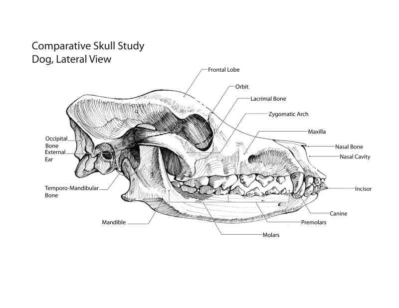
Canine Skull Anatomy
Orbital Bones: Surround the eye.
Nasal bones- contain the sinuses
Maxilla/Mandible: Upper/lower jaw.
Various bone features important for identification.
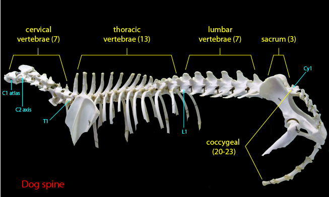
Vertebral Column
Cervical: C1-C7 (Atlas, Axis).
Thoracic: Attaches to ribs, limited movement.
Lumbar: Greater movement than thoracic.
Sacrum: Fused, rigid.
Coccygeal: Tail vertebrae.
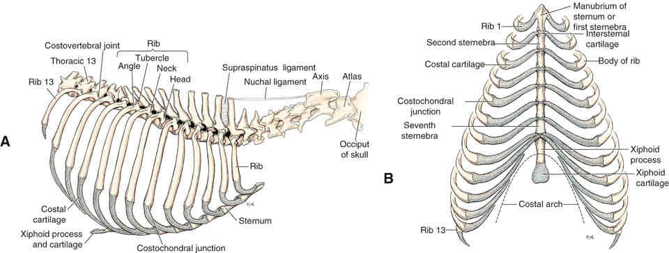
Ribs and Sternum
Ribs: True, false, and floating categories.
Sternum: Attachment for ribs and muscles.
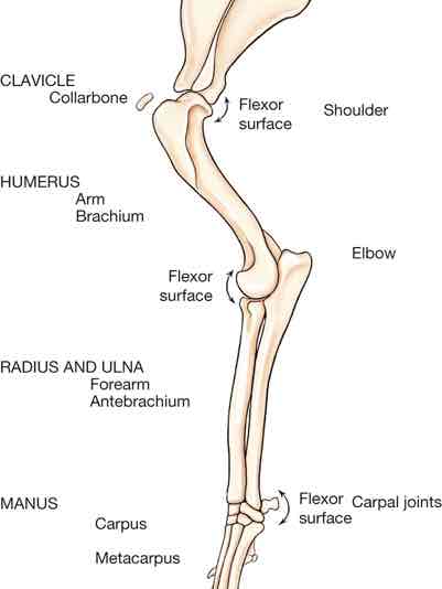
Limb Anatomy
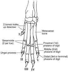
Forelimb: Humerus, Radius, Ulna, Carpus, Digits.
Hindlimb: Femur, Tibia, Fibula, Tarsals, Digits.
Dewclaw: Underdeveloped toe.
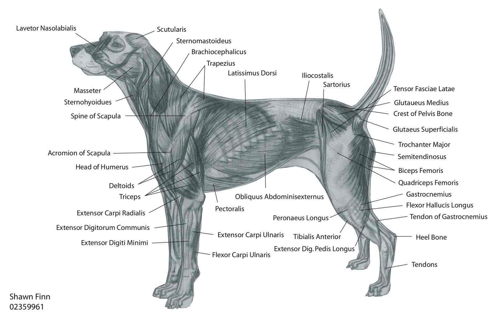
Muscle Types
Skeletal Muscle: Voluntary, striated muscles for movement (neuromuscular junction).
Cardiac Muscle: Involuntary, makes up the heart.
Smooth Muscle: Involuntary, found in internal organs (controlled peristalsis).
Additional Resources
Video links provided in lessons to aid understanding of complex concepts.
References
Sirois, M. (2021). Elsevier's Veterinary Assisting Textbook, 3rd Edition.