Muscle & Integumentary System
Unit 9 - Muscle & Integumentary System
- Muscles
- Nearly ½ body’s weight.
- ~600 human skeletal muscles.
- Specialized for one purpose → transforming ATP into mechanical energy of motion.
- Types:
- Cardiac
- Skeletal
- Smooth muscle
- Overview
1. Muscle
• Muscle Cells and Types of Muscle
• Muscle Tissue
• Muscles of the Head
• Muscles of the Thoracic and Abdominal Regions
• Muscles of the Shoulder and Upper Limb
• Muscles of the Hip and Lower Limb
2. Integument
• Anatomy of the Skin
• Anatomy of Hair, Nails, and Glands
- Muscular Tissue - Muscle Characteristics
- Muscle characteristics
- Excitability/responsiveness
- To chemical signals, stretch, and electrical signals.
- Conductivity
- Waves of excitation through muscle fibre.
- Contractility
- Shortens when stimulated.
- Extensibility
- Capable of being stretched between contractions.
- Elasticity
- Returns to its original rest length after stretching.
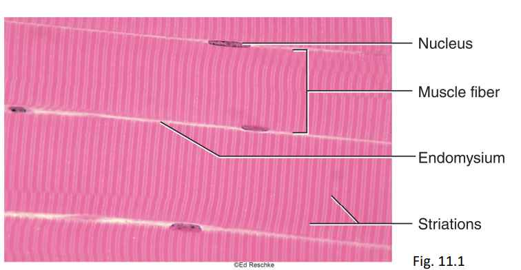
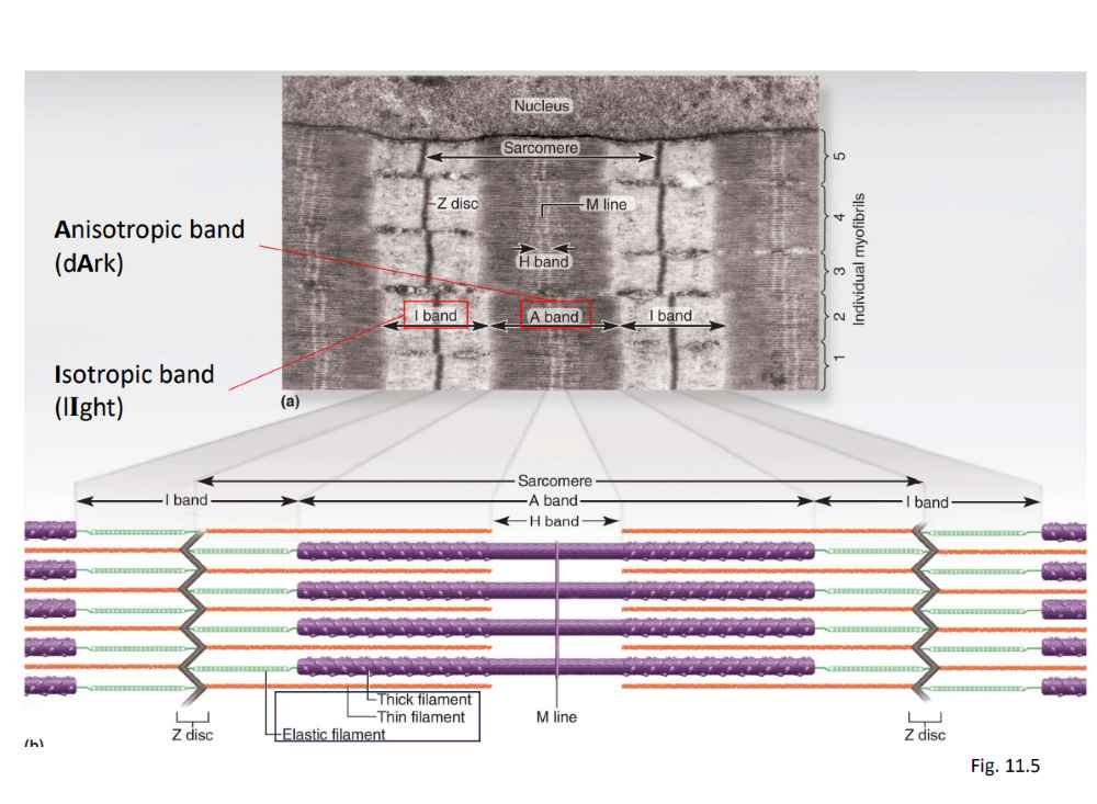
- Skeletal muscle
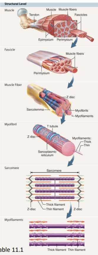
- Striated muscle attached to bone:
- Striations from arrangement of internal contractile proteins.
- Voluntary.
- Muscle cell = muscle fibre (myofibre):
- Can be 30cm long!
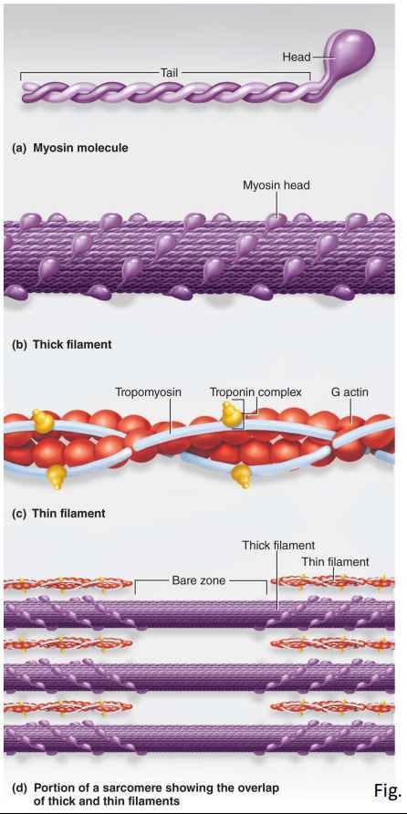
- Myofilaments
- Fibrous proteins that carry out the contraction:
- Thick:
- Hundreds of gold-club shaped myosin molecules.
- Heads directed outward in a helical array.
- Thin:
- Two intertwined strands of fibrous actin:
- String of globular actin subunits that can bind to head of myosin molecule.
- Elastic:
- Made of a huge springy protein called titin:
- Run through the core of each thick filament and anchor it.
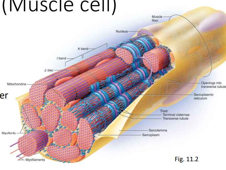
- Muscle Fiber (Muscle cell)
- Sarcolemma
- Plasma membrane of a muscle fibre.
- Plasma membrane of a muscle fibre.
- Sarcoplasm
- Cytoplasm of muscle fiber.
- Myofibrils
- Glycogen
- Myoglobin (Binds to 1 oxygen)
- Cytoplasm of muscle fiber.
- Multiple nuclei
- Many Mitochondria
- Sarcoplasmic reticulum (SR)
- Smooth ER forms a network around each myofibril.
- T (transverse) tubules - tubular infoldings of the sarcolemma which penetrate through the cell and emerge on the side.
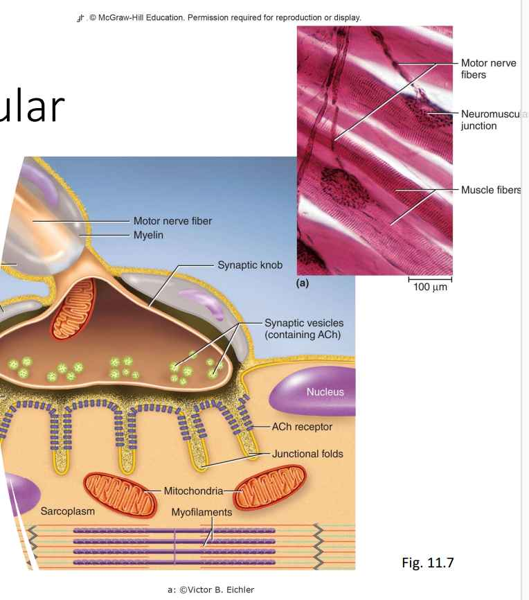
- Neuromuscular Junction
- Skeletal muscle only contracts when stimulated by a nerve.
- Denervation atrophy is shrinkage of a paralyzed muscle when a nerve is disconnected.
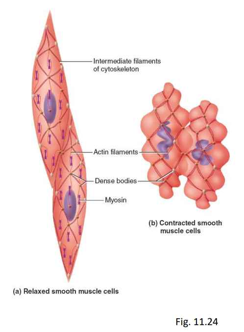
- Smooth Muscle
- Slower than other muscle types:
- Can remain contracted for a long time without fatigue.
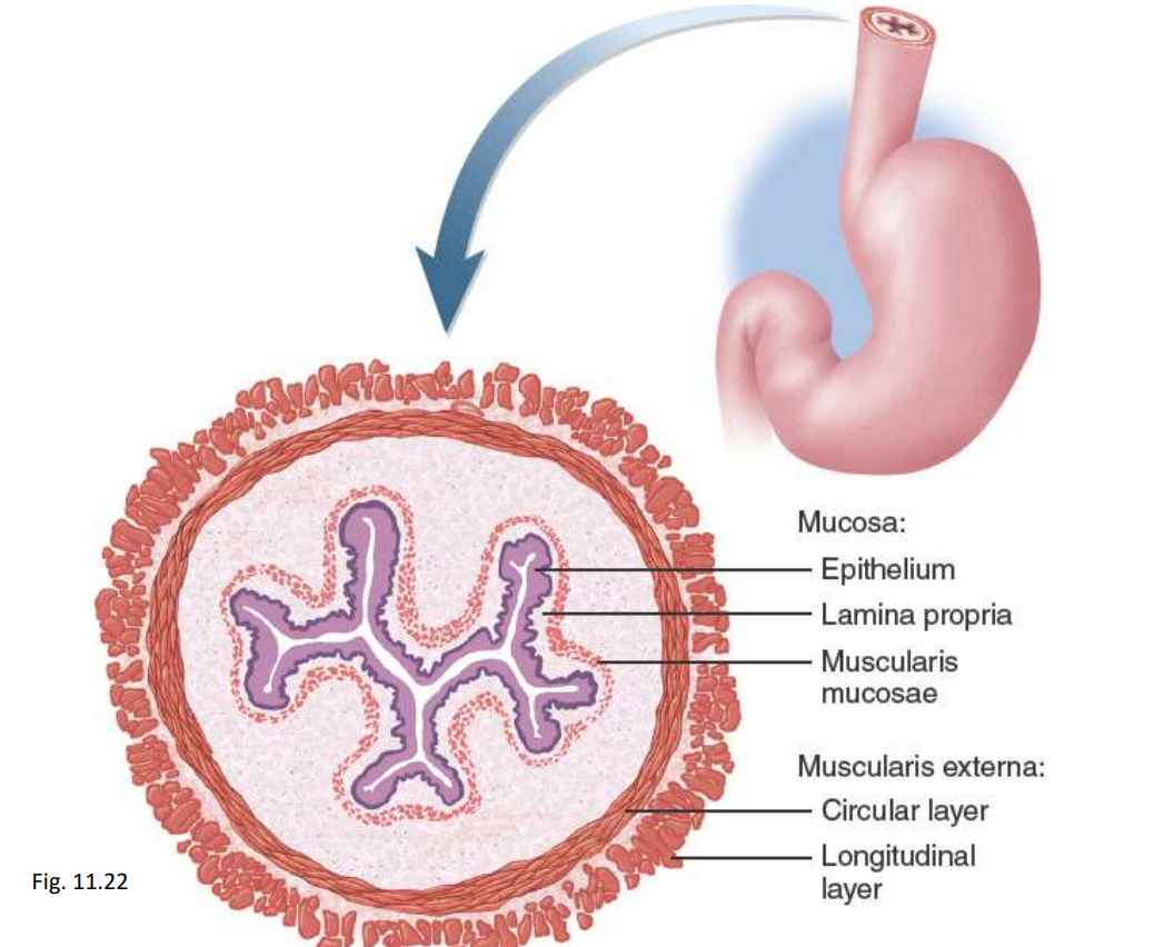
- Forms layers within walls of hollow organs.
- Can provide fine control in some locations:
- Piloerector muscles.
- Iris smooth muscle controls pupil size.
- Myocyte:
- Fusiform shape with one nucleus.
- Have thick and thin filaments, but not aligned with each other (no striations).
- Types of Smooth Muscle
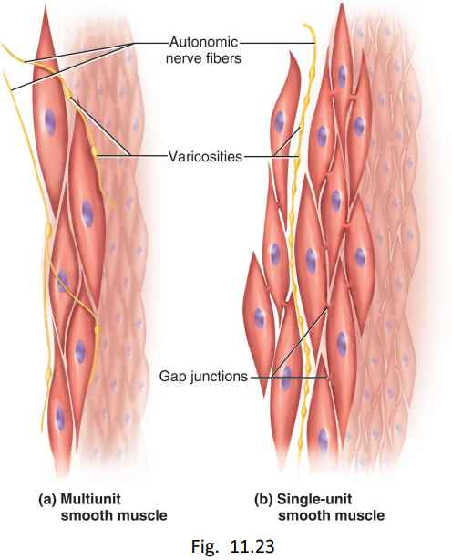
- Multiunit smooth muscle
- Occurs in some of the largest arteries and air passages, piloerector muscles, iris of eye.
- Autonomic innervation forms motor units:
- Each motor unit contracts independently.
- Single-unit smooth muscle:
- More common.
- Called visceral muscle, two layers, inner circular and outer longitudinal.
- Cells are electrically coupled via gap junctions.
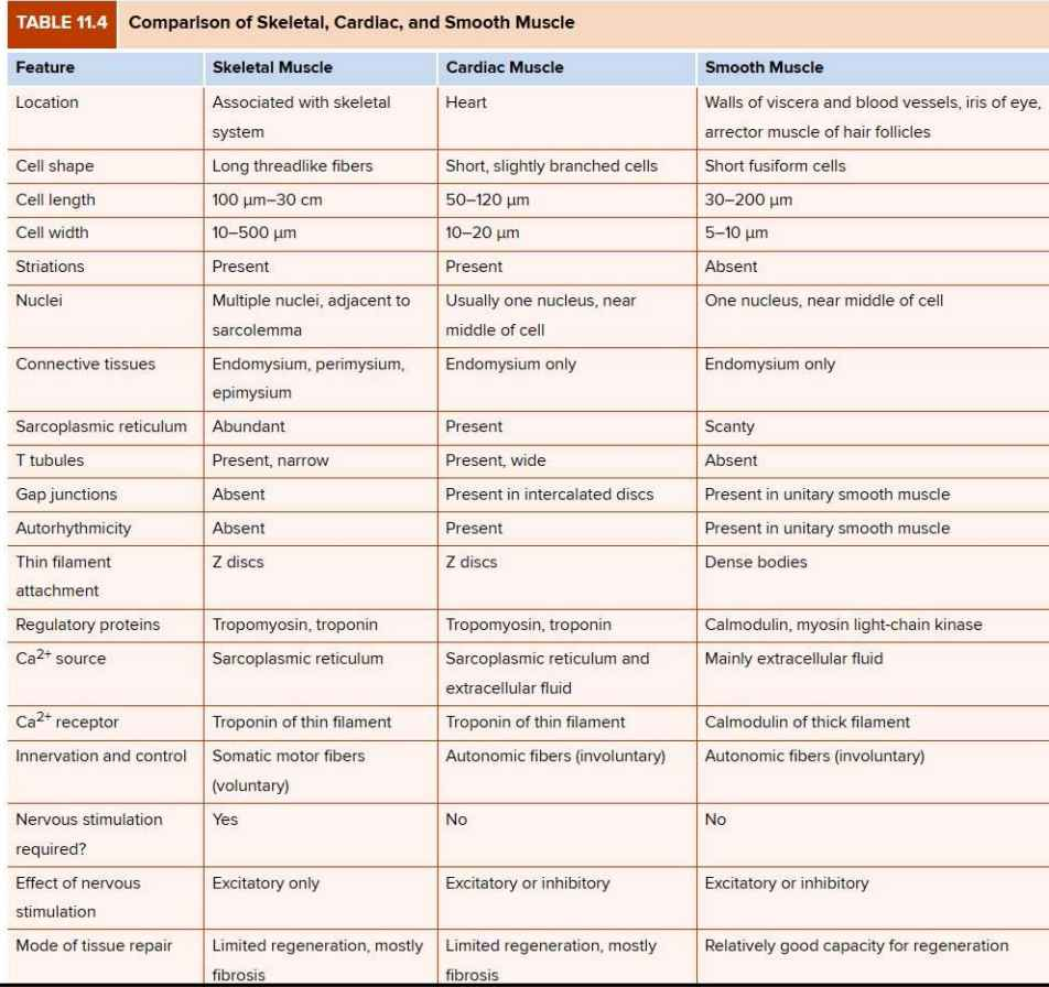
- Comparison of Skeletal, Cardiac, and Smooth Muscle:
- Functions of Muscle
- Movement
- Physically moving, moving body contents in breathing, circulation, and digestion.
- Communication (speech, writing, expressions)
- Stability
- Maintain posture by preventing unwanted movements, stabilize joints, and presence of antigravity muscles.
- Control of body openings and passages
- Sphincters are internal muscular rings that control movement of food, blood, etc.
- Heat production
- Skeletal muscle produces as much as 85% of our body heat.
- Glycemic control
- Absorb and store glucose, which helps regulate blood sugar.
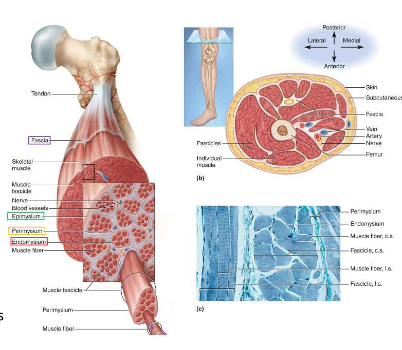
- Connective Tissues and Fascicles
- Endomysium
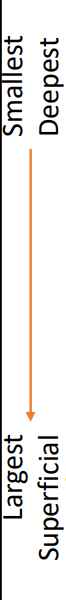
- Thin sleeve of loose connective tissue around each fibre.
- Perimysium
- Thicker layer of connective tissue that wraps fascicles (bundles of muscle fibres).
- Epimysium
- Fibrous sheath surrounding entire muscle.
- Fascia
- Sheet of connective tissue that separates neighbouring muscles or muscle groups from each other.
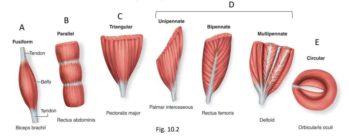
- Fascicles and Muscle Shapes
- Fusiform (thick in the middle, tapered at each end)
- Parallel (uniform with parallel fascicles)
- Triangular (convergent, fan-shaped)
- Pennate (feather-shaped), which has three types:
- Unipennate (muscle on one side of tendon)
- Bipennate (muscle on both sides of tendon)
- Multipennate (multiple “feathers”)
- Circular (sphincters, which form certain body openings)
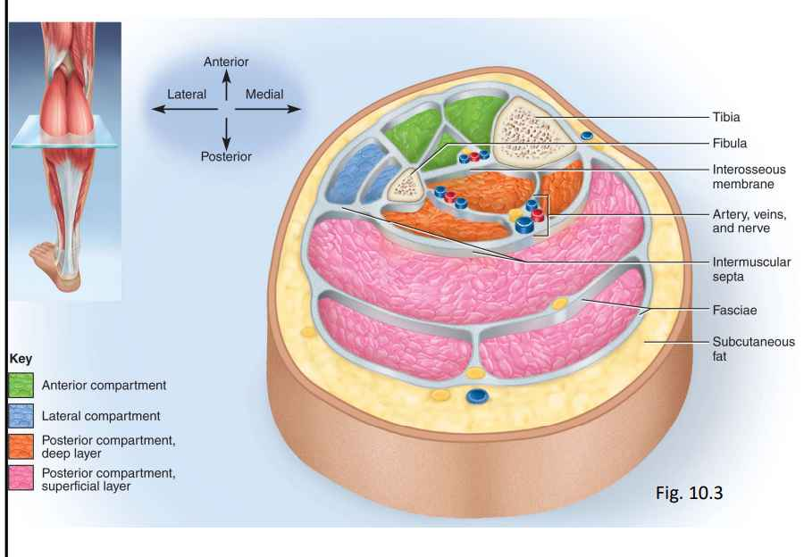
- Compartments
- Group of functionally related muscles enclosed by fascia:
- Contains nerves and blood vessels that supply the muscle group.
- Intermuscular septa:
- Very thick fascia that separate one compartment from another.
- Muscle Attachments
- Muscles attach to bones through extension of connective tissue components:
- Indirect:
- Tendons connect muscle to bone.
- Collagen fibers of the endo-, peri-, and epimysium continue into the tendon and from there into the periosteum and matrix of bone.
- Direct (fleshy):
- Little separate between muscle and bone.
- Muscle seems to emerge directly from bone (but there's a small gap between muscle and bone).
Indirect:

Direct:
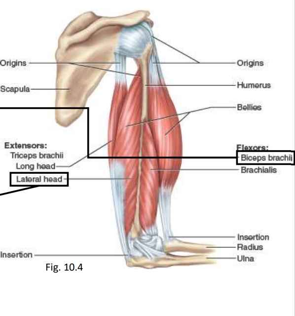
- Muscle Origins and Insertions
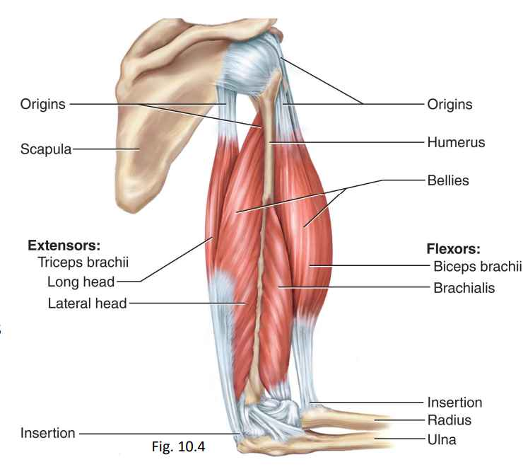
- Origin
- Bony attachment at stationary end of muscle.
- Belly
- Thicker, middle region between origin and insertion.
- Insertion
- Bony attachment to mobile end of muscle.
- Some anatomists prefer proximal vs distal or superior vs inferior.
- Functional Groups of Muscles
- An action is the effect produced by a muscle, whether it is to produce or prevent movement.
- Muscles seldom act independently, movement is the combined action of multiple muscles.
- Action of muscle depends on what other muscles are doing.
- Gastrocnemius usually flexes the knee.
- If quadriceps (anterior thigh) prevents knee flexion, gastrocnemius flexes the ankle, causing plantar flexion.
- Categories of Muscle Action
- Prime Mover (Agonist):
- Muscle that produces most of force during joint action.
- Synergist:
- Muscle that aids prime mover.
- Antagonist:
- Muscle that opposes prime mover.
- Often prevents overextension of joint.
- Muscle that opposes prime mover.
- Fixator
- Prevents a bone from moving.
- Rhomboids prevents scapula from moving.
- Prevents a bone from moving.
- For elbow flexion:
- Prime mover-brachialis.
- Synergist-biceps brachii.
- ANtagonist-triceps brachii.
- Fixator-rhomboids (hold scapula in place).
- Naming
- Latin names.
- Describes distinctive aspects of the structure, location, or action of a muscle.
- i.e - depressor labii inferioris. (Depressing the inferior part of labii)
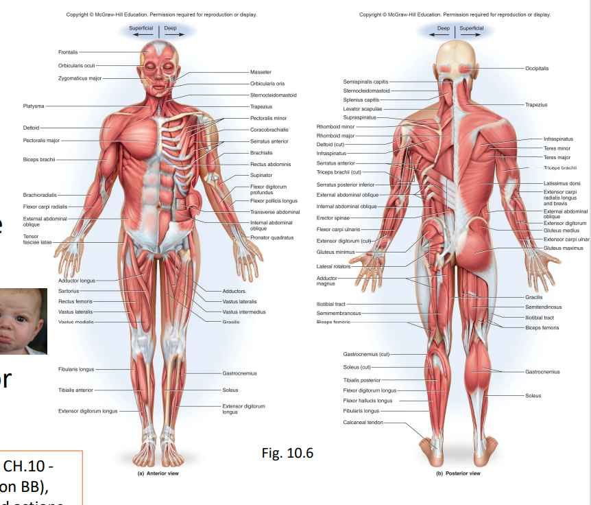
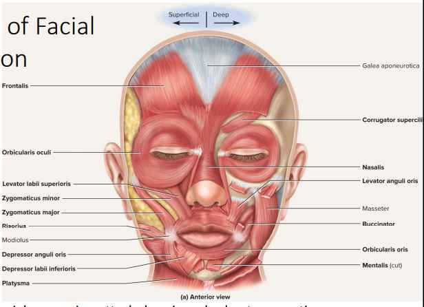
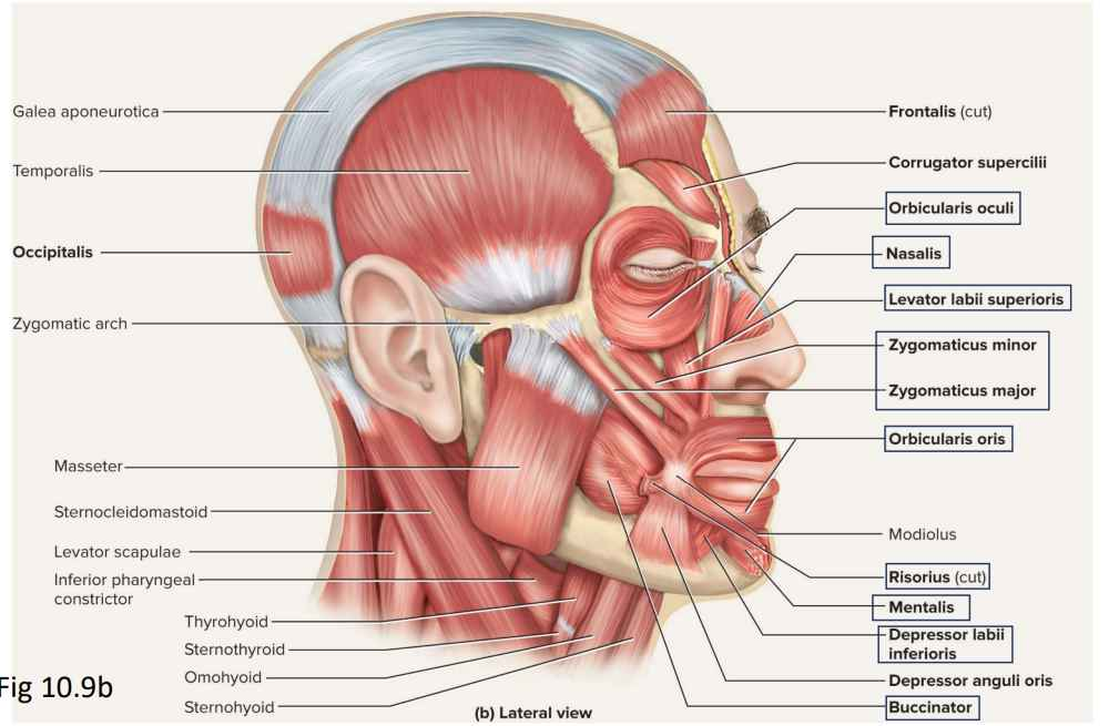
- Muscles of Facial Expression
- Muscles of facial expression attach dermis and subcutaneous tissues.
- Tense skin and produce facial expressions.
- Innervated by facial nerve (CN VII)
- Paralysis cause face to sag.
- Found in scalp, forehead, around the eyes, nose, and mouth, and in the neck.
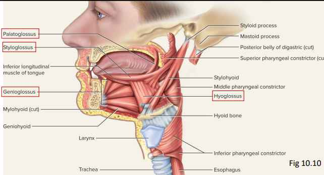
- Muscles of Chewing and Swallowing
- Extrinsic muscles of the tongue (Table 10.2):
- Tongue is very agile organ.
- Pushes food between molars for chewing (mastication).
- Forces food into the pharynx for swallowing (deglutition).
- Crucial importance to speech.
- Intrinsic muscle of tongue:
- Vertical, transverse, and longitudinal fascicles.
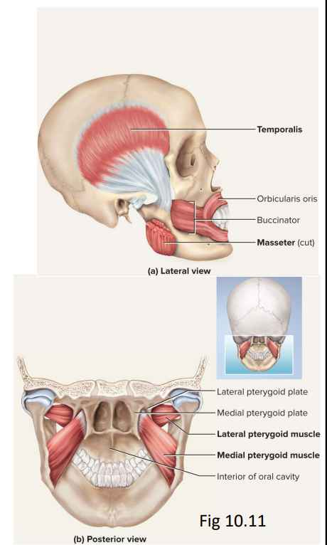
- Muscles of Chewing
- 4 pairs of muscles produce biting and chewing movements of mandible.
- Depression: to open mouth
- Elevation: biting and grinding
- Protraction: incisors can cut
- Retraction: make rear teeth meet
- Lateral and medial excursion: grind food.
- Temporalis
- Masseter
- Medial pterygoid
- Lateral pterygoid
- Innervated by mandibular nerve, a branch of the trigeminal (CN V)
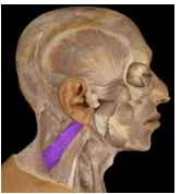
- Muscles Acting on the Head
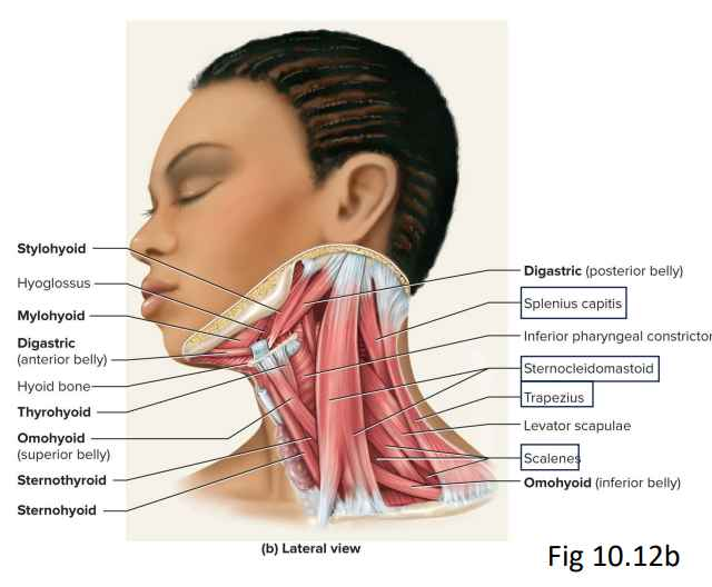
- Attachments:
- Inferior: vertebral column, thoracic cage, and pectoral girdle.
- Superior: cranial bones.
- Actions:
- Flexion (tipping head forward)
- Sternocleidomastoid
- Scalenes
- Extension (holding head erect)
- Trapezius
- Splenius capitis
- Semispinalis capitis
- Flexion (tipping head forward)
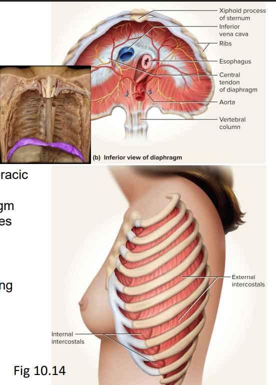
- Muscles of Respiration
- Breathing requires use of muscles enclosing thoracic cavity
- Diaphragm
- Muscular dome between thoracic and abdominal cavities.
- Contraction flattens diaphragm.
- Diaphragm rises when relaxes.
- External intercostals
- Elevate ribs.
- Expand thoracic cavity.
- Create partial vacuum causing inflow of air.
- Internal intercostals
- Depresses and retracts ribs.
Compresses thoracic cavity. - Expelling air.
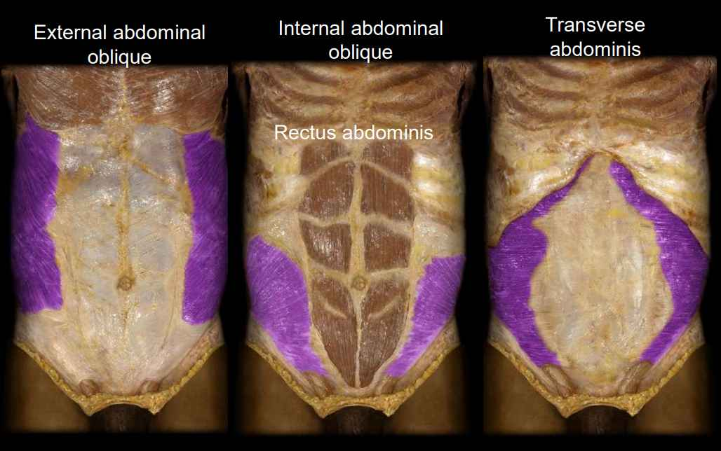
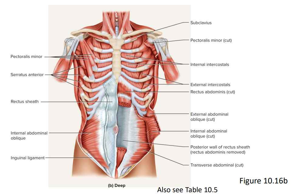
- Muscles that Support the Anterior Abdominal Wall
- External abdominal oblique:
- External layer of lateral abdominal muscles.
- Supports viscera, breathing, unilateral contraction causes contralateral rotation (twist) of waist.
- Internal abdominal oblique:
- Intermediate layer of lateral abdominal muscles.
- Unilateral contraction causes ipsilateral rotation (bend) of waist.
- Transverse abdominal:
- Deepest of lateral abdominal muscles.
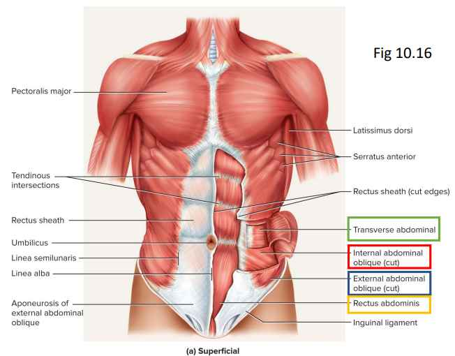
- Horizontal fibres.
- Compresses abdominal contents.
- Similar action to the external oblique.
- Deepest of lateral abdominal muscles.
- Rectus abdominis:
- Flexes lumbar vertebral column.
- Produces forward bending at waist.
- Extends from sternum to pubis.
- Segments (“six pack”).
- Lateral Abdominal Muscles
- Muscles of the Anterior Abdominal Wall
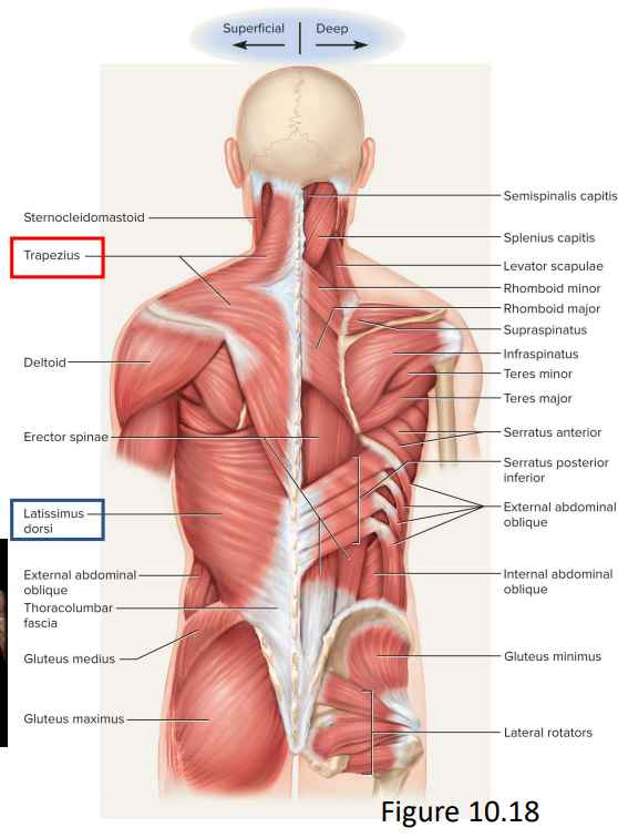
- Muscles of the Back
- Actions of back muscles involve:
- Extension, rotation, lateral flexion of vertebral column.
- Upper limb movement.
- Most prominent superficial back muscles:
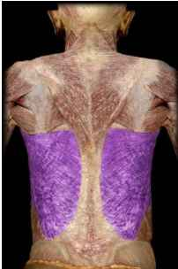
- Latissimus dorsi
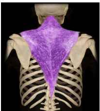
- Trapezius
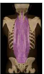
- Muscles Acting on Vertebral Column
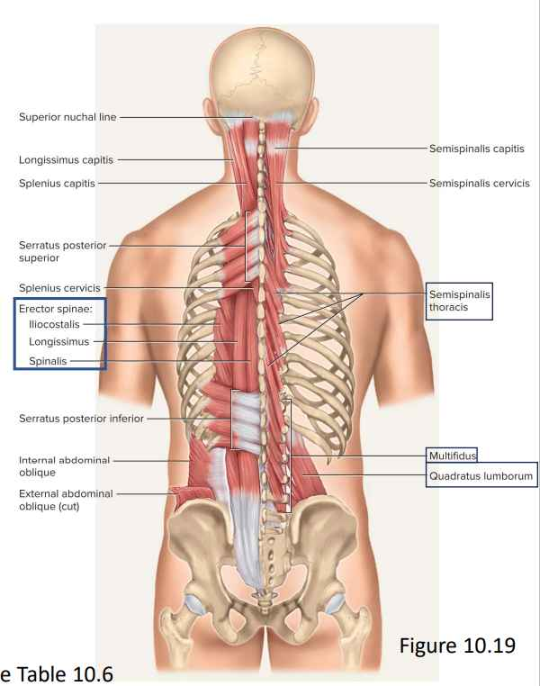
- Deep muscles of the back:
- Erector spinae
- Iliocostalis
- Longissimus
- Spinalis
- From cranium to sacrum.
- Extension and lateral flexion of vertebral column (sitting and standing erect, straighten back after one bends at waist).
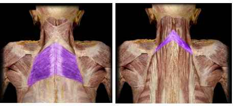
- Muscles Acting on the Shoulder
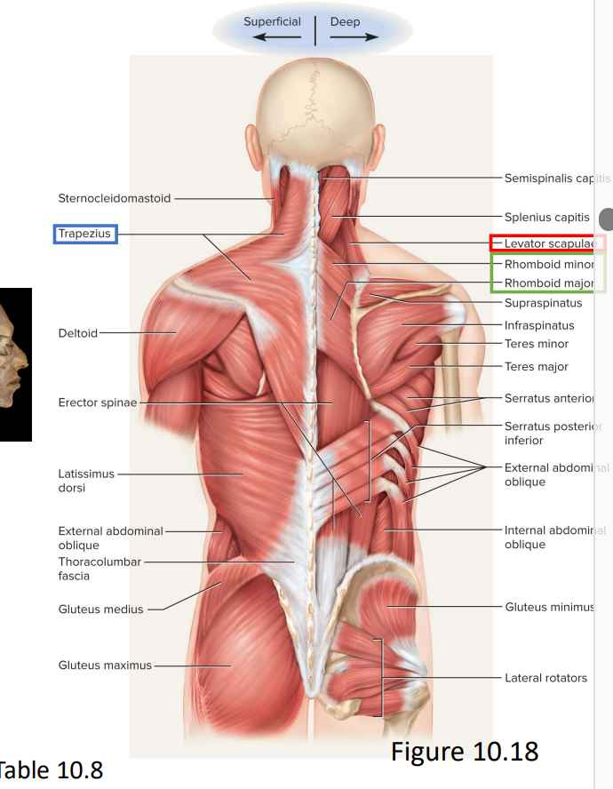
- Posterior Compartment of Pectoral Girdle
- Trapezius
- Levator Scapulae
- Rhomboids

- Muscles Acting on the Shoulder
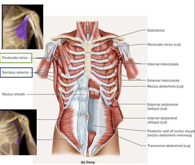
- Anterior compartment of pectoral girdle
- Pectoralis minor
- Ribs 3 to 5 coracoid process of scapula
- Draws scapula laterally
- Serratus anterior
- All ribs to medial border of scapula
- Draws scapula laterally and forward
- Prime mover for reaching and pushing
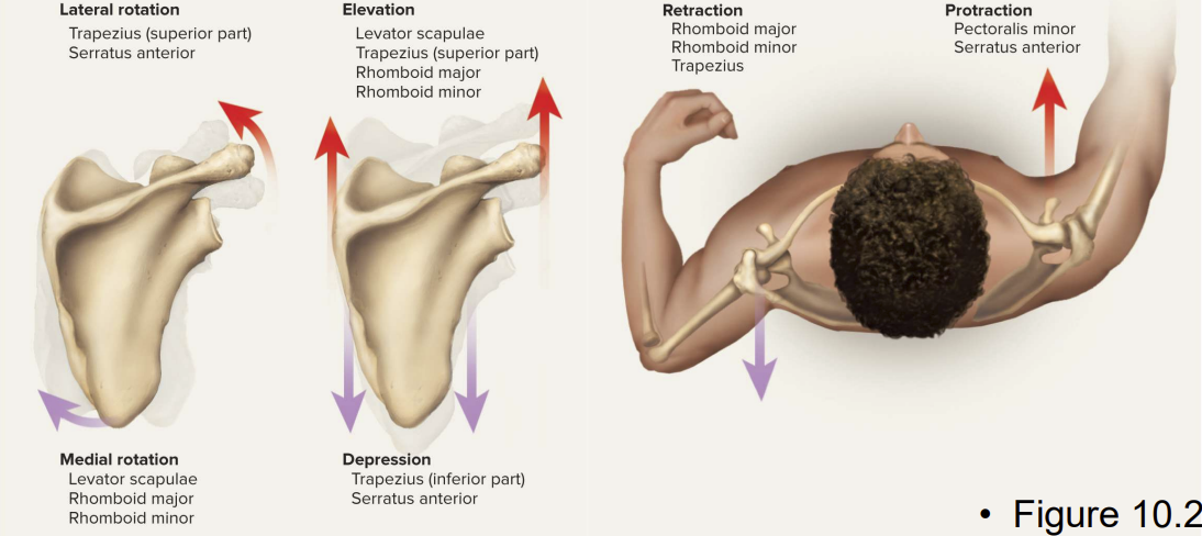
Muscles Acting on the Shoulder
- A group of muscles attach on the axial skeleton and also on clavicle or scapula
- Scapula loosely attached to thoracic cage
- Capable of great movement
- Rotation, elevation, depression, protection, retraction
- Clavicle braces the shoulder and moderates movement
Muscles Acting on the Shoulder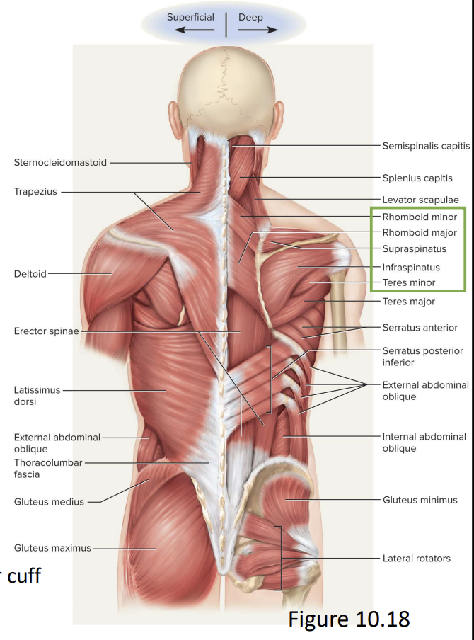
- Latissimus Dorsi
- Trapezius
- Deltoid
- Rhomboids
- Supraspinatus
- Infraspinatus
- Teres minor
- Erector spinae
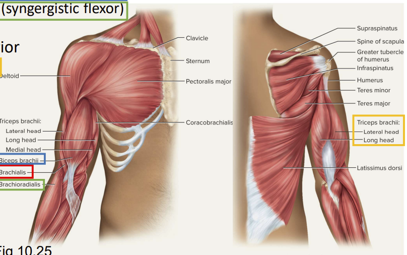
Muscles Acting on the Arm
- 9 muscles cross shoulder joint and attach to humerus
- 2 axial muscles attaching on axial skeleton
- Pectoralis major:
- Flexes, adducts, medially rotates humerus
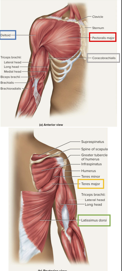
- Latissimus dorsi:
- Adducts and medially rotates humerus
- 7 muscles with scapular attachments
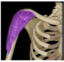
- Deltoid
- rotates and abducts arm
- Intramuscular injection site
- Teres major
- Extension and medial rotation of humerus

- Coracobrachialis
- Flexes and medially rotates arm
- Remaining 4 = rotator cuff,
- Reinforce shoulder joint
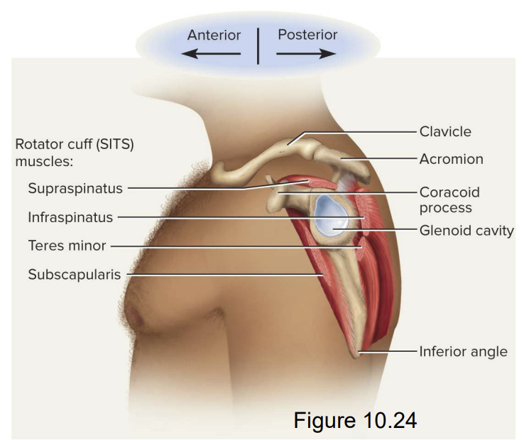
Muscles Acting on the Arm
Rotator Cuff Muscles
- Acronym “SITS muscles”
- Supraspinatus
- Infraspinatus
- Teres minor
- Subscapularis
- Tendons merge with joint capsule of shoulder as cross on route to humerus
- Holds head of humerus into glenoid cavity
- Supraspinatus tendon easily damaged
Muscles Acting on Elbow and Forearm
- Elbow and forearm capable of Flexion, Extension, Pronation, Supination
- Carried out by muscles in both brachium (arm) and anytebrachium, (forearm)
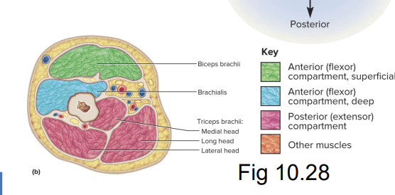
- Elbow Flexors: Anterior
- Brachialis (prime mover)
- Biceps brachii (synergistsic flexor)
- Brachioradialis (synergistic flexor)
- Elbow Extensor: Posterior
- Triceps brachii
- Elbow Flexors: Anterior
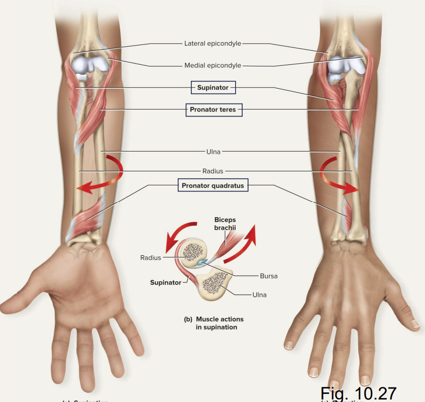
Actions of the Rotator Muscles on the Forearm
Supinator:
- Supinates forearm
Pronator quadratus:
- Pronates forearm
Pronator teres:
- Supports pronator quadartus in pronation
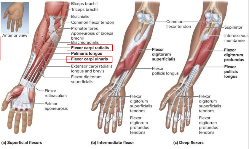
Flexor Muscles Acting on the Hand
Flexors
- Flexor carpi radialis
- Flexor carpi ulnaris
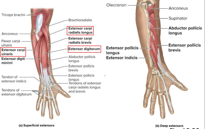
Extensor Muscles Acting on the Hand
Extensors
- Extensor digitorum
- Extensor carpi radialis longus
- Extensor carpi ulnaris
Overview
Muscles of thehip and lower limb
Muscles Acting on the Hip and Lower Limb
- Bodys largest muscles found in lower limb
- For strength needed to stad, maintain balance, walk, and run
- Several cross and act on two or more joints
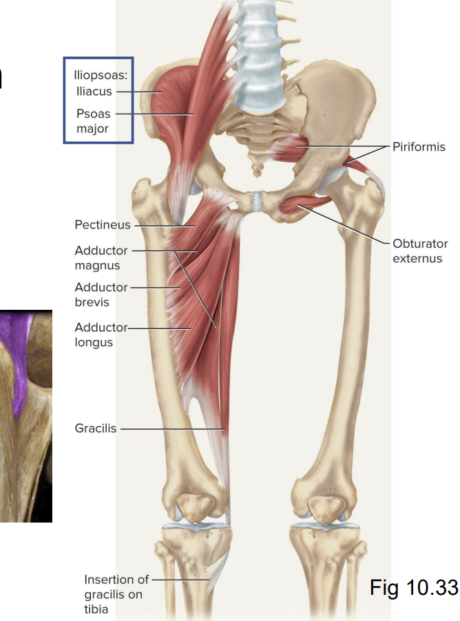
Muscles Acting on Hip and Femur:
Anterior muscles of hip:
- Iliacus
- Flexes thigh at hip
- I;iacus portion arises from iliac crest and fossa
- Psoas major
- Flexes thigh at hip
- Arises from lumbar vertebrae
- Common tendon on femur
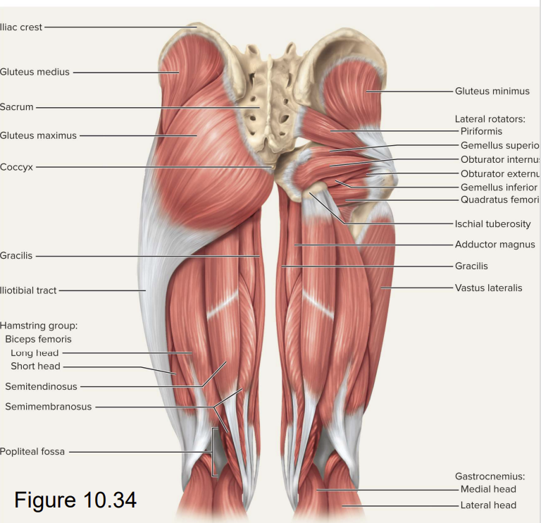
Muscles Acting on the Hip and Femur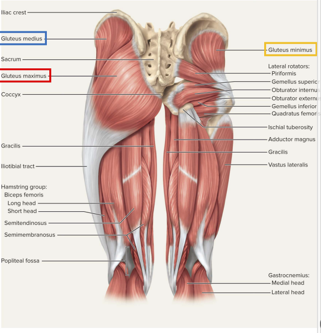
Lateral and posterior muscles of hip
- Gluteus maximus
- Forms mass of the buttock
- Prime hip extensor
- Provides most of lift when you climb stairs
- Gluteus medius and minimus
- Abduct and medially rotate thigh
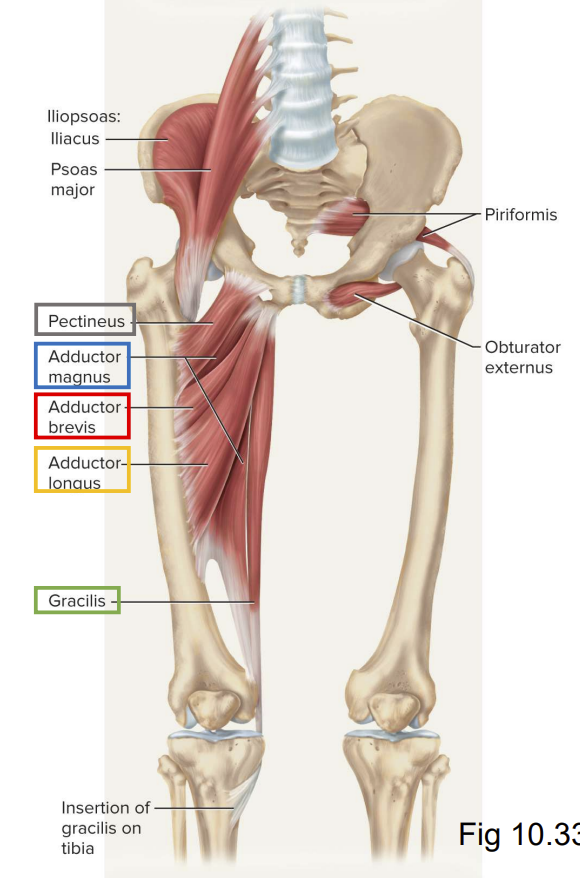
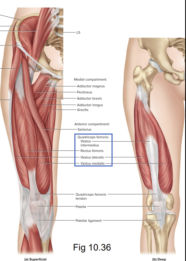
Muscles Acting on the Hip and Femur
- Medial (adductors) of thigh:
- Adductor brevis
- Adducytor longus
- Adductor magnus
- Gracilis
- pectineus
Muscles Acting on Knee and Leg
- Amnteiro (extensor) of thigh
- Contains large quadriceps femoris muscle
- Prime mover of knee extension
- Most powerful muscle in body
- 4 heads- rectus femoris, vastus lateralis, vastus medialis, and vasrus intermedius
- Converge on single (patellar) tendon
- Extends to patella
- Continues as patellar ligament to attach attach to tibial tuberosity
- Sartorius: longest muscle in the body
Muscles Acting on Knee and Leg
- Psoterioir (knee flexor) compartment of thigh
- Contains hamstring muscles

- Lateral to medial:
- Contains hamstring muscles
- Biceps femoris
- Semitendinosus
- Semimebranosus
Muscles Acting on Foot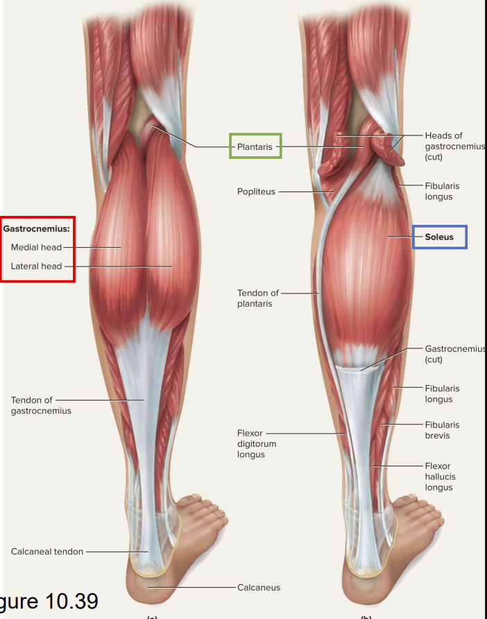
Posteriori compartment
- Superficial group:
- Gastrocnemius:
- Plantar flexes foot
- Flexes knee
- Soleus:
- Plantar flexes foot
- Plantaris:
- Weak synergist
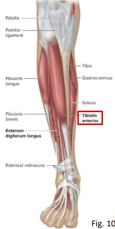
Muscles Acting on the Foot
Anterior compartment of the leg:
- Tibialis anterior
- Dorsiflexes the foot at the ankle
- Helps resist backward tipping
Common Athletic Injuries
- Muscles an tendons vulnerable to sudden and intense stress
- Common injuries include:
- Compartment syndrome
- Pulled hamstings
- Pulled groin
- Rotator cuff injury
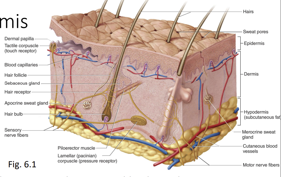
Anatomy of the Skin
Integumentary System
Consists of:
- Skin and accessory organs
- Hair
- Nails
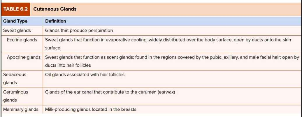
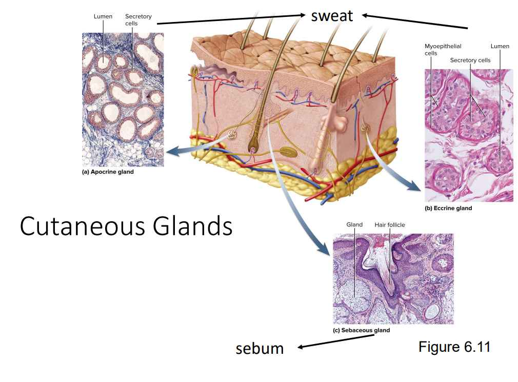
- Cutaneous glands
- Most vulnerable organ
- Exposed to radiation, trauma, infection, injurious chemicals
Structure of Skin and Subcutaneous Tissue
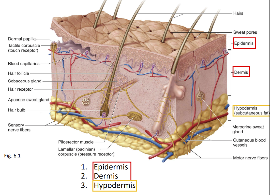
The Epidermis
- Kertinized stratified squamous epithelium
- Includes dead cells at skin surface packed with tough keratin protein
- avascualr - dpends on diffusion of nutrients from underlying connective tissue
- Spare nerve endings for touch and pain
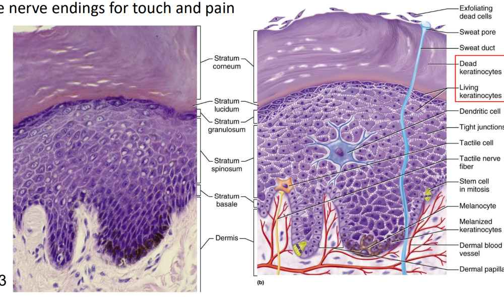
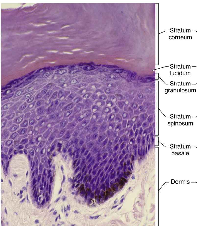
Layers of the Epidermis
- Stratum basale (deepest epidermal layer)
- Single layer of stem cells and keratinocytes
- Stratum spinosum
- Several layers of keratinocytes
- Named for appearance of cells after histological preparation (spiny)
- Stratum ghranulosukm
- 3-5 layers of flat kertinocytes
- Cells contain dark-staining keratohyalin
- Stratum lucidum
- Thin, pale layer found only in thick skin
- Keratinocytes packed with clear protein
- Stratum corenum (surface layer)
- Several layers (up to 30) of dead, scaly, keratinized cells
- Reisys abradion, penetration, water loss
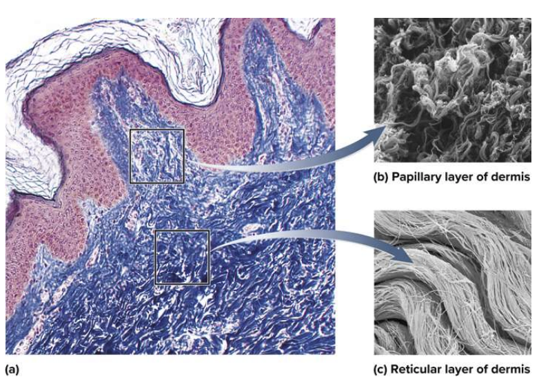
Layers of Dermis
Wavy, conspicuous boundrary with the superficial epidermis
- Dermal papillae- upwards, finger-like extensions of dermis
- Epidermal ridges are downward waves of dermis
- Prominent waves on fingers produce friction ridges (fingerpirnts)
- Papillary layer - superficial layer of dermis
- Thin zone near dermal papilla
- Allows for mobility of leukocytes
- Rich in small blood vessels
- Reticular zone- deeper and thicker layer of dermis
- Dense, irregular connective tissue
- Stretch marks (striae) - tears in collagen fibers caused by stretching of skin.
- Papillary layer - superficial layer of dermis
- Prominent waves on fingers produce friction ridges (fingerpirnts)
Hypodermis
- Common site of drug injection due to many blood vessels
- Subcutaneous tissue
- Pads body and binds skin to underlying tissues
- Subcutaneous fat
- Energy reservoir
- Thermal insulation
- Thicker in women, thinner in infants, elderly
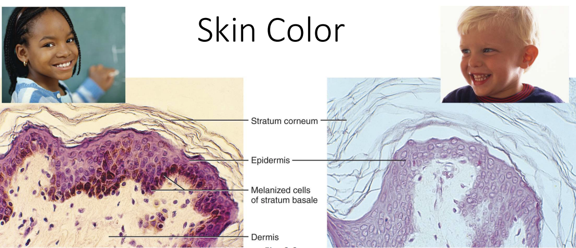
Skin Colour
Melanin: produced by melanocytes, accumulates in keratinocytes
Darker skin
- Melanocytes produce more melanin and breaks down more slowly
- Melanin granules more spread out in keratinocytes and seen throughout epidermis
Lighter Skin
- Melanin clumped near keratinocyte nucleus
- Little melanion seen beyond stratum basale
Anatomy of Hair, Nails, and Glands
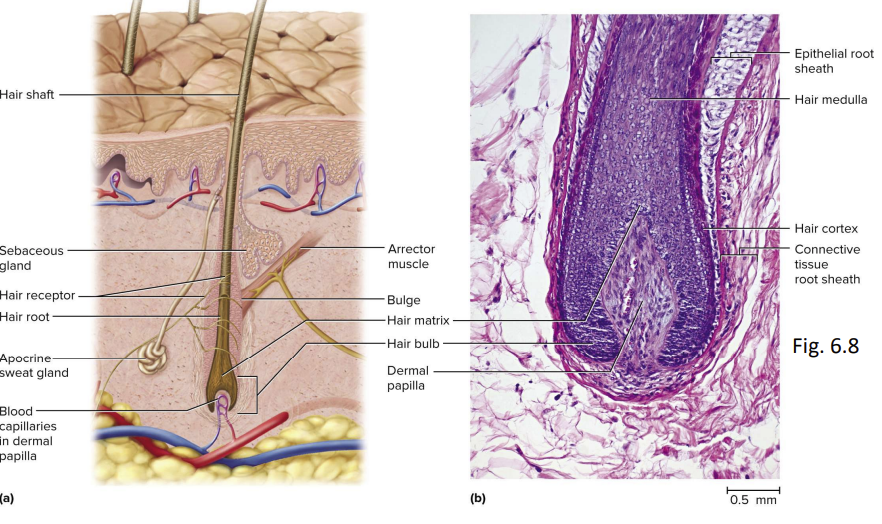
Structure of the Hair and its Follicle
- Mostly dead, keratinized cells
- Pliable soft keratin makes up starum coreneu, of skin
- Compact hard keratin makes up hair and nails
- 3 zones
- Bulb
- Root
- Shaft
Follicle = diganole tube that contains hair root
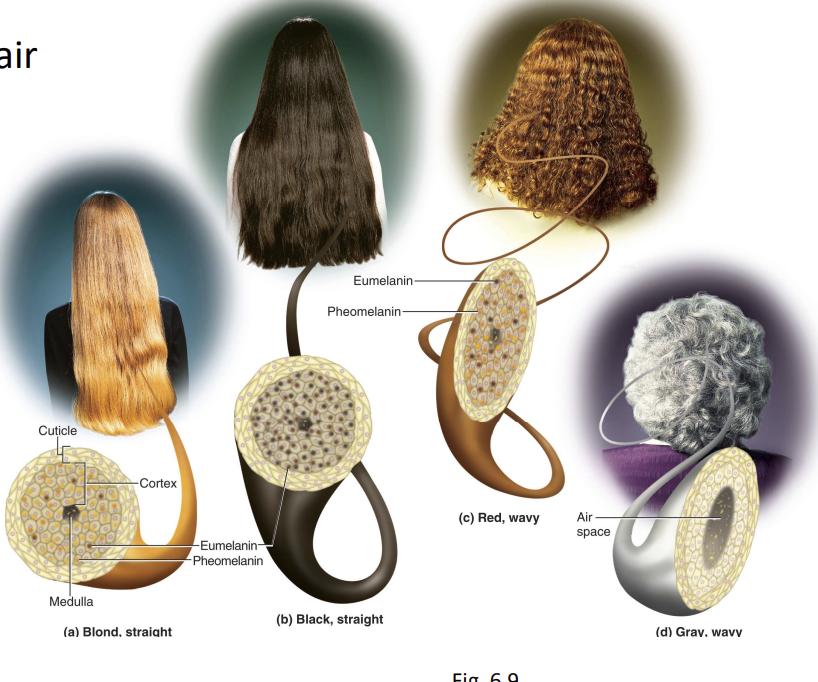
Hair Colour and Texture
- Texture from cross sectional shape of hair
- Straight = round
- Wavy = oval
- Curly = flat
- Colour from pigment granules in cortex
- Brown/black = eumelanin
- Red= pheomelanin
- Blond = some pheomelanin and little eumelanin
- Gray/white = little or no melanin
- Nails
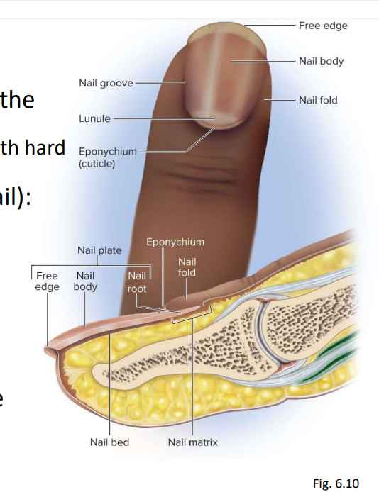
- Clear, hard derivatives of the stratum corneum.
- Thin, dead cells packed with hard keratin.
- Nail plate (hard part of nail):
- Free edge
- Nail body
- Nail root
- Nail fold
- Nail groove
- Nail bed
- Nail matrix - growth zone
- Eponychium (cuticle)
- Cutaneous Glands