knowt logo

L14 trafficking 1

Protein Synthesis and Ribosomes

  • Proteins are synthesized by ribosomes

    • Typical mammalian cells contain up to 10,000 different proteins.

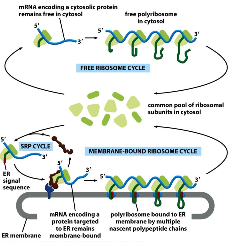
    • Majority synthesized by free cytosolic ribosomes.

    • 1/3 of proteins are synthesized by ribosomes on the endoplasmic reticulum (ER) membrane.

Synthesis of Proteins on Membrane-Bound vs. Free Ribosomes

  • Types of Ribosomes

    • 1/3rd of the human proteome is synthesized on the rough ER.

      • Produces:

        • Secreted proteins

        • Integral membrane proteins

        • Soluble proteins of organelles

    • Proteins synthesized on free ribosomes:

      • Cytosolic proteins

      • Cytosolic peripheral membrane proteins

      • Nuclear proteins

      • Proteins targeted to mitochondria, chloroplasts, and peroxisomes.
Protein Sorting Mechanisms

  • Key Components

    • Outer nuclear membrane

    • mRNA

    • Ribosomes

    • Inner nuclear membrane with nuclear pore

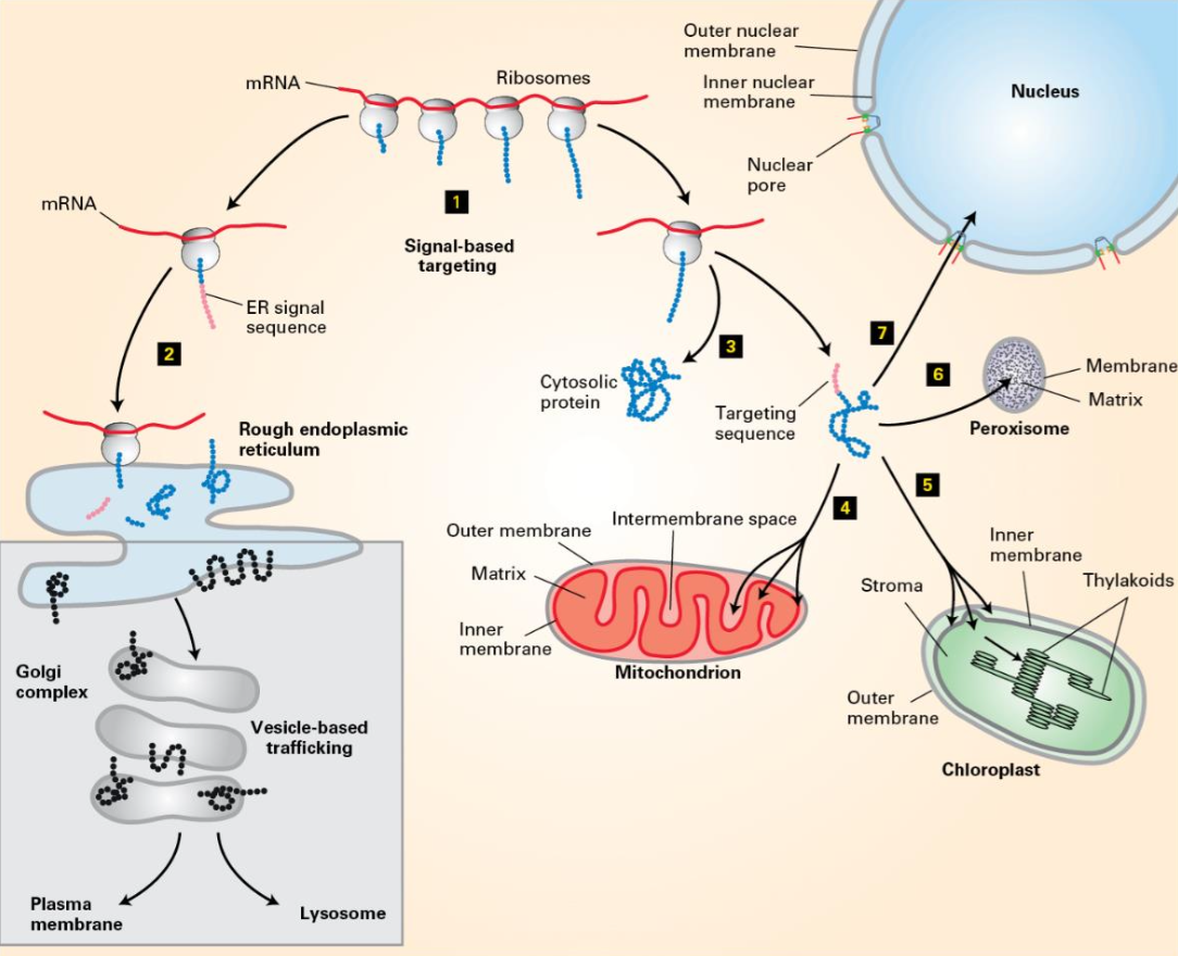
  • Signal-based targeting involves various sequences.

    • Rough ER sequence

    • Peroxisome sequence

  • Vesicle-based trafficking mechanisms illustrated.
Experimental Methods to Study Protein Sorting/Transport

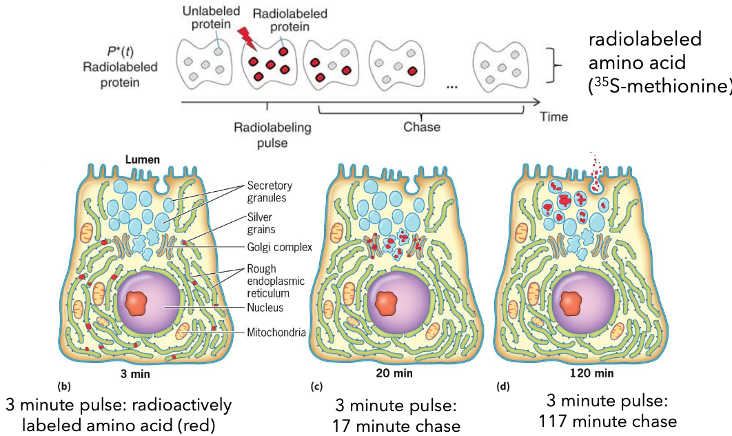
  • Pulse Chase Experiment: This technique involves labeling newly synthesized proteins with a radioactive or fluorescent tag, followed by a chase period where the label is removed, allowing researchers to track the movement and processing of these proteins over time.

    • Radiolabeled amino acid: 35S-methionine

    • Pulse phase (3 minutes): Radioactively labeled amino acid.

    • Chase phase: 17-minute and 117-minute chases to observe transport.
GFP-based Protein Tracking

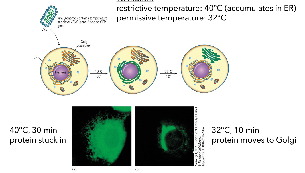
  • Temperatures influence protein tracking:

    • Restrictive temperature (40°C): Protein accumulates in ER.

    • Permissive temperature (32°C): Protein moves to Golgi
Subcellular Fractionation

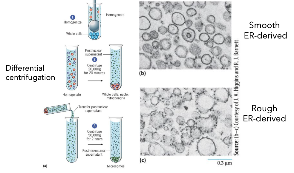
  1. Homogenize cells into a whole cells homogenate.

  2. Differential centrifugation (20,000g for 20 minutes) yields postnuclear supernatant.

  3. Another centrifugation (50,000g for 2 hours) creates post-microsomal supernatant containing fractions.
Analysis of Genetic Mutants

  • Nobel Prize 2013: Research on Saccharomyces cerevisiae (budding yeast).

    • sec12: Shows vesicle formation at the ER with expanded ER.

    • sec17: Causes accumulation of vesicles in the cell.

Page 11: Protein Sorting/Protein Targeting

  • Signal-based targeting directly targets newly synthesized proteins to organelles.
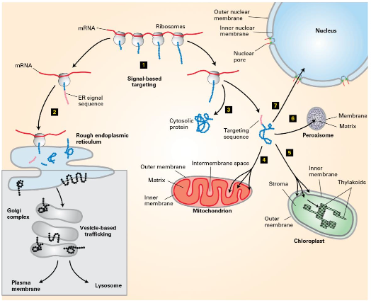
  • Vesicle-based targeting (secretory pathway) noted.
Sorting Signals

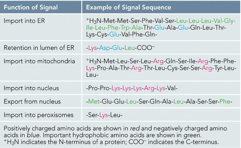
  • Signal sequence

    • Encoded in amino acid sequence or attached carbohydrates.

    • Recognized by receptors:

      • For soluble proteins: Receptors can be integral membrane proteins.

      • For transmembrane proteins: Receptors are coat components.

  • Targeting can occur during or after protein synthesis.

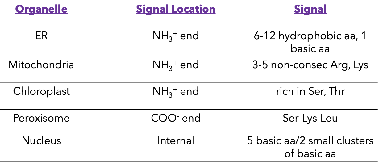
Organelle Signal Locations

  • Signaling mechanisms for targeting proteins:
Signal Sequences Examples
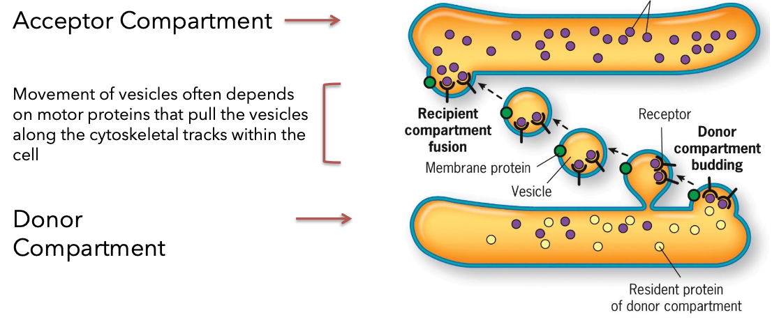
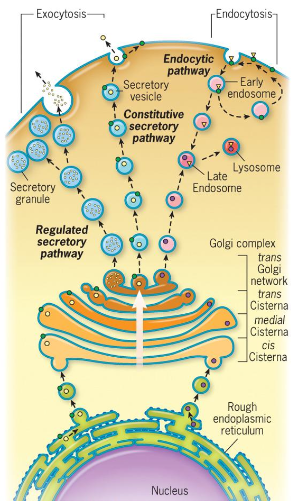
Vesicle based trafficking

  • Endomembrane system include: ER, Golgi complex, endosomes, lysosomes, vacuoles, seretory vesicles and granules

  • Biosynthetic pathway: Synthesis, modification, and transport of proteins.

  • Secretory pathway: where proteins are discharged (secreted) fro the cell.

    • Constitutive secretion: in a Continuous fashion. important for forming PM and extracellular matrix.

    • Regulated secretion: In response to stimuli, important for cellular signaling.

  • Overview of Vesicle based trafficking

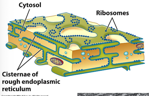
Endoplasmic Reticulum Structure
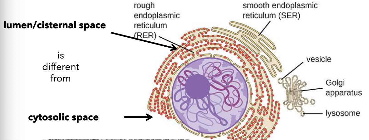
  • Types: Rough ER (RER) and Smooth ER (SER).

  • Differences between SER and RER in function and structure in conjunction with Golgi apparatus.

  • The Smooth ER

    • synthesis of steroid hormones in endocrine cells

    • detoxification in the liver of various organic compounds

    • sequetration of calcium ion from cytoplasm of muscle cells (has high conc. of calcium binding proteins)

      • Leydig cell example: Extensive SER for steroid hormone synthesis.

  • The Rough ER

  • Immunofluorescence techniques used to visualize rough ER.

  • Continuous with outer membrane of the nuclear envelope.

  • Serves as the starting point for the biosynthetic/secretory pathway.

Membrane Biosynthesis in the ER

  • Membranes arise from pre-existing membranes.

  • Most lipids synthesized in ER, which are asymmetric facing cytosol and luminal/ extracellular face .

Evidence of Secretory Proteins in ER

  • The Pulse Chase Experiemnt: radiolabeled amino acids

    • Newly synthesized proteins radioactively labeled.

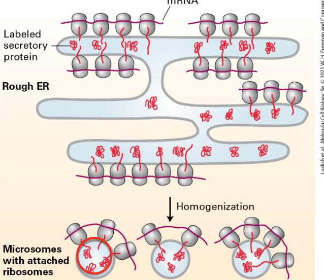
    • Homogenizing the cell leads to small vesicles called microsomes.

    • Ribosomes found on the outside of microsomes.
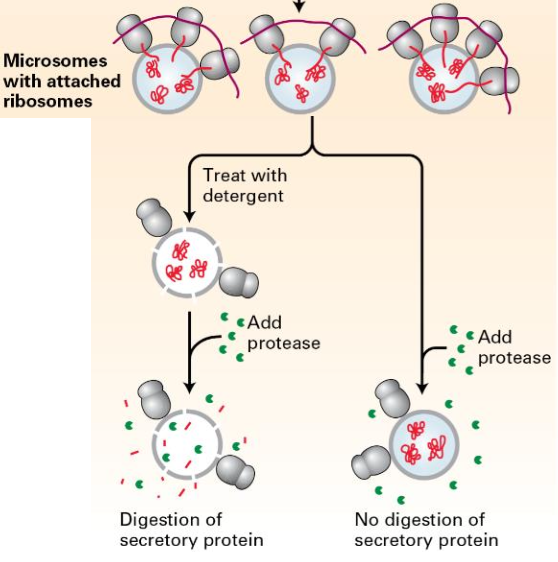
  • Protease experiment:

    • Sample 1: Add protease, Proteins inside the microsomes protected.

    • Sample 2: Detergent added before protease; secretory proteins degraded.
Cotranslational Translocation

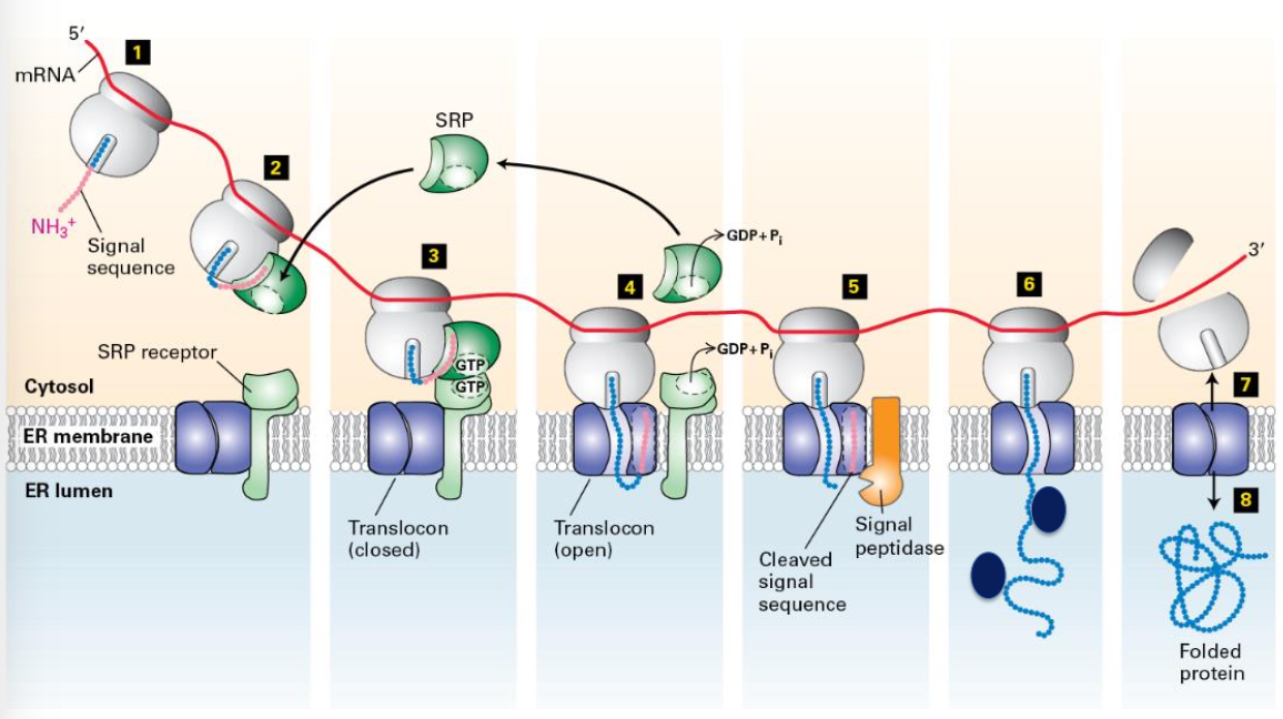
  • Proteins are incorporated into microsomes during translation (cotranslational translocation). Transport of most secretory proteins into the ER lumen begins while the nascent protein is still bound to the ribosome.

Synthesis of Secretory Proteins on rough ER

  1. mRNA binds to free ribosomes. Translation begins and the signal sequence is created.

    • continuous stretch of 6-12 amino acids at N terminus.

    • one of more positively charged amino acids next to this hydrophobic core

    • this hydrophobic core of the signal sequence is critical for interacting with the machinery that targets the protein to the ER membrane

  2. Signal sequence recognized by a signal recognition particle and translation is temporarily arrested.

  3. SRP binds SRP receptor (associated with translocon pore) .

  4. Ribosome interacts with translocon. SRP dissociates from ribosome and receptor. Protein synthesis resumes.

  5. Polypeptide translocates through the pore and enters the ER lumen. Signal sequence is cleaved by signal peptidase.

  6. Signal sequence is cleaved by signal peptidase and is bound by BiP (chaperone protein) after translocation.
Functions of BiP

  • Dual role in protein processing; acts upon binding and release of ADP/ATP.

    • guards translocon pore when ADP bound

      • ADP bound: high substrate affinity

      • ATP bound: low substrate affinity

Integral Membrane Protein Synthesis

  • Orientation of integral membrane proteins determined during synthesis, influencing plasma membrane orientation.

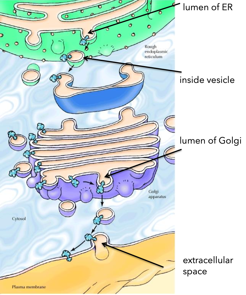
    • the end that faces or is inside the ER lumen will face the extracellular space

Topological Equivalence

  • Orientation concept: lumen of ER = Golgi lumen = extracellular space.
Insertion Mechanism of Membrane Proteins
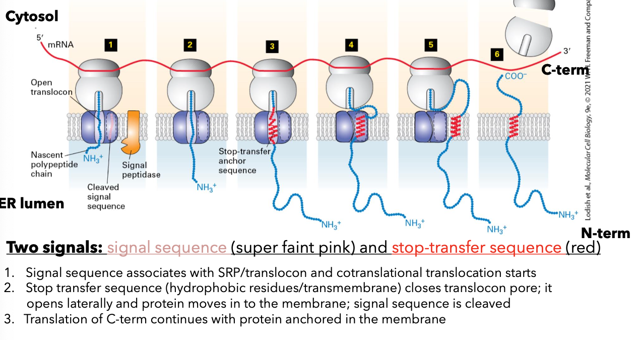
  • Signal sequences (an internal signal anchor sequence) is in the middle of the protein or at the COO- end (not at the NH3 end)

    • signal sequence targets the ribosome and peptide to the ER

  • Rjbsosome is transferred to open translocon.

    • NH3 end is in the cytosol

    • signal anchor sequence sticks in translocon

  • peptide elongated into ER lumen; carbohydrate is added

  • ribosome dissociated from the translocon; protein diffuses out of the translocon
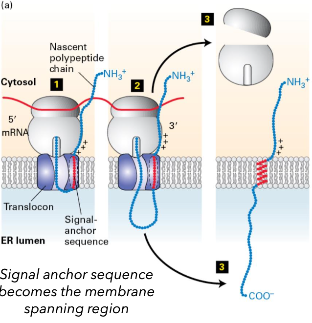
Insertion of Multipass Proteins

  • Membrane spanning regions pass from the translocon into the membrance cotranslationally in order they energy from the ribosome

  • orientation of the first transmembrane segment is first established

    • initial engagement with the translocon in SRP/SRP receptor dependent manner

  • subsequent transmembrane segment assumes the opposite orientation

    • independent of SRP

Processing of Newly Synthesized Proteins

  1. Specific Proteolytic cleavages in the ER, golgi complex, and secretory vesicles for protein maturation.

    1. signal peptidase: cleaves signal sequence

  2. Formation of Disulfide bond for stability and folding in the ER.

    • covalent bonds formed by the oxidative linkage of sulfhydryl groups, on two cysteine residues in the same or different polypeptide chains

    • functions: protein folding, increases stability of native structure

    • protein disulfide isomerase: promotes proper disulfide bond formation

  3. Glycosylation: covalent addition of carbohydrates

    • Covalent addition of carbohydrates produces glycoproteins, facilitating structural stability and interactions.

      • functions incldue proper folding of proteins, structural stability, and produces an array of chemically distinct molecules at the cell surafce that are the basis of specific molecular interactions used in cell to cell adhesion and communication

  4. Proper folding of polypeptide chains and assembly of multi-subunit proteins in the ER

    • Chaperone proteins like BiP play crucial roles in promoting protein folding and multi-subunit assembly.

Quality Control in the ER

  1. Modification of glycoproteins involves the removal of glucose by glucosidase II. Retain only a single glucose.

  2. Glucose recognized and bound by ER chaperone calnexin.

  3. Glucosidase II removes remaining glucose

  4. Conformation sensing enzyme UGGT to determine if properly folded.

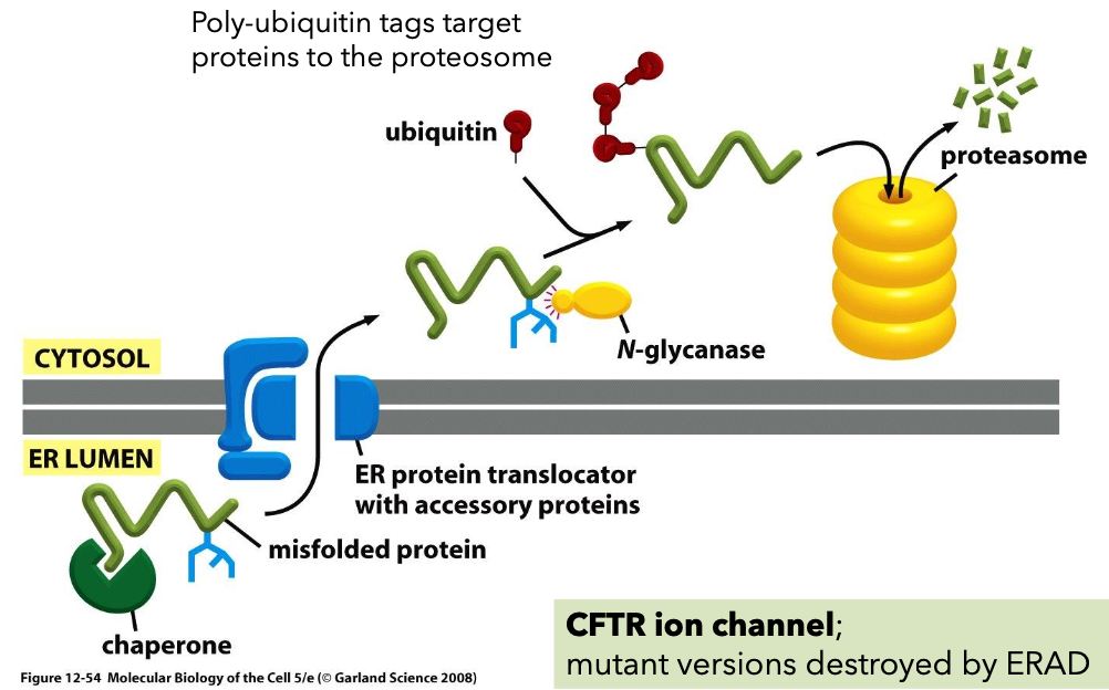
  5. Unfolded proteins are targeted for degradation if improperly folded.

ER associated degradation Mechanisms

  • ER-associated degradation (ERAD) with poly-ubiquitin tags targeting proteins to the proteasome for disposal.
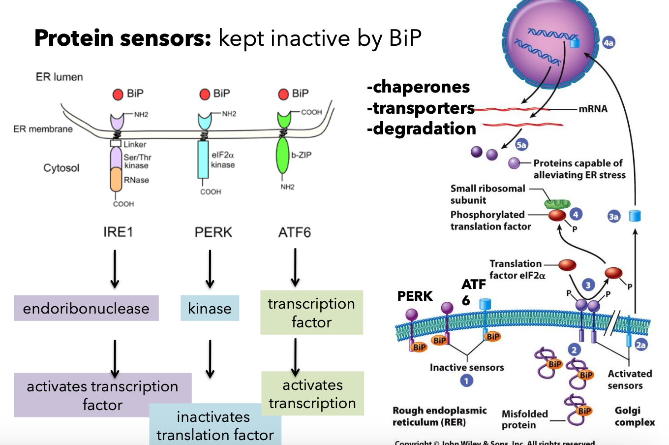
Unfolded Protein Response (UPR)

  • Activation of transcription factors in response to unfolded proteins, modulating chaperones and degradation pathways.
RM

L14 trafficking 1

Protein Synthesis and Ribosomes

  • Proteins are synthesized by ribosomes

    • Typical mammalian cells contain up to 10,000 different proteins.

    • Majority synthesized by free cytosolic ribosomes.

    • 1/3 of proteins are synthesized by ribosomes on the endoplasmic reticulum (ER) membrane.

Synthesis of Proteins on Membrane-Bound vs. Free Ribosomes

  • Types of Ribosomes

    • 1/3rd of the human proteome is synthesized on the rough ER.

      • Produces:

        • Secreted proteins

        • Integral membrane proteins

        • Soluble proteins of organelles

    • Proteins synthesized on free ribosomes:

      • Cytosolic proteins

      • Cytosolic peripheral membrane proteins

      • Nuclear proteins

      • Proteins targeted to mitochondria, chloroplasts, and peroxisomes.

Protein Sorting Mechanisms

  • Key Components

    • Outer nuclear membrane

    • mRNA

    • Ribosomes

    • Inner nuclear membrane with nuclear pore

  • Signal-based targeting involves various sequences.

    • Rough ER sequence

    • Peroxisome sequence

  • Vesicle-based trafficking mechanisms illustrated.

Experimental Methods to Study Protein Sorting/Transport

  • Pulse Chase Experiment: This technique involves labeling newly synthesized proteins with a radioactive or fluorescent tag, followed by a chase period where the label is removed, allowing researchers to track the movement and processing of these proteins over time.

    • Radiolabeled amino acid: 35S-methionine

    • Pulse phase (3 minutes): Radioactively labeled amino acid.

    • Chase phase: 17-minute and 117-minute chases to observe transport.

GFP-based Protein Tracking

  • Temperatures influence protein tracking:

    • Restrictive temperature (40°C): Protein accumulates in ER.

    • Permissive temperature (32°C): Protein moves to Golgi

Subcellular Fractionation

  1. Homogenize cells into a whole cells homogenate.

  2. Differential centrifugation (20,000g for 20 minutes) yields postnuclear supernatant.

  3. Another centrifugation (50,000g for 2 hours) creates post-microsomal supernatant containing fractions.

Analysis of Genetic Mutants

  • Nobel Prize 2013: Research on Saccharomyces cerevisiae (budding yeast).

    • sec12: Shows vesicle formation at the ER with expanded ER.

    • sec17: Causes accumulation of vesicles in the cell.

Page 11: Protein Sorting/Protein Targeting

  • Signal-based targeting directly targets newly synthesized proteins to organelles.

  • Vesicle-based targeting (secretory pathway) noted.

Sorting Signals

  • Signal sequence

    • Encoded in amino acid sequence or attached carbohydrates.

    • Recognized by receptors:

      • For soluble proteins: Receptors can be integral membrane proteins.

      • For transmembrane proteins: Receptors are coat components.

  • Targeting can occur during or after protein synthesis.

Organelle Signal Locations

  • Signaling mechanisms for targeting proteins:

Signal Sequences Examples

Vesicle based trafficking

  • Endomembrane system include: ER, Golgi complex, endosomes, lysosomes, vacuoles, seretory vesicles and granules

  • Biosynthetic pathway: Synthesis, modification, and transport of proteins.

  • Secretory pathway: where proteins are discharged (secreted) fro the cell.

    • Constitutive secretion: in a Continuous fashion. important for forming PM and extracellular matrix.

    • Regulated secretion: In response to stimuli, important for cellular signaling.

  • Overview of Vesicle based trafficking

Endoplasmic Reticulum Structure

  • Types: Rough ER (RER) and Smooth ER (SER).

  • Differences between SER and RER in function and structure in conjunction with Golgi apparatus.

  • The Smooth ER

    • synthesis of steroid hormones in endocrine cells

    • detoxification in the liver of various organic compounds

    • sequetration of calcium ion from cytoplasm of muscle cells (has high conc. of calcium binding proteins)

      • Leydig cell example: Extensive SER for steroid hormone synthesis.

  • The Rough ER

  • Immunofluorescence techniques used to visualize rough ER.

  • Continuous with outer membrane of the nuclear envelope.

  • Serves as the starting point for the biosynthetic/secretory pathway.

Membrane Biosynthesis in the ER

  • Membranes arise from pre-existing membranes.

  • Most lipids synthesized in ER, which are asymmetric facing cytosol and luminal/ extracellular face .

Evidence of Secretory Proteins in ER

  • The Pulse Chase Experiemnt: radiolabeled amino acids

    • Newly synthesized proteins radioactively labeled.

    • Homogenizing the cell leads to small vesicles called microsomes.

    • Ribosomes found on the outside of microsomes.

  • Protease experiment:

    • Sample 1: Add protease, Proteins inside the microsomes protected.

    • Sample 2: Detergent added before protease; secretory proteins degraded.

Cotranslational Translocation

  • Proteins are incorporated into microsomes during translation (cotranslational translocation). Transport of most secretory proteins into the ER lumen begins while the nascent protein is still bound to the ribosome.

Synthesis of Secretory Proteins on rough ER

  1. mRNA binds to free ribosomes. Translation begins and the signal sequence is created.

    • continuous stretch of 6-12 amino acids at N terminus.

    • one of more positively charged amino acids next to this hydrophobic core

    • this hydrophobic core of the signal sequence is critical for interacting with the machinery that targets the protein to the ER membrane

  2. Signal sequence recognized by a signal recognition particle and translation is temporarily arrested.

  3. SRP binds SRP receptor (associated with translocon pore) .

  4. Ribosome interacts with translocon. SRP dissociates from ribosome and receptor. Protein synthesis resumes.

  5. Polypeptide translocates through the pore and enters the ER lumen. Signal sequence is cleaved by signal peptidase.

  6. Signal sequence is cleaved by signal peptidase and is bound by BiP (chaperone protein) after translocation.

Functions of BiP

  • Dual role in protein processing; acts upon binding and release of ADP/ATP.

    • guards translocon pore when ADP bound

      • ADP bound: high substrate affinity

      • ATP bound: low substrate affinity

Integral Membrane Protein Synthesis

  • Orientation of integral membrane proteins determined during synthesis, influencing plasma membrane orientation.

    • the end that faces or is inside the ER lumen will face the extracellular space

Topological Equivalence

  • Orientation concept: lumen of ER = Golgi lumen = extracellular space.

Insertion Mechanism of Membrane Proteins

  • Signal sequences (an internal signal anchor sequence) is in the middle of the protein or at the COO- end (not at the NH3 end)

    • signal sequence targets the ribosome and peptide to the ER

  • Rjbsosome is transferred to open translocon.

    • NH3 end is in the cytosol

    • signal anchor sequence sticks in translocon

  • peptide elongated into ER lumen; carbohydrate is added

  • ribosome dissociated from the translocon; protein diffuses out of the translocon

Insertion of Multipass Proteins

  • Membrane spanning regions pass from the translocon into the membrance cotranslationally in order they energy from the ribosome

  • orientation of the first transmembrane segment is first established

    • initial engagement with the translocon in SRP/SRP receptor dependent manner

  • subsequent transmembrane segment assumes the opposite orientation

    • independent of SRP

Processing of Newly Synthesized Proteins

  1. Specific Proteolytic cleavages in the ER, golgi complex, and secretory vesicles for protein maturation.

    1. signal peptidase: cleaves signal sequence

  2. Formation of Disulfide bond for stability and folding in the ER.

    • covalent bonds formed by the oxidative linkage of sulfhydryl groups, on two cysteine residues in the same or different polypeptide chains

    • functions: protein folding, increases stability of native structure

    • protein disulfide isomerase: promotes proper disulfide bond formation

  3. Glycosylation: covalent addition of carbohydrates

    • Covalent addition of carbohydrates produces glycoproteins, facilitating structural stability and interactions.

      • functions incldue proper folding of proteins, structural stability, and produces an array of chemically distinct molecules at the cell surafce that are the basis of specific molecular interactions used in cell to cell adhesion and communication

  4. Proper folding of polypeptide chains and assembly of multi-subunit proteins in the ER

    • Chaperone proteins like BiP play crucial roles in promoting protein folding and multi-subunit assembly.

Quality Control in the ER

  1. Modification of glycoproteins involves the removal of glucose by glucosidase II. Retain only a single glucose.

  2. Glucose recognized and bound by ER chaperone calnexin.

  3. Glucosidase II removes remaining glucose

  4. Conformation sensing enzyme UGGT to determine if properly folded.

  5. Unfolded proteins are targeted for degradation if improperly folded.

ER associated degradation Mechanisms

  • ER-associated degradation (ERAD) with poly-ubiquitin tags targeting proteins to the proteasome for disposal.

Unfolded Protein Response (UPR)

  • Activation of transcription factors in response to unfolded proteins, modulating chaperones and degradation pathways.

robot