Lecture 09 2024 10 03 ECM
Learning Objectives
Define and characterize the extracellular matrix (ECM).
Delineate the roles of different ECM components.
Identify the roles of Integrins in mammalian cells.
Differentiate between models for cellular adhesion.
Identify and differentiate between classes of molecules that mediate cell-cell interactions.
The Extracellular Matrix
Extra: Means "outside" or "beyond."
Matrix: Refers to a foundational structure or substance that other elements form from or develop within.
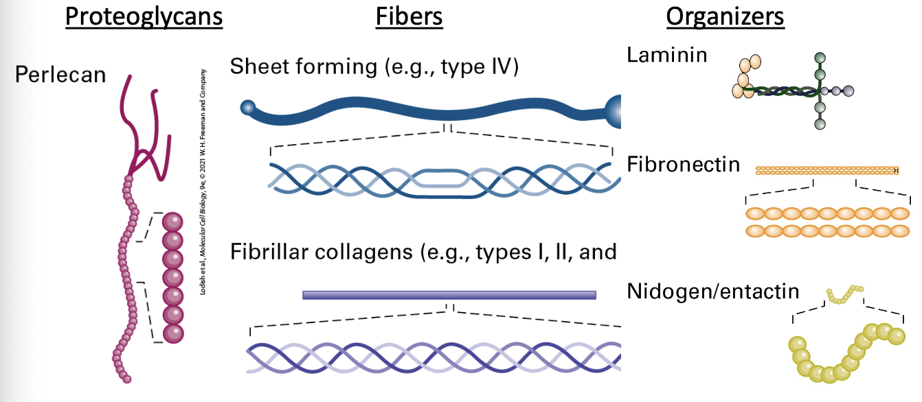
Major Components of the ECM
Proteoglycans: Large molecules that play critical roles in ECM structure.
Fibers: Provide mechanical strength.
Organizers: Help to arrange matrix components.
Examples: Laminin, Perlecan, Type IV Collagen, Fibronectin, Fibrillar collagens (e.g., types I, II), Nidogen/entactin.
Functions of the ECM
Anchoring and surrounding cells to maintain solid-tissue three-dimensional architecture and define tissue boundaries
2. Determining the biomechanical properties (stiffness/elasticity, porosity, shape) of the extracellular environment
3. Controlling cellular polarity, survival, proliferation, differentiation, and fate (e.g., asymmetric division of stem cells; see Chapter 22), and thus embryonic and neonatal development and adult function and responses to the environment and to disease
4. Inhibiting or facilitating cell migration (e.g., serving as either a barrier to movement or, conversely, as a “track” along which cells — or portions of cells — can move)
5. Binding to and acting as a reservoir of growth factors; in some cases, the ECM (a) helps generate an extracellular concentration gradient of the growth factor, (b) serves as a co-receptor for the growth factor, or (c) aids in proper binding of the growth factor to its receptor (ECM component and growth factor jointly serve as a receptor’s combined ligand)
6. Activating cell surface signaling receptors
Tissue Variations of ECM
The ECM’s composition varies significantly across different tissue types.
Ex: Connective tissue vs tightly packed epithelial cells and their associated fibroblasts.
Plant Comparative Matrix
Cell walls constitute the supportive matrix in plant tissues. Cell walls = their supportive matrix
Cellulose and Pectin are primary components.
Arabidopsis thaliana serves as a model organism in these studies.
Cell Adhesions
Connections between cells are vital for multicellularity.
Different adhesions serve distinctive roles, making multicellularity feasible.
Animal Tissues and Their Functions
Epithelial Tissue: Covers and protects; includes squamous, glandular types.
Muscular Tissue: Involved in movement, includes striated (skeletal, cardiac) and non-striated types.
Connective Tissue: Provides support; includes blood, lymph, adipose, cartilage, and bone.
Nervous Tissue: Responsible for communication and coordination.
Epithelial Cell Model
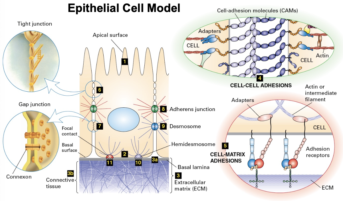
Cellular Structures
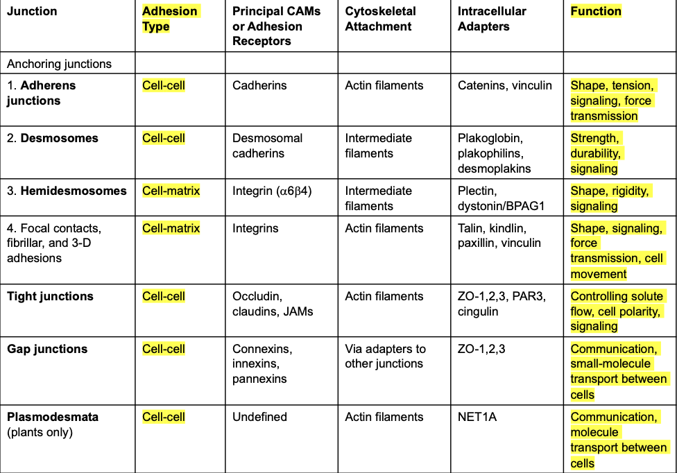
Models display various cell adhesion molecules (CAMs), including tight junctions, adherens junctions, desmosomes.
Features of cell-cell adhesion mechanics visualized through cellular interactions.
Adhesion Receptors
Cell surface molecules, known as adhesion receptors, play a critical role in cell communication. 2 types::
Cell-Cell Adhesion
Cell-Matrix Adhesion
CAM is important for communication. No communication is bad for the system.
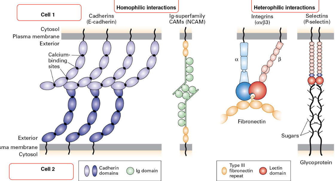
Major CAM families: Cadherins, Immunoglobulin (Ig) superfamily, Integrins, and surgar binding proteins called Lectins
Adherence Molecules
Homophilic interactions: CAD molecules interact between the same cell types.
Heterophilic interactions: CAD molecules interact with different cell types.
Key CAM examples include E-Cadherin and Selectins.
Cis/Trans Interactions
Cadherin molecules engage in both cis (within the same cell) and trans (adjacent cells) interactions, promoting cellular cohesion. The ability to mic and match them makes them a versatile family of proteins.
Page 14: Information Relay
Cell adhesion molecules connect to signaling pathways, affecting cell behavior based on adhesion status.
Page 15: Engagement Changes
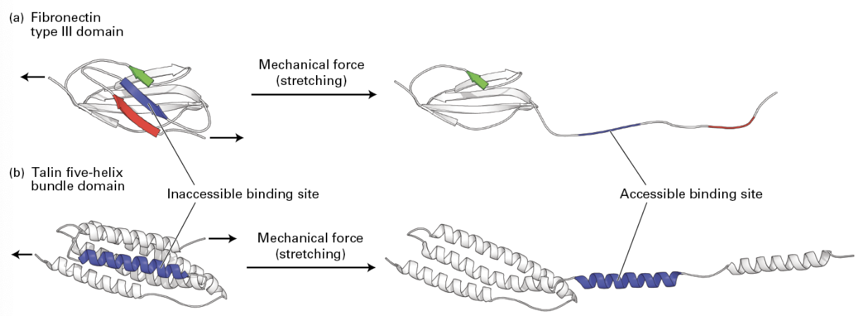
Adhesion molecules can transmit mechanical signals through conformational changes, recruiting other proteins for reinforcement.
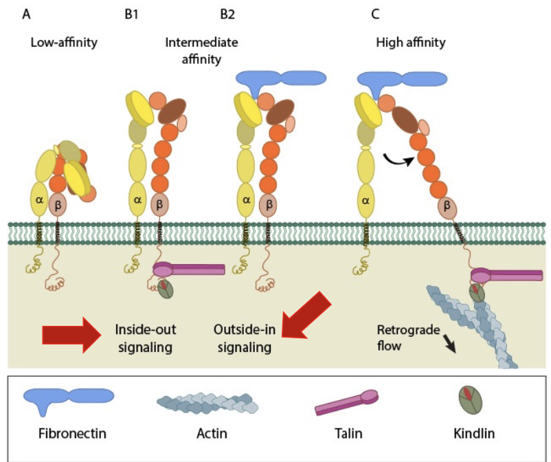
Adhesion molecules can be mechanotransducing. Fibronectin stretching in the ECM form cells nearby can cue recruitment of further proteins for reinforcement by revealing cryptic binding sites
Cell Junctions
Epithelial Cells
Function: Line, protect, absorb, secrete, cushion.
Classified as simple (one cell layer) or stratified based (2+ layers) on cell layers.
Epithelial Cell Interactions
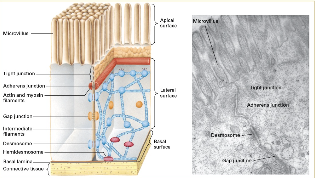
Cadherin Zippers
Adherens junctions utilize cadherin interactions to form zipper-like structures, enhancing cell adhesion. Adherens junctions connect the lateral membranes of adjacent epithelial cells near the apical surface, below tight junctions
Cadherin molecules interact with each other in the same cell (cis) or with adjacent cells (trans) to create a zipper
Individual interactions = weak, en masse = strong
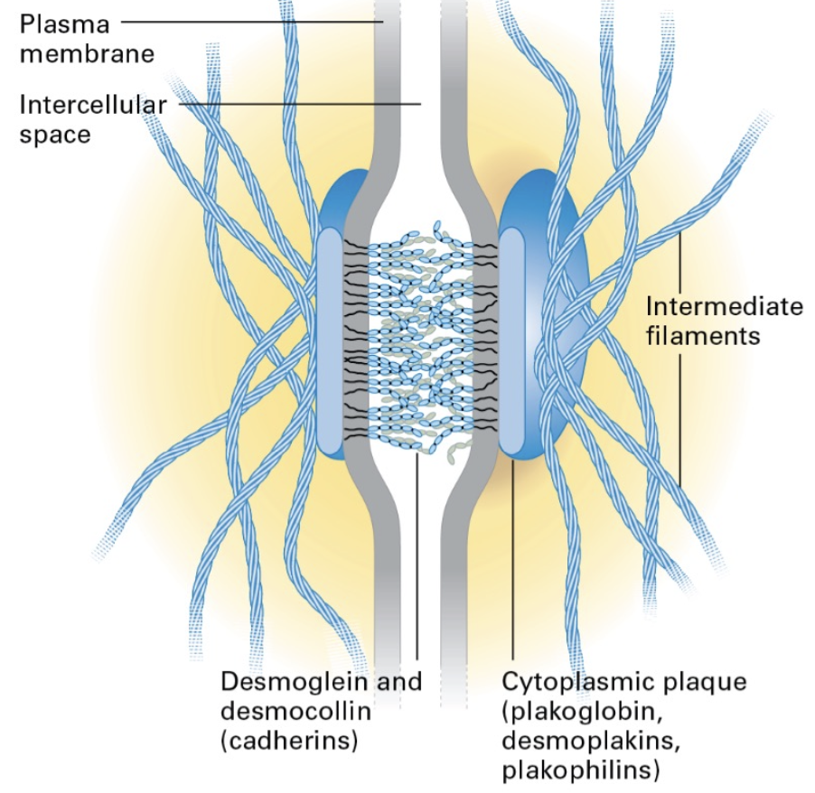
Desmosomes
Act as strong adhesion sites that connect cells through cadherin proteins, adding strength through intermediate filaments.
Important in diseases affecting cell-adhesion integrity. Characterized by defects in adhesion: cell - cell and/or cell-substrate
Page 22: Hemidesmosomes
Function to anchor epithelial cells to the basal lamina, dancing analogously to traditional desmosomes.
Kertain filament = intermediate filaments; spot welding to the basal lamina

Integrins Activated by Binding Target
integrin are a large family of alpha/beta heterodimeric cell surface proteins that play a crucial role in cell adhesion and signaling, facilitating the connection between the extracellular matrix and the cytoskeleton.
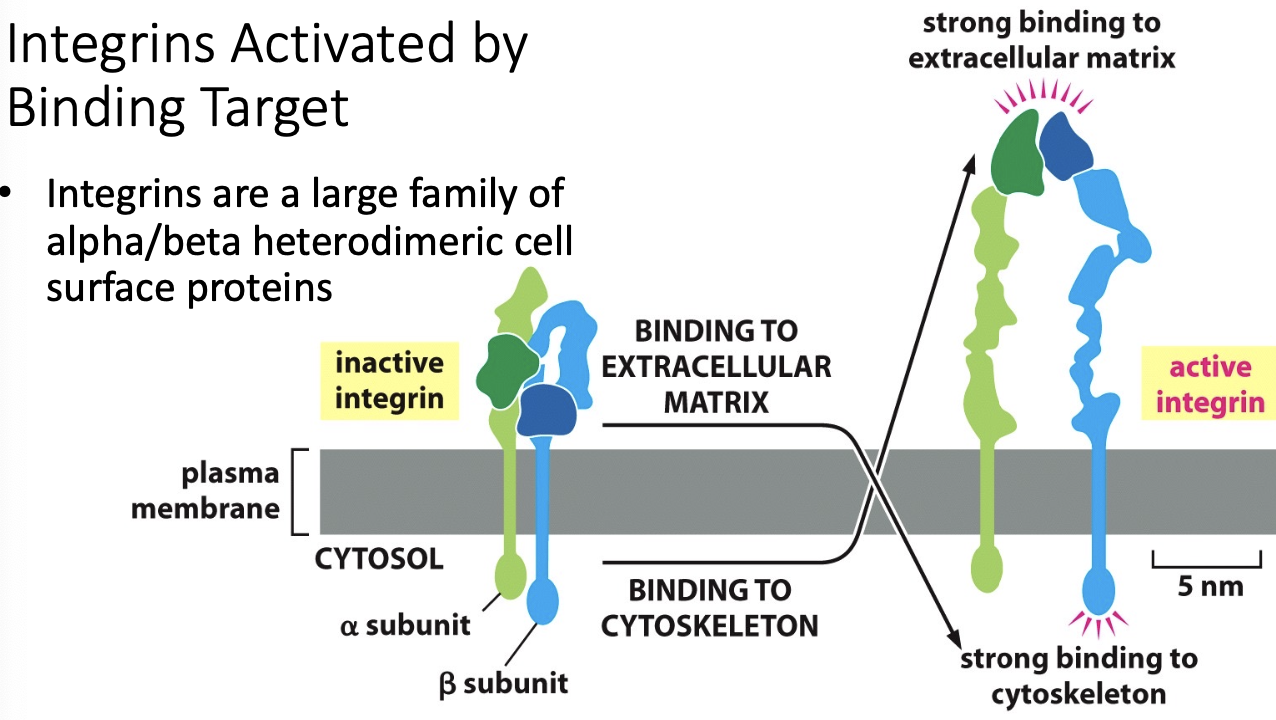
Dynamics of Integrin Function
Integrins modulate both cell-cell/matrix interactions and adapt conformation to engage with varying targets.
they exhibit differing conformational states that alter affinity for targets
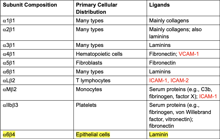
a6b4 is the main laminin binding one in epithelium
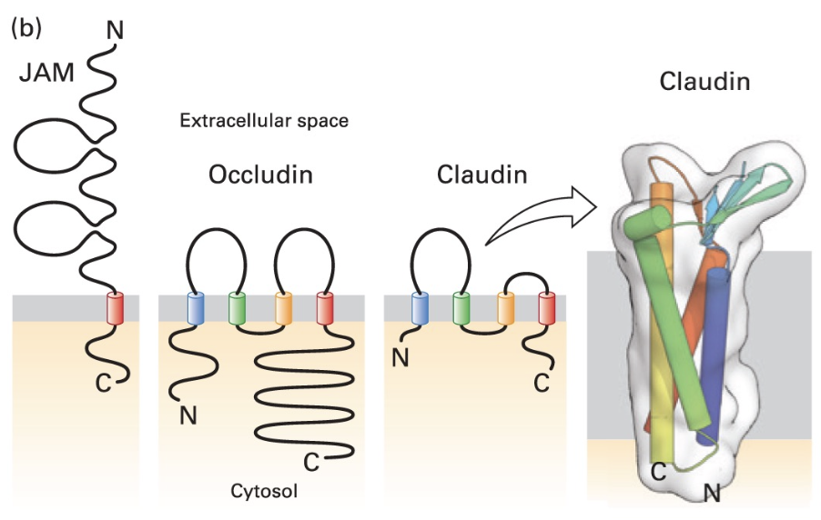
Tight Junctions Overview
Characteristics
Seal body cavities, restrict the diffusion of membrane components, important in maintaining barrier integrity in epithelial tissues. It keeps the fluids separated between the epithelial cell layer.
they form ring structures around the top of the cells. Rings are connected to adjacent cells rings.
Key Proteins
Many integral membrane proteins are involved in tight junctions.
Occludin, Claudin, and Tricellulin contribute significantly to forming tight junctions across epithelial cells.
Occludin and Claudin are highly studied
Tricellulin appears where three cells tight junctions meet
Junction Adhesion Molecules (JAM) so involved.
Tight Junction Interaction
Adapter Proteins
Adapter proteins interact with these proteins in tight junctions including Zona occludens proteins mediate transcellular transport, influencing solute movement between cells.
Gap Junctions
Large complexes found in junctions amidst the gaps
Facilitate cell communication through connexon channels that allow small molecules and ions to pass between cells.
6 protein TM connexon units form hemichannels that connect to adjacent cells.
Tunneling Nanotubes
Connect adjacent cells and facilitate the transfer of organelles and smaller molecules, enhancing intercellular communication.
Basal Lamina Structure
Layers of the basal lamina consist mainly of Type IV collagen, laminin, and nidogen, serving as a foundation for epithelial cells.
Mat like structure; sheets of epithelia sit on basal lamina
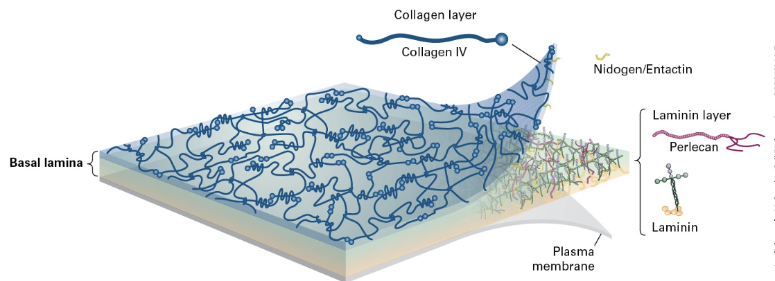
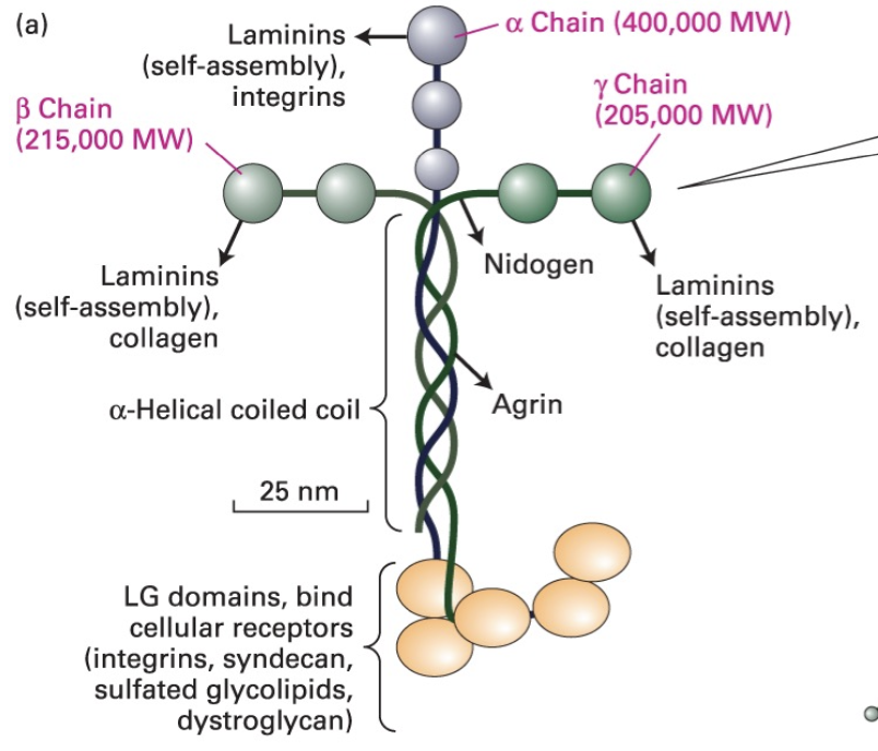
Laminins
Laminins are multi-adhesive proteins critical for the structure of the basal lamina and serve as binding sites for integrins.
has a cross like trimer (a, B, y)
they self assemble into large structures using their globular domains.
C terminal domains allow for binding by cell receptors, including some integrins.
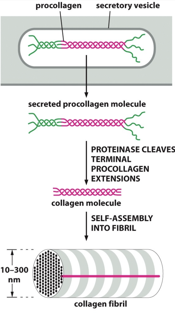
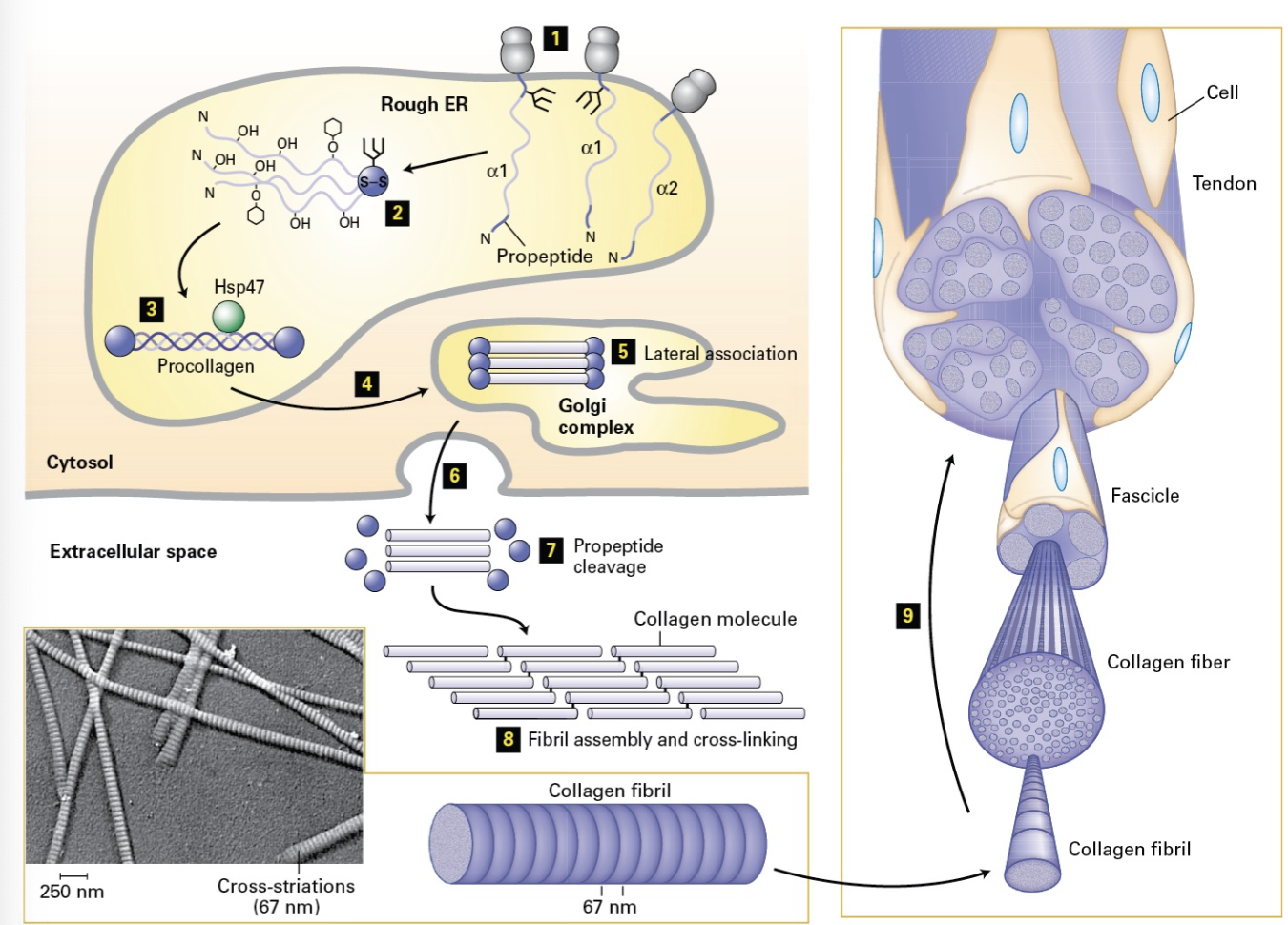
Collagen Structure
Collagen forms a triple-helix structure formed from left handed helical proteins and is the most abundant protein in animals, playing a vital role in tissue strength.
characteristic glycine-x-y repeats. the bundles form right hand helices.
Connective Tissue
Bone Structure
Matrix constitutes the majority of bone with collagen fibrils and calcium phosphate contributing to its structure and strength.
cells are dark spots
bands contain collagen fibrils
calcium phosphate also present
Collagen Bundles
Formation of collagen fibers (superstructure) occurs via fibroblasts and osteoblasts, crucial for maintaining connective tissue integrity. Built by:
fibroblasts - skin
osteoblasts -bone
exocytosis
Cross linked triple helix
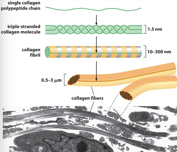
Collagen Synthesis
Collagen synthesis involves procollagen production (IC), secretory vesicle formed, and self-assembly into functional fibers.