Common Blood Chemistry Test
What are blood chemistry tests?
- Tests performed on blood sample to measure the amount of certain analytes in our body
- involve electrolytes, proteins, enzymes, hormones, vitamins, minerals, lipids and glucose
- provide important information on the function of kidneys, liver, heart, and other organs
- used for screening and diagnosis of various human diseases, as well as treatment monitoring
Lipids (fats)
- organic, nonpolar compounds
- mainly composed of carbon-hydrogen (C-H) bonds (hydrocarbon chains)
- rich source of energy & storage for excess calories
- integral part of cell membrane
- protection & insulation
- <<precursors for steroid hormones, prostaglandins, leukotrienes & lipoxins<<
Fatty acids
- occur in esterified form (^^as building blocks for lipids^^), and free fatty acid form
- linear hydrocarbon chains with terminal carboxyl group (-COOH)
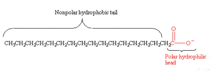
- amphipathic/amphiphilic molecule
- categorisation based on the length of hydrocarbon chain:
- short-chain FA: ≤6 carbons
- medium-chain FA: 8-12 carbons
- long-chain FA: 14-18 carbons
- very-long chain FA: ≥20 carbons
- most commonly found: palmitic acid (16C) & stearic acid (18C)
- categorisation based on the degree of saturation in the hydrocarbon chain:
- saturated FA
- unsaturated FA
Triglycerides (triacylglycerols)
- consists of ^^1 molecule of glycerol^^ esterified with ==3 fatty acids molecules==
- present in dietary fat; synthesised in the liver & adipose tissue
Cholesterol
- ==unsaturated== ==steroid alcohol==; amphipathic lipid (both hydrophilic and hydrophobic)
- almost exclusively synthesised by animals
- present in dietary fat; synthesised in liver
- precursor of steroid hormones & bile acids;
Lipoproteins
- consists of a non-polar core triglycerides & cholesteryl esters
- surrounded by a polar surface layer of phospholipids, cholesterol, & apolipoproteins
- classification based on different density fractions after ultracentrifugation
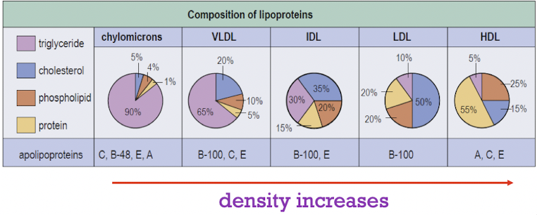
- LDL - associated with increased risk of cardiovascular disease
Lipids/Lipoproteins Analyses
Dyslipidemias
- diseases associated with abnormal lipid concentrations
- cause:
- genetic abnormalities
- environmental/lifestyle imbalances
- secondary to other diseases
- defined by clinical characteristics & laboratory test results
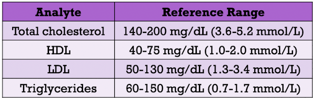
- laboratory analyses:
- total cholesterol
- HDL
- LDL
- triglycerides
- clinical reference ranges for lipids
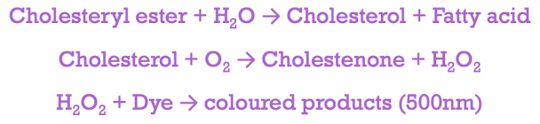
- Cholesterol measurement
- serum/plasma specimens are collected after fasting for at least 12 hours
- serum/plasma specimens can be refrigerated at 4C for several days
- HDL measurement
- two-step procedure with manual pretreatment
- precipitation reagent aggregates non-HDLs which are sedimented via centrifugation
- HDL is then quantified by enzymatic assays
- LDL measurement
- via Friedewald calculation
- total cholesterol, triglycerides and HDL are quantified
- VLDL is estimated as [triglyceride level/5] in mg/dL unit
- LDL = total cholesterol - HDL - VLDL
- Triglyceride measurement
- perform in conjunction with total cholesterol
Lipid Disorders
- [[Arteriosclerosis[[
- deposition of lipids (esterified cholesterol) in artery walls
- results in fatty streaks → plaques (smooth muscle cells, extracellular lipid, calcification & fibrous tissue) → partial or complete occulsion of blood flow
- LDL → initiate & promote plaque formation
→ every 1% decrease in LDL concentration leads to 2% decrease in arteriosclerosis risk
- Hypercholesterolemia
- associated with genetic abnormalities [i.e., familial hypercholesterolemia (FH)]
- homozygotes
- total cholesterol ~800-1000mg/dL (20-26 mmol/L)
- first heart attack in teenage years
- heterozygotes
- ~300-600 mg/dL (8-15 mmol/L)
- symptomatic for heart disease in 20s-50s
- Hypertriglyceridemia
- due to genetic abnormalities (e.g., FH)
- consequence of secondary causes (e.g., hormonal abnormalities, diabetes mellitus or nephrosis)
- imbalance between synthesis & clearance of VLDL
- many coronary heart disease patients have moderately elevated triglycerides & decreased HLDL level
Summary (Part 1)
- [ ] Different types of lipids
- [ ] Laboratory analyses for lipids/lipoproteins
- [ ] Development of arteriosclerosis
Carbohydrates
- Large macromolecules containing C, H & O atoms
- Generic formula:

- Contain C=O and -OH functional groups
- Grouping based on the number of carbon:
- trioses (3C)
- tetroses (4C)
- pentoses (5C)
- hexoses (6C)
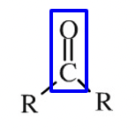
- Grouping based on the location of the C=O functional group
- aldehyde group (O=CH-)
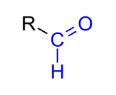
- ketone group (O=C)
- Saccharide - basic unit structure
- Classification:
- monosaccharide (e.g., glucose, fructose, galactose)
- disaccharide (e.g., lactose, maltose, sucrose)
- oligosaccharide (e.g., oligofructose, maltotriose)
- polysaccharide (e.g., amylose, glycogen)
Glucose
- Major energy supply
- primarily stored as glycogen in liver & muscle
- disease states:
- hyperglycaemia
- hypoglycaemia
Hyperglycaemia
- hyper (high) + glykys (sweet/sugar) + haima (blood)
- an increase in blood glucose level
- defined as blood glucose level (>125mg/dL during fasting)
- Causes:
- reduced insulin secretion
- decreased glucose utilisation
- increased glucose production
- Laboratory findings (blood specimen)
- increased glucose level in plasma
- increased serum osmolality
- ketones in serum (ketonemia)
- decreased blood pH (acidosis)
- electrolyte imbalance
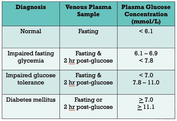
Diabetes Mellitus
- metabolic disorder characterised by chronic hyperglycemia
- arise from defects in the secretion, or action of insulin, or both
- clinically defined as high plasma glucose concentrations at which there is an increased risk of retinopathy, nephropathy & neuropathy
- type 1 diabetes mellitus
- pancreatic islet B-cell destruction
- tendency to ketoacidosis
- type 2 diabetes mellitus
- insulin resistance
- insulin secretory defect
- gestational diabetes mellitus
- glucose intolerance
- metabolic & hormonal changes during pregnancy
- Laboratory diagnosis
- measurement of glycated haemoglobin
- ‘time-weighted’ average plasma glucose concentration over the past 2-3 months
- in vivo glycation of haemoglobin is proportional to plasma glucose concetration
- average plasma glucose concentration during the last 30 days accounts for 50% of the HBAlc concentration (glycated haemoglobin)
- expressed as a proportion of total haemoglobin (in percentage)
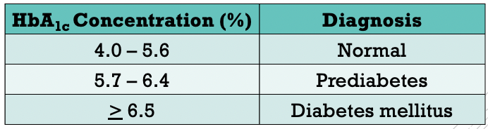
- Prediabetes
- higher than normal blood glucose level, but insufficiently high to be considered as diabetes
- increased risk of type 2 diabetes mellitus, heart disease & stroke
- <<reversible<<
- criteria:
- 126mg/dL > fasting glucose level ≥ 100 mg/dL
- 200mg/dL > 2hr OGTT level ≥ 140 mg/dL
- HBAlc concentration of 5.7-6.4%
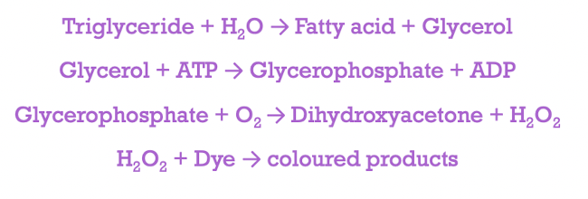
Glucose Assay
- serum/plasma sample
- enzymatic approach - glucose oxidase, hexokinase, glucose dehydrogenase
- generate measurable coloured product proportional to glucose concentration
Glucometer
- medical device for point-of-care measurement of blood glucose concentration
- rapid & easy home-based blood glucose monitoring, particularly among individuals with Type 1, 2 & gestational diabetes mellitus
- requires only a small amount of blood sample (from fingertip)
Hypoglycaemia
- Blood glucose level is lower than the standard range
- fasting blood sugar ≤ 70 mg/dL or 3.9 mmol/L
- common among
- particularly in type 1 diabetic patients
- type 2 diabetic patients who are on insulin/medication
- rare in individuals with normal glucose metabolism
- diagnosis should be made only if the individual demonstrates:
- hypoglycaemic symptoms
- low plasma glucose concentration <50 mg/dL
- symptoms are relieved by administering glucose/glucagon
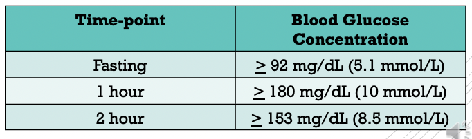
Gestational Diabetes Mellitus
- Recommendation: all nondiabetic pregnant women should be screened at 24-28 weeks of gestation
- one-step approach: 2-hr oral glucose tolerance test (OGTT) by using 75g glucose load
- requires overnight fasting of at least 8hr
==Complete Blood Count==
- to evaluate overall health
- to screen, diagnose & monitor certain health disorders, e.g., anaemia, infection & leukemia
- Measurement and/or counting of:
- red blood cells
- white blood cells
- haemoglobin
- haematocrit
- platelets
- mean corpuscular volume (MCV)
Red Blood Cells
- measure the number of red blood cells, which carry oxygen from the lungs to the rest of our body
- normal count (male):
- 4.35-5.65 million cells/ul
- normal count (female):
- 3.92-5.13 million cells/ul
- increased level may indicate:
- polycythemia
- respiratory distress
- high altitude
- decreased level may be a sign of:
- iron anemia
- vitamin B12, vitamin B6 and/or folic acid anaemia
- haemorrhage
- liver & renal dysfunction
White Blood Cells
- measure the total number of WBCs (without differential)
- measure the number of each of the 5 basic white blood cell types (differential)
- neutrophils, eosinophils, basophils, lymphocytes & monocytes
- expressed as percentage or absolute value
- elevated white blood cells count may indicate inflammation or infection
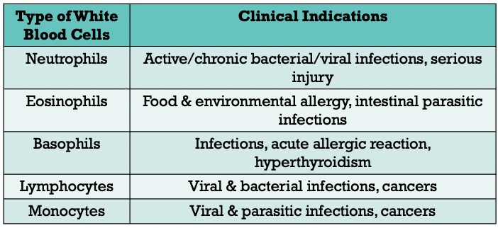
- low cells count may indicate autoimmune diseases, bone marrow disorders or cancers
- normal: 3.49-9.6 billion cell/litre
Haemoglobin
- protein that carries oxygen in the red blood cells
- normal (male): 132-166 g/L
- normal (female): 116-150 g/L
- increased level may indicate dehydration, emphysema, asthma, polycythemia
- decreased level may indicate iron deficiency anaemia, microscopic internal bleeding, digestive inflammation
Hematocrit
- known as packed cell volume
- percentage of the total occupied by packed red blood cells when a given volume of whole blood is centrifuged
- normal (male): 38.3% - 48.6%
- normal (female): 35.5% - 44.9%
- abnormal value indicates the similar clinical conditions as haemoglobin
Platelets
- responsible for blood coagulation and vascular integrity
- reference (male): 135-317 billion/L
- reference (female): 157-361 billion/L
- increased level: atherosclerosis, rheumatoid/inflammatory arthiritis, certain cancers
- decreased level: idiopathic thrombocytopenia, blood loss
Means Corpuscular Volume (MCV)
- the volume in cubic microns occupied by an average single red blood cell
- ↑ level (macrocytic RBCs): vitamin B12/folic acid anaemia, dehydration
- ↓ level (microcytic RBCs): iron anaemia, microscopic internal bleeding
- other RBCs-related markers:
- MCH (mean corpuscular haemoglobin)
- MCHC (mean corpuscular haemoglobin concentration)
Summary (Part 2)
- [ ] Hyperglycaemia and hypoglycaemia
- [ ] complete blood count & its clinical indications