U3-Electrocardiogram
Depolarization: state of stimulation that precedes contraction; electrical activation of heart cells; causes heart to contract
Repolarization: state of cellular recovery after contraction; cells return to a resting state; heart relaxes, allowing for the refilling of chambers
Polarization: cells are at peak resting energy
Isoelectric line/ baseline: The line on the ECG that remains flat
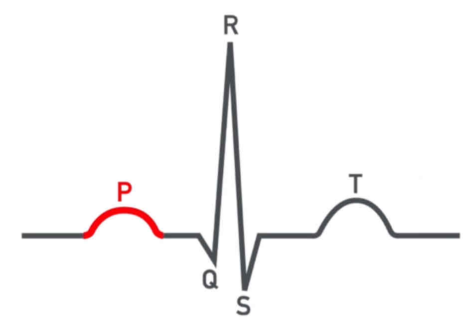
P wave: atria depolarization; first positive deflection
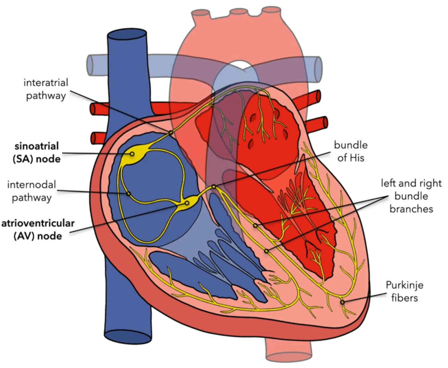
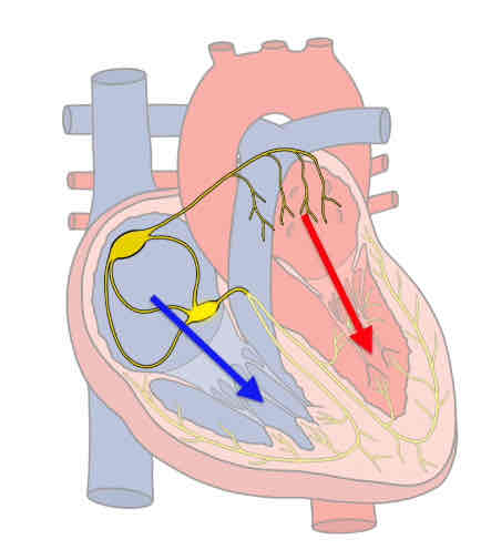
Signals travel through the SA node, through the internodal pathways, into the AV node as well as the interatrial pathways causing depolarization of the atria
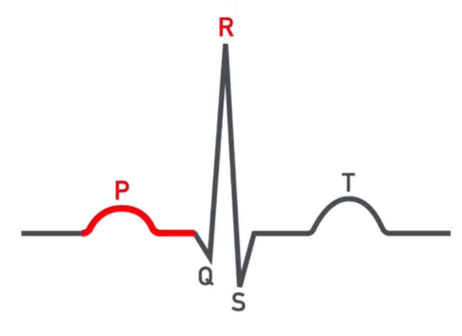
PR interval: atria are going to contract . Send blood from the right atrium into the ventricle. & the blood from the left atrium into the left ventricle
The contractions of the atria will last early in the P wave
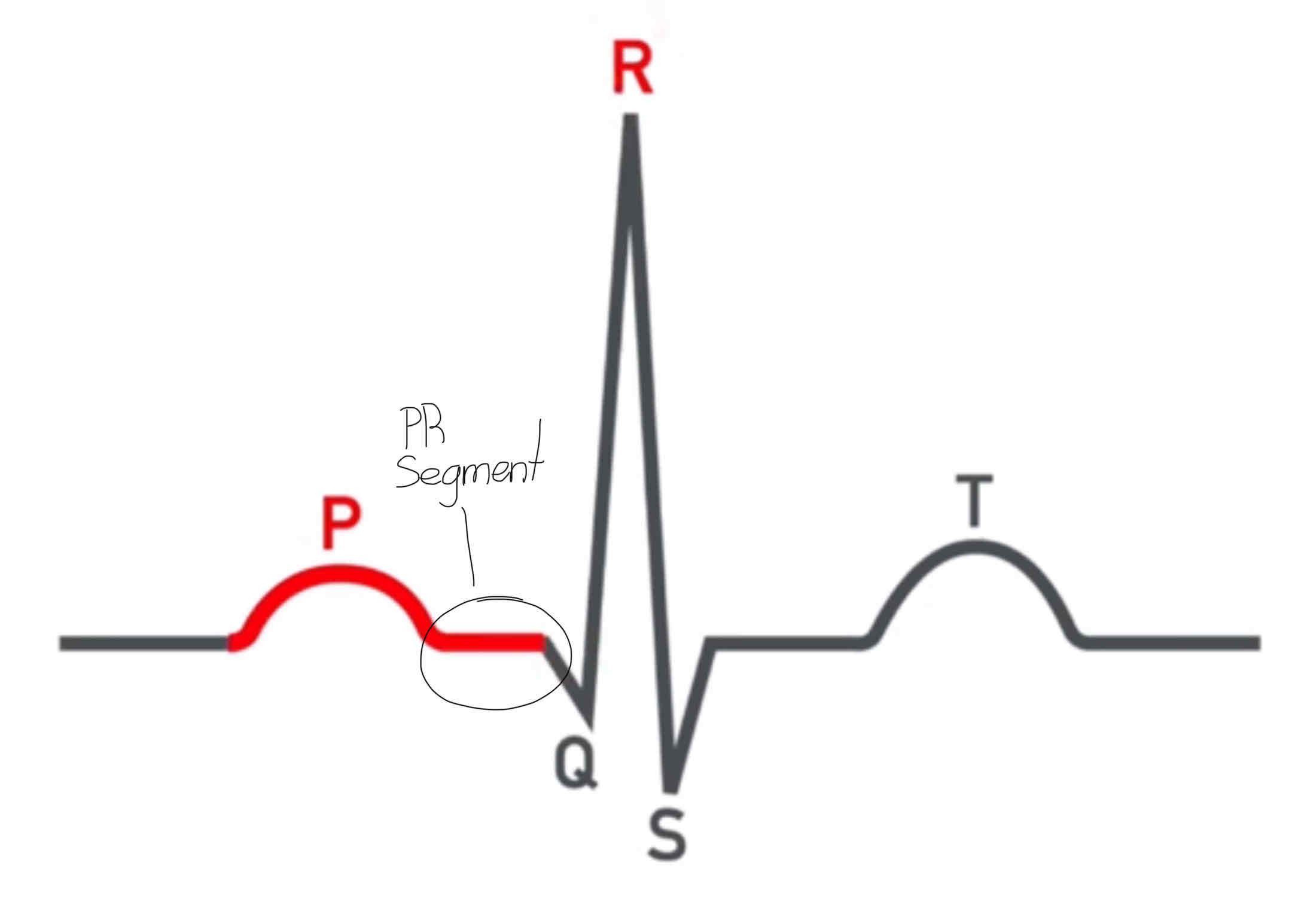
The PR segment: is where AV nodal delay will occur; normal length is 0.12- 0.20

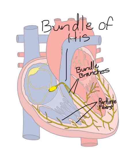
QRS complex: The R spike is a signal passing through the bundle of His, bundle branches & the purnkinje fibers that stimulates all the cardiac muscle. This process causing depolarization of the ventricles ; as well as the atria will repolarize right after
Electric activity will be much higher in the ventricles because the ventricles have more cardiac tissue
Ventricles will pump blood into the lungs, aorta & basically the whole body. In term needing a really strong contraction from the ventricles. I.E the ventricles working harder.
The Q wave: represents conduction of impulse down the interventricular septum; first negative deflection
The R wave: represents conduction of electrical impulse to the left ventricle
The S wave: negative deflection; represents conduction of electrical impulse through both ventricles
ST Interval: ventricles will contract causing blood to be pumped through the aorta, as well as blood being pumped through the pulmonary artery. The forceful contraction will begin at the end of the QRS complex & will last until the T wave is complete.
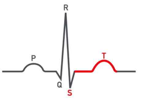
The T wave is where repolarization of the ventricles & stopping the contraction. The ventricles will be contracting during the ST interval.
ST Segment: indicates end of ventricular depolarization and beginning of ventricular repolarization; elevation or depression may be an indication of ischemia
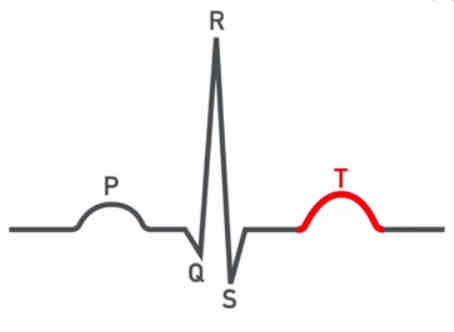
T Wave: The repolarization of the ventricles. Or in other words,the turning off of ventricular contraction.
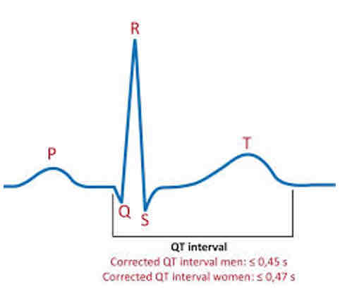
The QT interval: is the time required for ventricular depolarization and repolarization to occur; normally measures less than one R-R interval
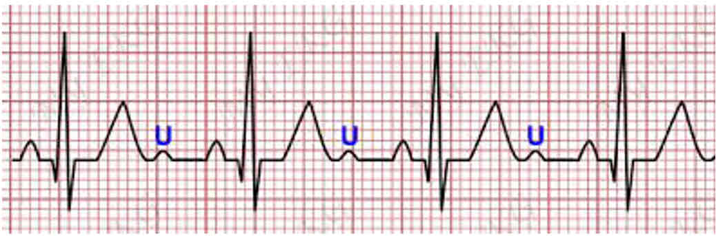
The U wave: occurs after the T wave; represents repolarization of bundle of His & purkinje fibers; presence can indicate electrolyte balance.
R-R interval: used to calculate heart rate
The J point: represents end of the QRS complex & ventricular depolarization; QRS complex measures normally 0.06- 0.1 seconds