Unit 10 - Nervous System, Spinal Cord, Spinal Nerves
- Nervous Tissue (Ch. 12)
- Overview of Nervous System (12.1)
- Properties of Neurons (12.2)
- Supportive Cells (12.3)
- 2. The Spinal Cord, Spinal Nerves, (Ch. 13)
- The Spinal Cord (13.1)
- Spinal Nerves (13.2)
- Internal Communication
- Endocrine and nervous system maintain internal coordination
1.Endocrine system 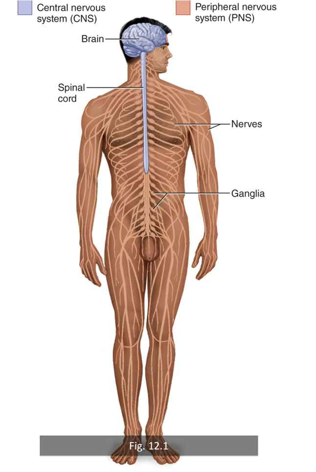
- uses hormones secreted into blood
- “long-distance” communication
- Slow
2.Nervous system
- Uses electrical and chemical means
- sends message from cell to cell
- Very quick
- Nervous system components
- Central nervous system (brain and spinal cord)
- Enclosed by cranium and vertebral column (very protected).
- Peripheral nervous system (everything else)
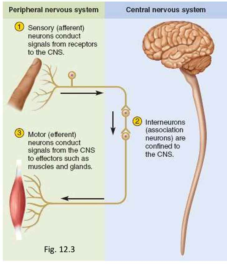
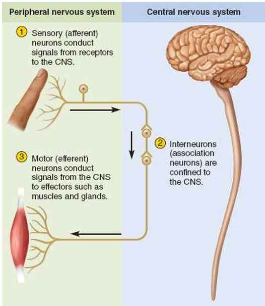
- Sense organs receive information and transmit coded messages to the CNS (brain, spinal cord)(sensory/afferent).
- CNS processes information, determines appropriate response.
- CNS issues commands to muscles and gland cells to carry out response (motor/efferent).
Visceral: heart, stomach, bladder
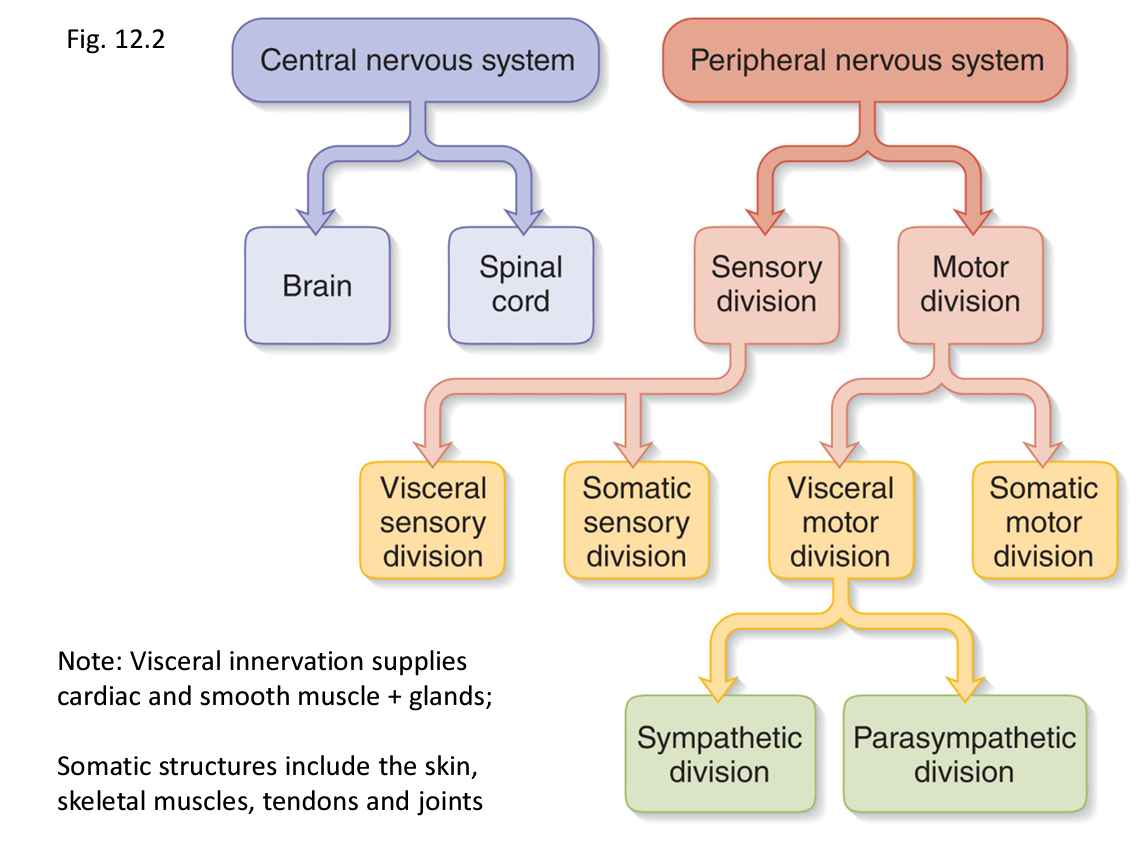
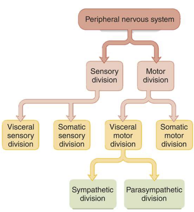
- Peripheral Nervous System
1. Sensory (afferent) division – carries signals from receptors to
CNS [Receptors]
- Somatic sensory division
- Signals from skin, muscles, bones, and joints
- Visceral sensory division
- Signals from the viscera (heart, lungs, stomach, and urinary bladder)
2. Motor (efferent) division – carries signals from CNS to
effectors (glands/muscles) [Effectors]
- Somatic motor division
- Carries signals to skeletal muscles
- Visceral motor division(autonomic nervous system)
- Signals to glands, cardiac, and smooth muscle
- Involuntary responses are visceral reflexes.
- Visceral Motor Division (a.k.a Autonomic Nervous System)
- Subdivisions:
- Sympathetic Division
- Arouses body for action (“fight or flight”)
- Increases heart rate and respiration
- Inhibits digestive and urinary systems
- Parasympathetic Division
- Calming effect (“rest and digest”)
- Slows heart rate and breathing
- Stimulates digestive and urinary systems
- Properties of Neurons
- Neurons (nerve cells) all have 3 fundamental properties:
- Excitability
- Will respond to stimuli (environmental changes)
- Conductivity
- Produce electrical signals that are quickly conducted to other cells at distant locations
- 3.Secretion
- When signal reaches the end of a nerve fiber, a neurotransmitter will be released that crosses the gap and influences the next cell.
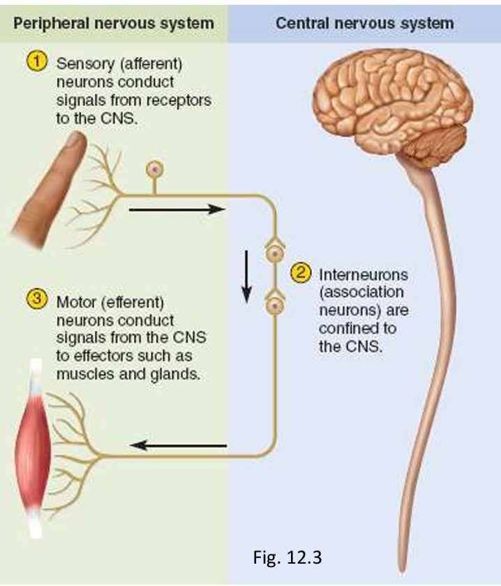
- Functional Classes Neurons
- Afferent (sensory) neurons
- Detect stimuli and transmit the information toward the CNS
- Interneurons
- ~90% of all neurons connect neurons together
- Receive signals from many neurons and carry out integrative functions (make decisions)
- Efferent (motor) neurons
- Sends signals to muscles and/or gland cells.
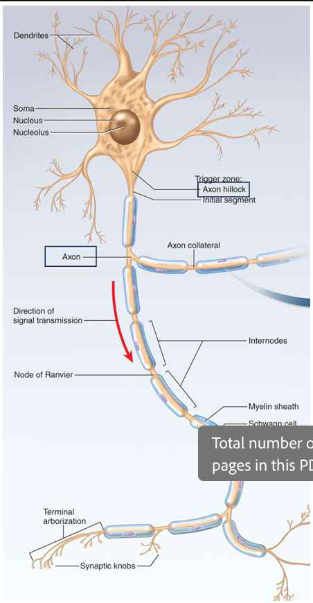
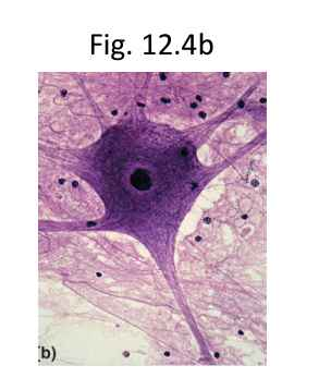
- Neuron Structure
- Soma
- Control center of neuron
- a.k.a. Neurosoma or cell body
- Dendrites
- Branches off of the soma.
- Receiving signals from other neurons.
- Axon
- Originates from a mound on the soma called the axon hillock
- For rapid conduction of signals to distant points
- Axoplasm - cytoplasm of axon
- Axolemma - plasma membrane of axon
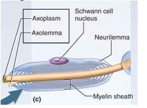
- Myelin sheath may enclose it.
- Distal end has terminal arborization.
- Synaptic knob (terminal button) is swelling that forms junction (synapse) with next cell.
- Contains synaptic vesicles full of neurotransmitter.
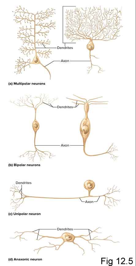
- Multipolar neuron
- One axon, multiple dendrites
- Most common neuron, most neurons in CNS are this kind
- Bipolar neuron
- One dendrite and one axon
- Olfactory cells, retina, inner ear
- Unipolar neuron
- Single process leading away from soma
- Sensory cells from skin and organs to spinal cord
- Anaxonic neuron
- Many dendrites, no axon
- Retina, brain, and adrenal gland
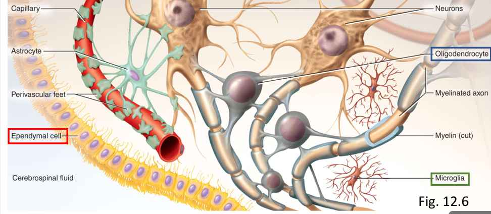
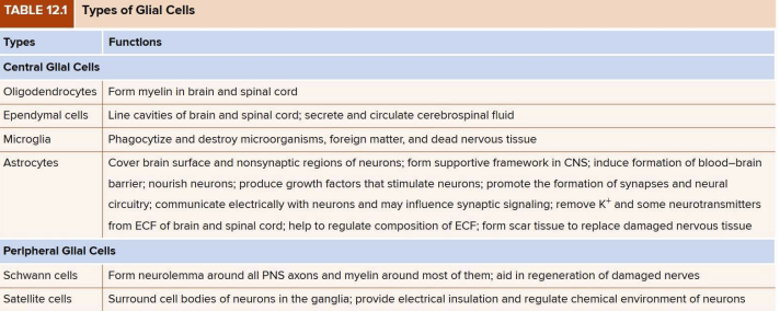
- Neuroglia (Supportive Cells)
- Outnumber neurons 10:1
- Protect neurons and help them function
- Bind neurons together and form framework for nervous tissue
- 4 Types in CNS
- Oligodendrocytes
- Form myelin sheaths that speed conduction.
- Ependymal cells
- Line internal cavities of brain, secrete and circulate cerebrospinal fluid (CSF)
- Cuboidal epithelium
- Microglia
- Wander through CNS looking for damage/debris
- For debris and foreign matter
- Found in places fighting infection, injury
- Astrocytes
- Most abundant in CNS, clever entire brain surface and most non-synaptic regions of neurons.
- Neuroglia Types - CNS
- Astrocytes
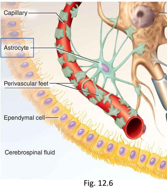
- Functions:
- Form supportive framework.
- Perivascular feet (extensions) contact blood capillaries to form seal (blood-brain barrier).
- Convert glucose to lactate for neurons.
- Secrete nerve growth factors
- Communicate electrically with neurons.
- Absorb excess neurotransmitters and ions.
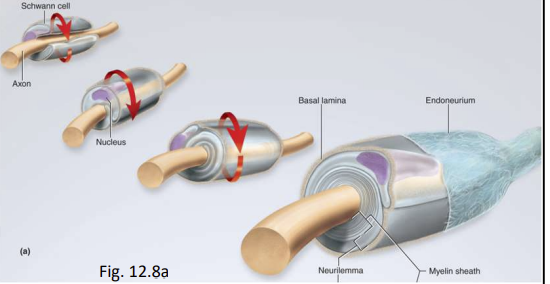
Neuroglia in PNS:
- Schwann cells
- Envelop nerve fibers and produce a myelin sheath in PNS
Assist in regeneration of damaged fibres
- Satellite cells
- Surround neurosomas in ganglia of PNS and provide electrical insulation around soma
- Regulate chemical environment of neurons
Blood Brain Barrier, Clinical Insight
- The blood-brain barrier is a tightly packed layer of cells that line the blood vessels in the brain and spinal cord. Separates the capillary blood from the surrounding interstitial fluid.
- Prevents large molecules (most drugs), immune cells, and disease-causing organisms such as bacteria and viruses form passing from the bloodstream, into the central nervous system (CNS)
- Protective, semi-permeable and highly selective. This also plays a role in homeostasis
Brain Tumors- Gliomas
- A tumor is a mass of rapidly dividing cells. Most adult brain tumors are composed of glial cells
- Glial cells usually grow rapidly and are highly malignant
- Because of the blood-brain barrier, they usually do not yield to chemotherapy and must be treated with radiation or surgery
Myelin
- Myelin sheath is insulation around nerve fibre
- Formed by:
- Oligodendrocytes in CNS
- Schwann cells in PNS
- Consists of the plasma membrane of glial cells
- Begins in fetal development but proceeds rapidly in infancy.
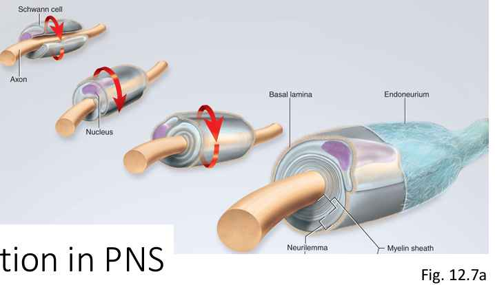
Myelination in the PNS
- Schwann cell spirals repeatedly around single nerve fibre
- as many as 100 layers of membrane
- no cytoplasm between membranes
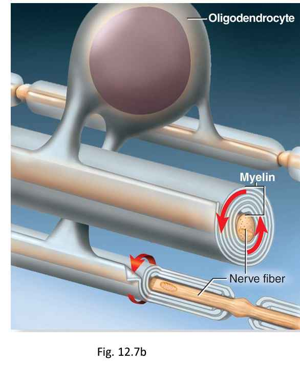
Myelination in the CNS
- Oligodendrocyte
- Myelinates several nerve fibres in immediate vicinity
- Since it is anchored to multiple nerve fibers, it cannot migrate around any of them
- Must push newer layers of myelin under the older ones
- myelination spirals inward toward nerve fibre
Myelin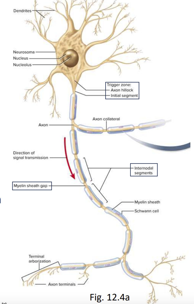
- Many schwann cells or oligodendrocytes needed to cover one nerve to fibre
- Sheath is segmented:
- Nodes of ranvier (myelin sheath gap)
- Internodes (intermodal segments)
- Myelin covered segments from one gap to the next
- Initial segment (in trigger zone)
- Short section of nerve fibre between one axon hillock and the first glial cell
Multiple Sclerosis
- Degenerative disorder of the myelin sheath
- The oligodendrocytes and myelin sheaths of the CNS deteriorate and are replaced by hardened scar tissue, especially between the ages of 20 and 40. Fatal from 25-30 years after diagnosis
- Nerve conduction are disrupted, with effects that depend on what part of the CNS is involved- double vision, blindness, speech defects, neurosis, tremors, or numbness, for example.
- The cause of MS remains uncertain; autoimmune triggered by virus?
Concept Check: Fill-in-the-blank
- Neurons that convey information to the CNS are called sensory, or afferent neurons.
- To perform their role, neurons must have the properties of excitability, secretion, and conductivity
- In the CNS, myelin is produced by glial cells call oligodendrocytes.
Nervous System
2. The Spinal cord, Spinal nerves (CH.13)
- The spinal cord (13.1)
- Spinal nerves (13.2)
Spinal Cord
- Information highway that connects brain with lower body
- We study the spinal cord, as well as the spinal nevers that arise from each vertebral level
- The central and peripheral nervous systems are linked structurally and functionally
Functions:
- Conduction
- Nerve fibers conduct sensory and motor information up and down spinal cord (2 directions!)
- Neural integration
- Spinal neurons receive input from multiple sources, integrate it, and execute appropriate output (e.g., bladder control)
- Locomotion
- Spinal cord contains central pattern generators: groups of neurons that coordinate repetitive sequences of contractions for walking
- Reflexes
- Involuntary responses to stimuli vital to posture, coordination and protection
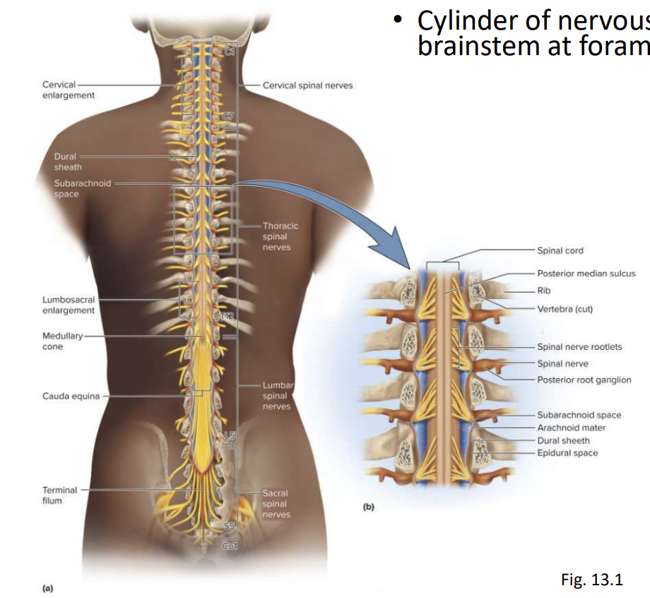
Spinal Cord Surface Anatomy
- Cylinder of nervous tissue that arises from brainstem at foramen magnum of the skull
- Occupies upper ⅔ of vertebral canal
- Inferior margin ends at L1 or slightly beyond in adults
- Gives rise to 31 pairs of spinal nerves
- 45 cm long
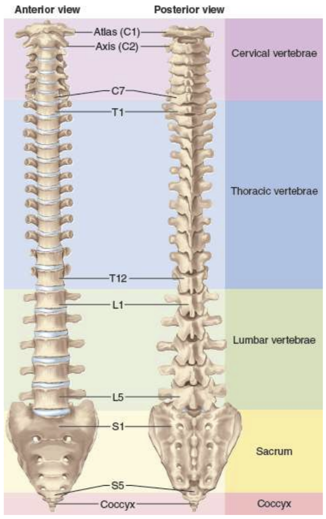
The Vertebral Column
Spinal Cord Surface Anatomy
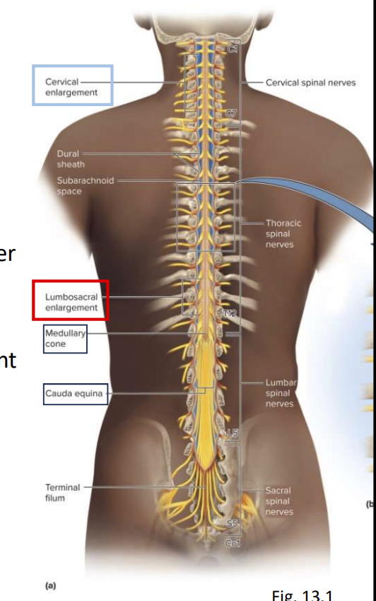
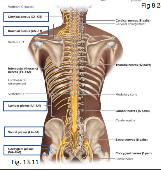
Spinal cord divided into 4 major regions:
- Cervical (7 vertebrae, 8 spinal nerve)
- Thoracic (12)
- Lumbar (5)
- Sacral (5)
Two enlargements where the cord is thicker:
- Cervical enlargement - nerves to upper limbs
- Lumbar enlargement - nerves to pelvic region and lower limbs
Medullary cone
- Cord tapers to a point inferior to lumbar enlargement
Cauda equina
- Bundle of nerve roots L2-S5
- Cauda = tail, Equine = horse
- Innervates the pelvic organs and lower limbs
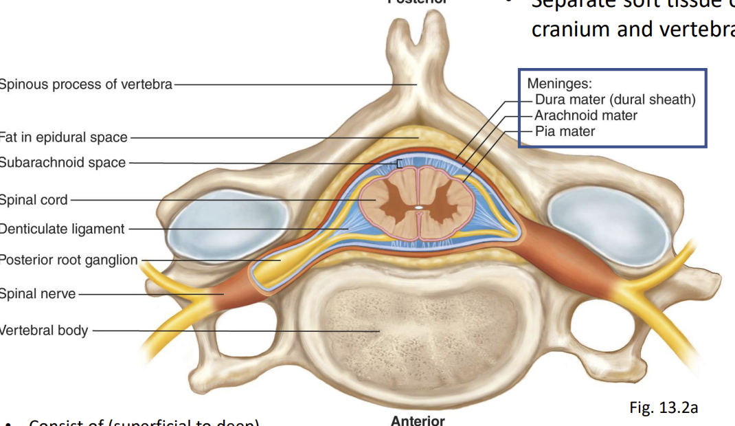
Meninges
- 3 fibrous membranes enclose braina nd spinal cord
- Seprate soft tissue of CNS fro, cranium and verteral canal
- Consist of (superficial to deep)
- Dura Mater - dura = tough outermost layer (dura- durable)
- Arachnoid mater looks like spider web (arachnoid=spider) 5-6 layers.
- Pia mater thinnest, 1-2 layers of squamous cubodial, innermost layer
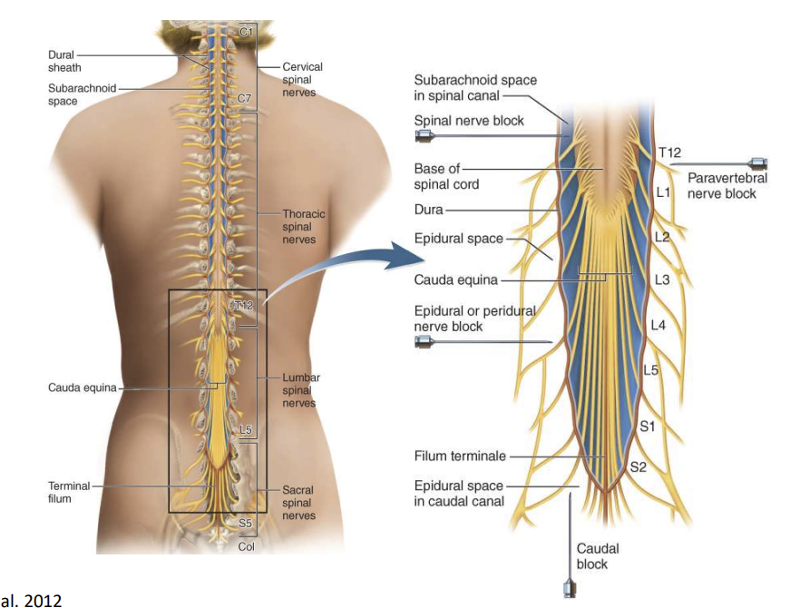
- Dural sheath surrounds spinal cord and is seperated from vertebrae by epidural space (fat, blood vessels, connective tissue): anesthetics can be introduced here and block pain signals during childbirth or surgery.
Arachnoid Mater
- Adheres to dura
- Separated from pia mater by subarachnoid space
- Filled with cerebrospinal fluid (CSF)
- Region inferior to medullary cone occupied by cauda equina and CSF
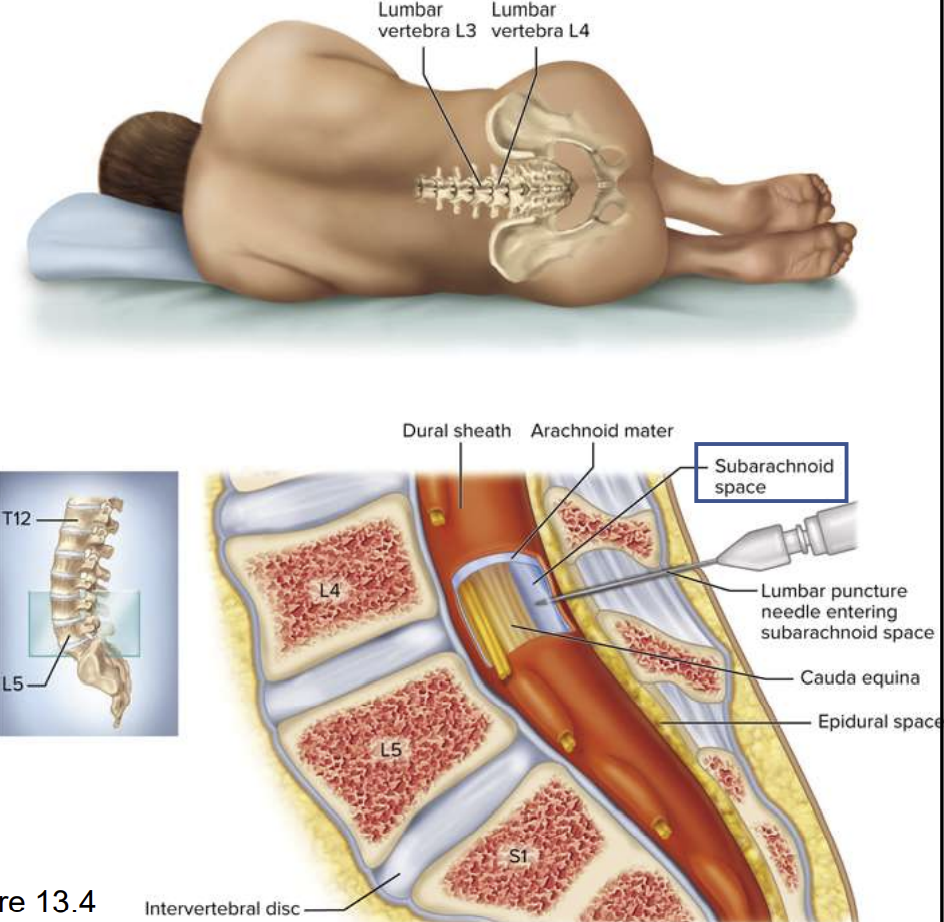
- Lumbar puncture (spinal tap) samples CSF
Pia Mater
- Membrane that follows contours of spinal cord
When a sample of CSF is needed for clinical purposes, it is taken from the lumbar cistern by a procedure called lumbar puncture (or colloquially, spinal tap). A spinal needle is insrted between two vertbrae at level L3/L4 or L4/L5, where there is no risk of accidntial injuryt to the spinal cord (which ends at L1 to L2). CSF drips fromt he spinal needle into a collection tube; usually 3 to 4 mL of CSF is collected.
Spinal Tap (Lumbar Puncture)
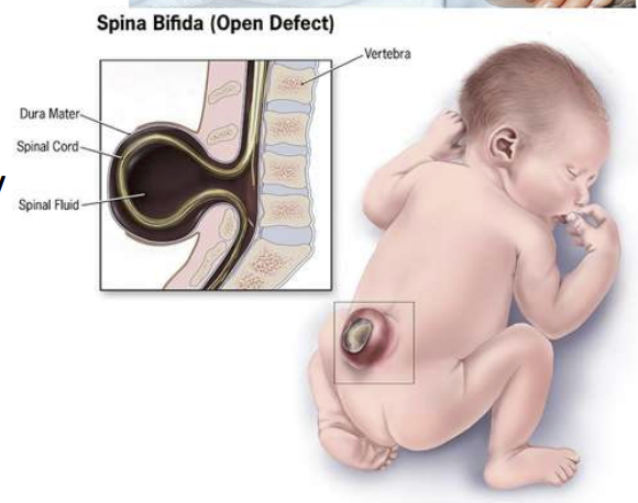
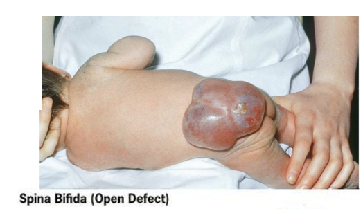
Spina Bifida
- Cogenital defect
- One or mor evertebrae fail to form complete vertebral arch for enclosure of the spinal cord
- Common in lumbosacral region
- Sac protrudes from spine and may contain meninges, CSF, parts of spinal cord and nerve roots
- Folic acid reduces incidence
- Start 3 months before conception as defcet in the first 4 weeks
Concept Check: Question
Which of these is not a region of the spinal cord?
- Cervical
- Thoracic
- Pelvic
- Lumbar
- Sacral
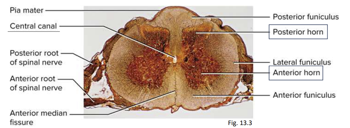
Cross-sectional Anatomy of the Spinal Cord
- Gray matter
- Central area
- Neuron cell bodies with little myelin (site of information processing, synaptic integration)
- White matter
- Surrounds gray matter
- Abundantly myelinated axons (carry signals in CNS)
- Lighter in colour because of this
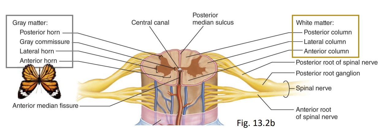
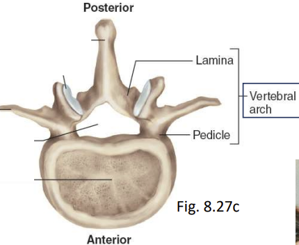
Gray Matter
- Pair of posteriori (dorsal) horns
- Posterior (dorsal) root of spinal nerve carries sensory nerve fibers
- Pair of thicker anteiroir (ventral) horns
- Anterior (ventral) root of spinal nerve carries only motor nerve fibers
- Lateral horn
- Visible from T2 through L1
- Contains neurons of sympathetic nervous system
- Gray commissure connects right and left sides
- Central canal lined with ependymal cells and filled with CSF
- Near its attachment to the spinal cord, a spinal nerve branches into a posterior (dorsal) root and anterior (ventral) root
- The posterior root carries sensory nerve fibers
- The anterior horns contain the large somas of the somatic motor neurons, which exit by way of the anteriori root of the spinal nerve and lead to the skeletal muscles
- A lateral horn is visible on each side of the gray matter from segments T2 through L1 of the cord. It contains neurons of the sympathetic nervous system
White Matter
- Surrounds gray matter
- Bundles of axons up and down cord providing communication between different levels of CNS
- Columns (funiculi):
- Three pairs of columns on each side
- Posterior (dorsal)
- Lateral
- Anterior (ventral)
- Each column consists of subdivisions = tracts or fasciculi
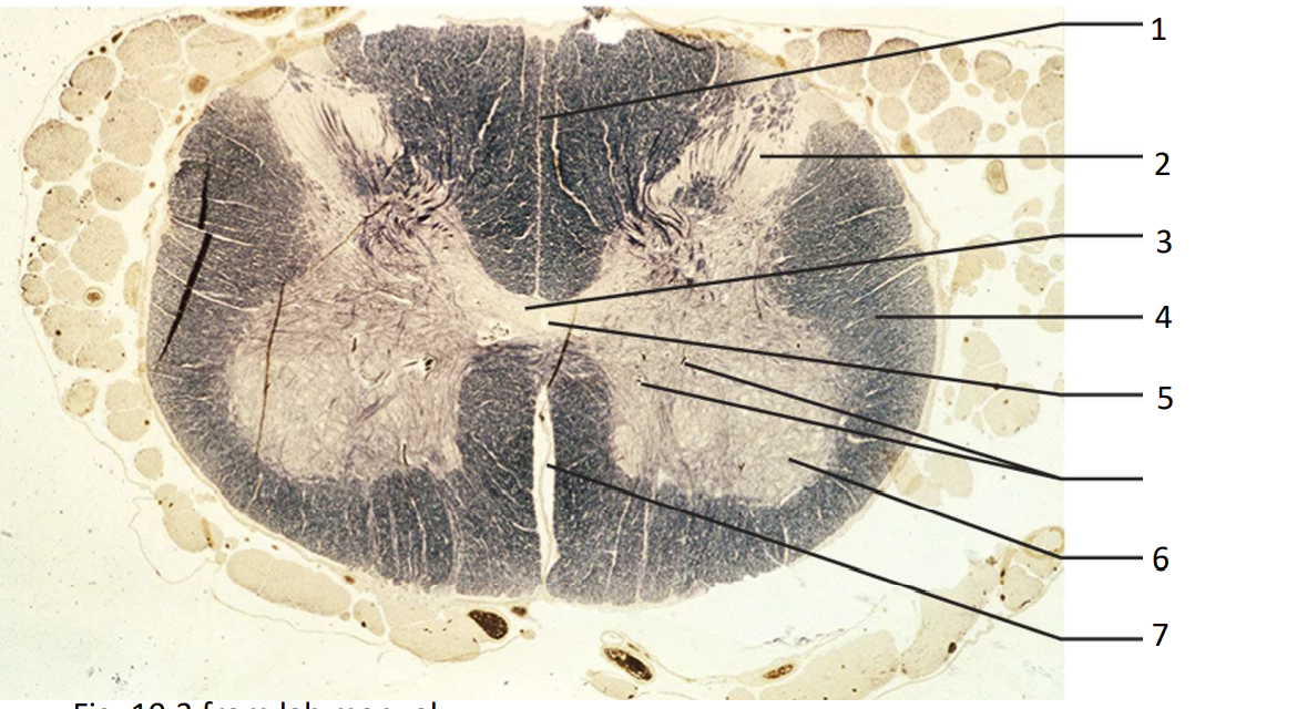
3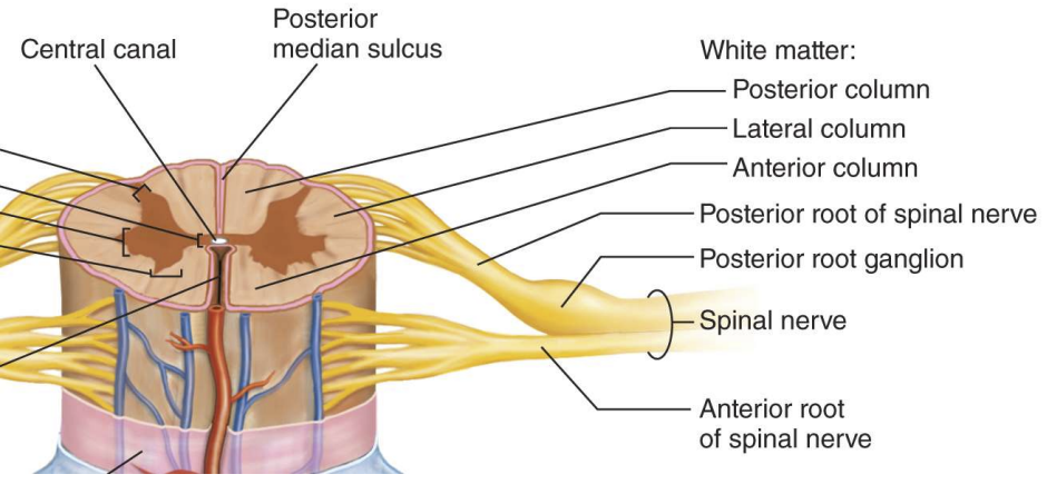
For your Practice- Label
Peripheral Nervous System
- Two divisions (sensory and motor), each with somatic and visceral subdivisions
- Composed of:
- Nerves
- Bundles of nerve fibers (axons) wrapped in fibrous conencitve tissue
- Ganglion
- Knot-like swelling in a nerve where neuron cell bodies are concentrated
- Plexus
- Interconnecting ganglia (network of interwoven nerves)
The Spinal Nerves
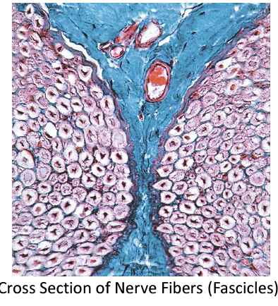
Nerve:
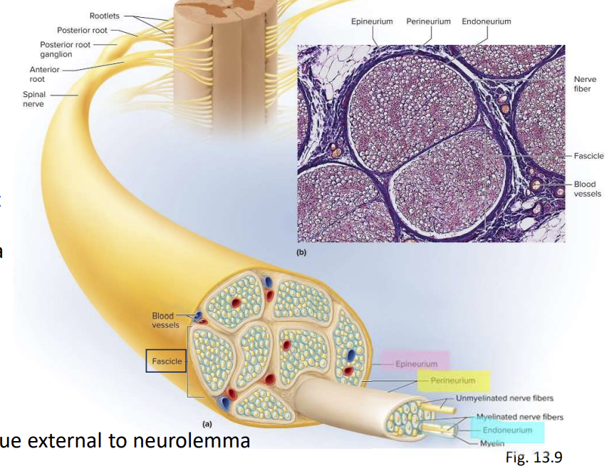
- Cord-like organ composed of numerous nerve fibres bound together by conenctibe tissue
- Mixed nerves
- Contain both afferent and efferent fibres
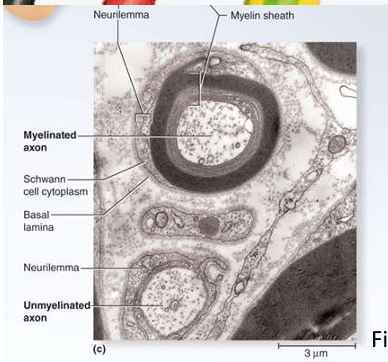
- Nerve fibres of PNS have a neurolemma (outmost sheath of Schwann cells) and myelin sheath from Schwann cells
- Endoneurium
- Loose connective tissue external to neurolemma
- Perineurium
- Layers of overlapping squamous cells that wrap fascicles (bundles of nerve fibres)
- Epineurium
- Dense irregular connective tissue that wraps entire nerve
- Blood vessels penetrate connectibve tissue coverings
Classification of Nerve Fibers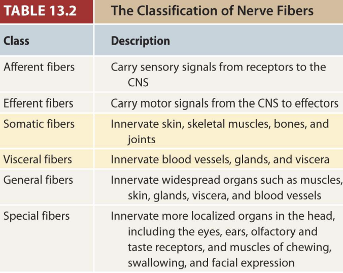
- Both afferent and efferent nerves can be:
- Somatic
- innervate skin, skeletal muscles, bones, and joints
- Visceral
- Innervates blood vesels, glands and viscera
Also:
- General
- innervate widespread organs such as msucles, skin, glands, viscera, and blood vessels
- Special
- Innervate more localized organs in the head (eyes, ears, olfactory and taste receptors, and muscles of chewing, swallowing, and facial expression)
Question
The outermost connectibve tissue wrapping of a nerve is called the:
- Epineurium
- Perineurium
- Endoneurium
- Arachnoid mater
- Dura mater
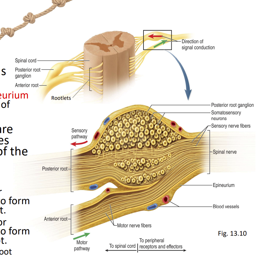
Ganglion
- Cluster of neurosomas outside the CNS
- Enveloped with epineurium continuous with that if nerve
- Among neurosomas are bundles of nerve fibres leading into and out of the ganglion
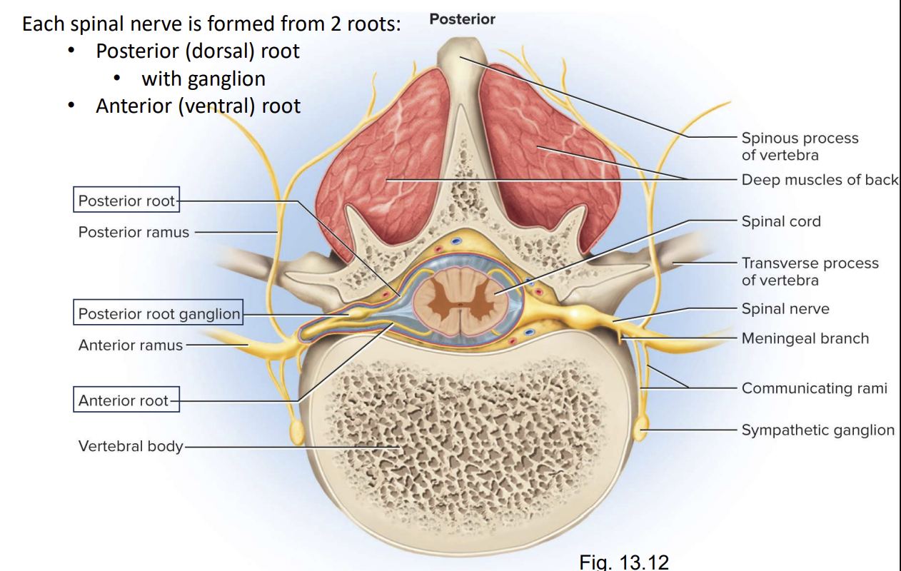
- Rootlets:
- Emerge from anterior surface → coverage to from anterior (ventral) root
- Emerge from posteriori surface → coverage to form posterior (dorsal) root
- Swells into Dorsal Root Ganglion
Proximal Branches of a Spinal Nerve
Distal Branches of a Spinal Nerve
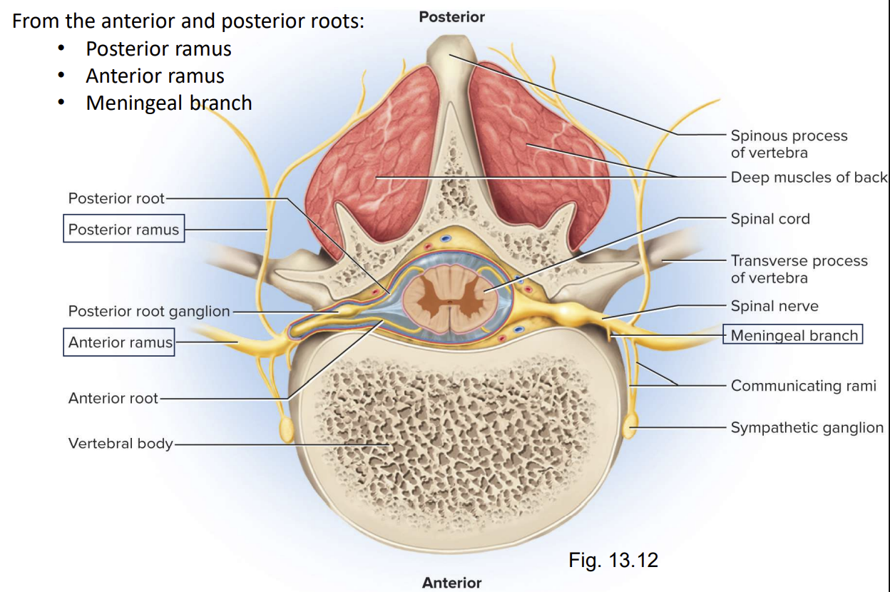
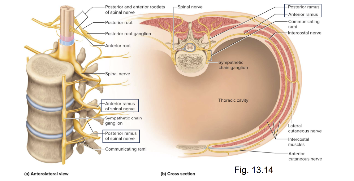
Rami of the Spinal nerves
Anterior ramus: in thoracic region, it gives rise to intercostal nerves in other regions, anterior rami form plexuses
Posterioir ramus: innervates the muscles and joints in that region of the spine and the skin of the back:
Meningeal branch: reenters the vbertbral canal and innervates the meninges, vertebrae, and spinal ligaments
- Spinal nerve plexuses:
- Except the thoracic region, nerves branch and merge repeatedly to form five webs = nerve plexuses
- 31 pairs of spinal nerves (mixed)
- 8 cervical
- 1st cervical exits between skull and atlas, all others at intervertebral foramina
- 12 thoracic
- 5 lumbar
- 5 sacral
- 1 coccygeal
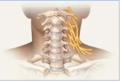
- Cervical plexus (C1-C5):
- Receives fibers from the anterior rami of nerves C1-C5 and gives rise to these nerves.
- Most important: phrenic nerves to diaphragm. C3,4,5 are important for breathing
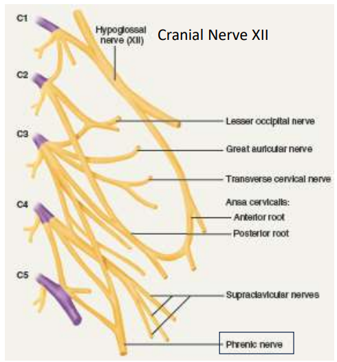
- Cervical plexus (C1-C5):
- Hypoglossal nerve (XII)
- Motor- innervates muscles of tongue
- Lesser occipital nerve
- Somatosensory- skin on parts of ear and neck
- Great auricular nerve
- Somatosensory- most of external ear, salivary gland
- Transverse cervical nerve
- Somatosensory- anterior and lateral neck, underside of chin
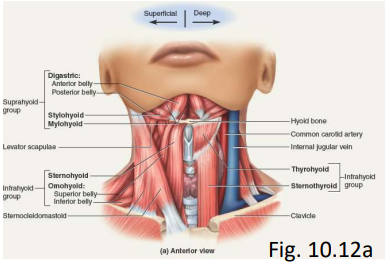
- Ansa cervicalis
- Motor- omohyoid, sternohyoid, and sternothyroid neck muscles
- Supraclavicular nerves
- Somatosensory- neck, shoulder, anterior chest
- Brachial plexus (C5-T1):
- Innervates upper limb and some muscles of neck and shoulder
- Median nerve → carpal tunnel
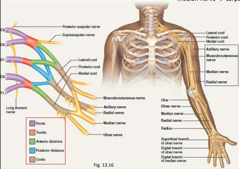
- Brachial plexus (C5-T1):
- Musculocutaneous: sensory: skin of anterolateral forearm; elbow joint; motor: brachialis, biceps brachii, coracobrachialis muscles
- Axillary: sensory: skin of lateral shoulder and arm; shoulder joint; motor: deltoid and teres minor muscles (rotator cuff of shoulder)
- Radial: sensory: skin of posterior arm; posterior and lateral forearm and wrist; joints of elbow, wrist, and hand; motor: extensor muscles of posterior arm and forearm
- Median: sensory: skin of lateral ⅔ of hand; tips of digits I-IV; joints of hand; motor: forearm flexors, thenar group and lumbricals I-II of hand
- Ulnar: sensory: skin on palm and med hand and digits II-V; joints of elbow and hand
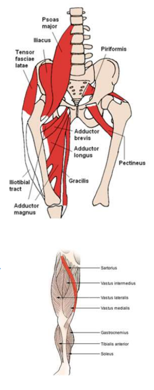
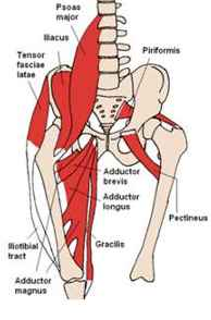
- Lumbar plexus (L1-L4):
- Innervates abdominal wall, anterior thigh, genitalia
- 5 roots and 2 divisions
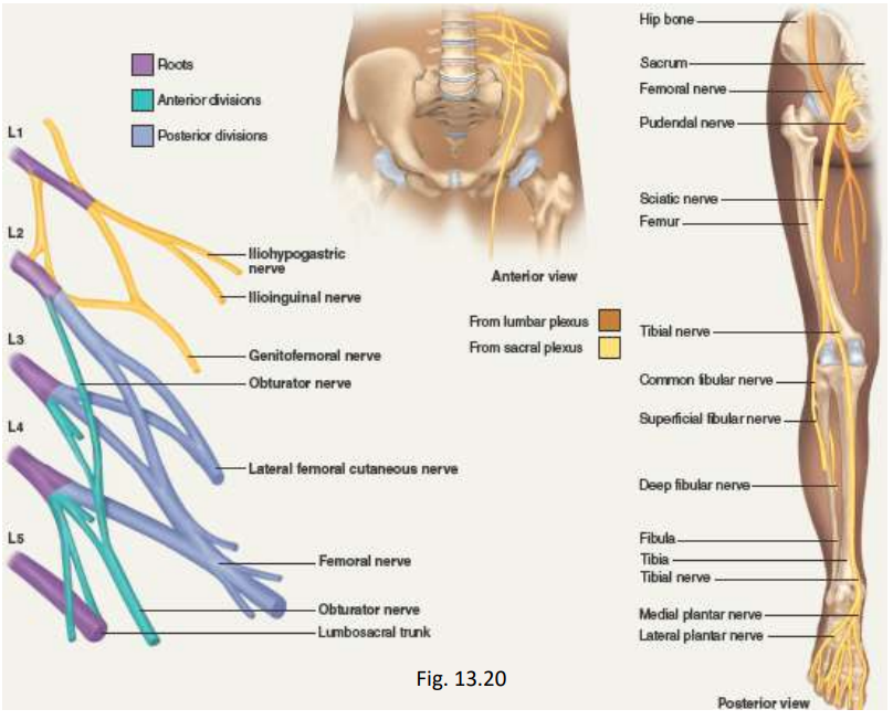
- Lumbar plexus (L1-L4):
- Iliohypogastric- sensory: skin of lower abdomen, gluteal region; motor: abdominal muscles
- Ilioinguinal- sensory: skin of upper thigh; scrotum and penis, labia; motor: abdominal muscles
- Genitofemoral- sensory: skin of thigh; scrotum, labia; motor: male cremaster muscle
- Lateral femoral cutaneous- sensory: skin of thigh;
- Femoral- sensory: skin of thigh and knee; skin of leg and foot; hip and knee joint; motor: muscles of hip, quadriceps
- Obturator- skin of thigh; thigh and knee joints; motor: obturator externus (pelvis); thigh muscles
- Sacral and Coccygeal Plexuses:
- Sacral (L4-S4):
- Innervates remainder of lower trunk and lower limb
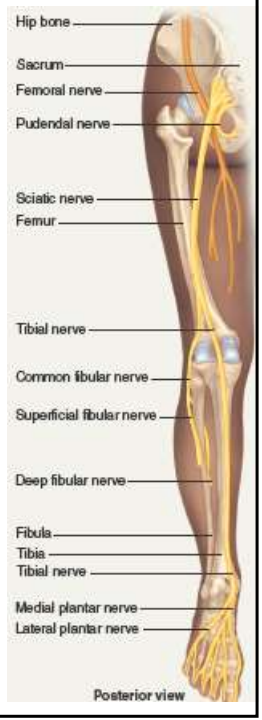
- Sciatic nerve; injury= 90% of cases result from herniated intervertebral disc or osteoarthritis of lower spine
- Coccygeal (S4-Co1):

- Sacral and Coccygeal Plexuses:
- Superior gluteal: motor: gluteus, hip muscle
- Inferior gluteal: motor: gluteus
- Posterior cutaneous: sensory: skin of gluteal region, perineum, posterior thigh and leg popliteal fossa
- Tibial: sensory: skin of posterior leg, plantar skin, knee and foot joints. Motor: hamstring muscles, posterior leg, foot
- Fibular: sensory: skin of anterior distal third of leg, dorsum of foot and toes, knee joint. Motor: biceps femoris muscle, anterior muscles of leg, extensor muscles of foot
- Pudendal: sensory: skin of penis and scrotum; clitoris, labia, lower vagina. Motor: muscle of perineum
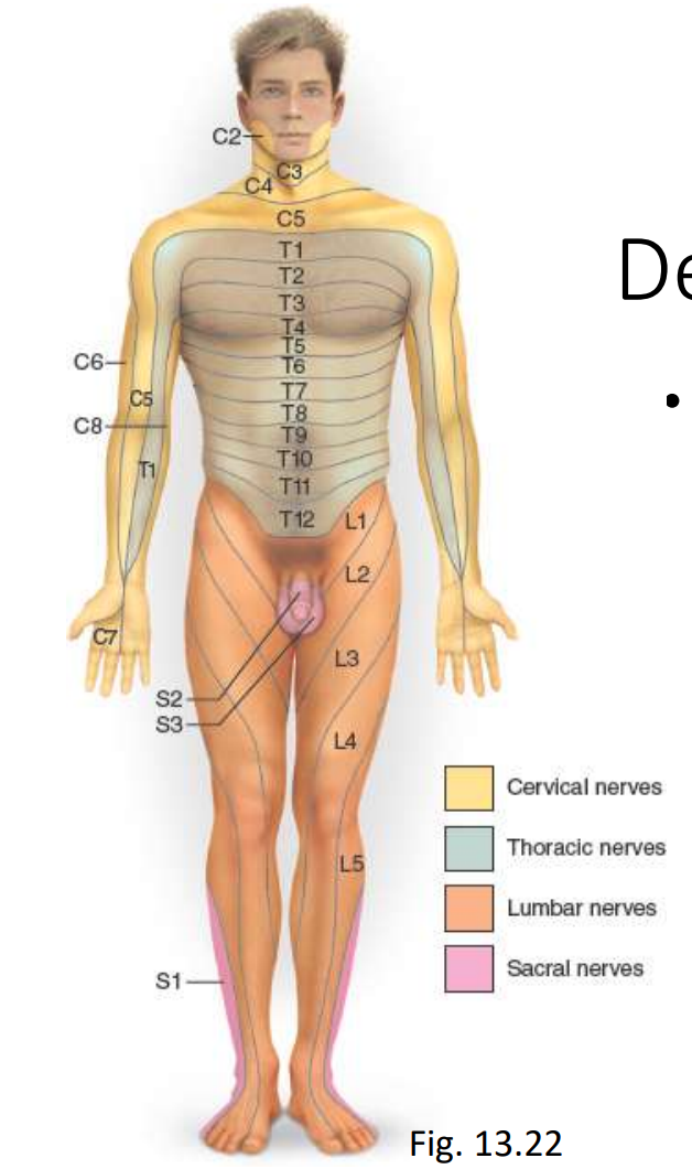
- Dermatome Map:
- Dermatome: 4 of them: cervical nerves, thoracic nerves, lumbar nerves, and sacral nerves.
- Specific area of skin that sends sensory input to a specific spinal nerve
- Oversimplified -50% overlap edges
- Necessary to sever or anesthetize three successive spinal nerves to produce total loss of sensation from one dermatome
- E.g. spinal nerve damage can be assessed by testing the dermatomes with pressure/pain and noting areas the pressure has no sensation
- Concept check: Fill-in-the-blank:
- 1. Outside the CNS, the somas of neurons are clustered in swellings called glanglioll?
- The phrenic nerves arise from the cervical plexus and innervates the diaphragm.
- 3. The sciatic nerve is a composite of two nerves, the fibular and tibia.
