Lecture 4: 🤍Necrosis and Apoptosis
notes are not finished.
Hypobiotic (regressive) Processes
Refers to damage in cells and tissues, resulting in a decline in their normal functions - with mostly changes in breakdown (catabolic) processes and structural changes in organs.
The severity of damage can depend on how strong and the type of harmful stimulus being applied, and the condition of tissue at the time of damage (metabolic state). Consider a stroke, the effects can vary between people who are regularly active and those who sit a lot.
Types of damage:
Degeneration: Reversible damage
Necrosis: Irreversible damage → cell death
Atrophy: Reversible or irreversible damage (decrease in size/function of cells)
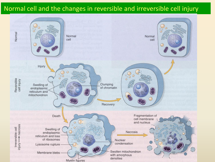
Reversible vs. Irreversible Cell Injury
Feature: | Reversible injury: | Irreversible injury: |
Cell & Organelle: | Generalized swelling | Increased swelling, disruption of lysosomes |
In plasma membranes: | Blebbing | Disruption |
Ribosomes: | Detachment from endoplasmic reticulum | — |
Nuclear chromatin: | Clumping | Profound changes |
Mitochondria: | — | Presence of large amorphous densities in swollen mitochondria |
Cellular membranes: | — | Disruption + nuclear changes. |
Myelin figures: | Myelin figures/laminated structures derived from damaged membranes of organelles + plasma membrane first appear during reversible → to transition of irreversible stage. | More pronounced in irreversible damaged cells. |
☠ Types of Cell Death
Necrosis (Oncosis): Cell swelling, oncotic cell death
Apoptosis: Cell shrinkage
What is Apoptosis?
form of programmed cell death. Can happen in two contexts:
Physiological Apoptosis: naturally happening, helping with organ development + maintaining proper tissue size.
Toxic Apoptosis: triggered by harmful external factors, like chemicals, pathogens, lack of O2 → cell death. Ex. Large scale death of thymocytes (type of immune cell) during stress or liver cells dying due to toxins.
During Apoptosis:
cells undergo a series of well-defined morphological changes.
Chell shrinkage occurs + chromatin in the nucleus condenses
The cell membrane
🔬 Ultrastructural Changes in Necrosis vs. Apoptosis
Feature | Necrosis | Apoptosis |
|---|---|---|
Initial Changes | Cytoplasmic blebs, digestion, and leakage of cellular components | Nuclear chromatin condensation and fragmentation |
Subsequent Events | N/A | Cytoplasmic budding and phagocytosis of the extruded apoptotic bodies |
🆚 Apoptosis vs. Necrosis: Key Differences
Characteristic | Apoptosis | Necrosis |
|---|---|---|
Affected Cells | Scattered individual cells | Massive and contiguous cells |
Chromatin | Marginates as large crescent aggregates | Marginates as small aggregates |
DNA Fragmentation | Ladder pattern (~200 bp) | Smear pattern |
Cytoplasm & Cell Volume | Decreases | Increases |
Organelles | Retain integrity | Swell (mitochondria, endoplasmic reticulum) |
Cell Integrity | Breaks into small fragments | Rupture |
Cell Contents | Cell fragments are phagocytized | Released |
Inflammation | No inflammation | Extensive inflammation |
🦠 Examples of Necrosis and Apoptosis
Right picture - Apoptosis: Pancreas, rat; individual acinar cells are shrunken with condensed chromatin.
Left picture - Necrosis and Apoptosis: Mouse hepatitis virus infection in the liver, causing hepatocyte death.
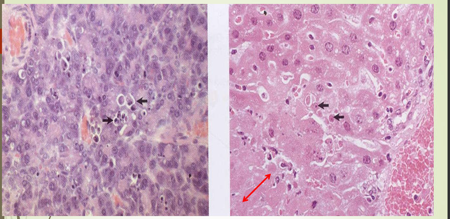
Rabbit Hemorrhagic Disease: Monocellular necrosis in the liver of a rabbit.
⏳ Necrosis Over Time
Necrosis: Intravital death of a cell, final irreversible state.
Necrobiosis: The process of degeneration and cell death. Morphological changes are visible after some time due to enzymatic effects.
💥 Factors Causing Cell Damage Leading to Necrosis
Poisons: Chemical poisons, toxins, or poisons from pathogenic microorganisms or certain plants.
Lack of Blood Supply: Deprivation of oxygen.
Lack of Nerve Supply: Deprivation of normal innervation.
Pressure: Long-continued pressure, often mild.
Mechanical and Thermal Injuries: Burns, freezing, crushing.
👁🗨️ Macroscopic Appearances of Necrotic Tissue
Paler than living tissue, unless filled with blood.
Little tensile strength.
Hemolysis of blood cells after death.
Slight inflammatory zone with leukocytes and hyperemia in the living tissue (hemorrhagic rim).
☢ Necrobiotic Changes on the Nuclei
Pyknosis: Nucleus decreases in size, becomes rounder, chromatin condenses, appears dark and homogenous. An early change.
Hyperchromatosis: Concentration of chromatin on the nuclear membrane, center of the nucleus is paler.
Karyorrhexis: Nuclear membrane ruptures, chromatin fragments into small, darkly staining aggregates.
Karyolysis: Dissolution of the nuclear material.
🧪 Cytoplasmic Changes
In some cases, the cytoplasm may show relatively little change, but if changes in the nucleus indicate death then the cell should be considered dead.
Following abnormalities of the cytoplasm may be present:
Acidophilia of Cytoplasm: Intracellular proteins denature, ribosomes disappear, increased eosin binding. Increased mitochondria appear as eosinophilic granules.
Cytoplasmolysis: Dissolution of cytoplasm and cytoplasmic membrane.
In tissues rich in fluid, lytic processes dominate, leading to vacuole formation. In tissues with low fluid content, cytoplasm becomes opaque, homogenous, glassy, and breaks down into small granules.
Changes on Extracellular Matrix: Change of staining, fibrillar mass dissolves to amorphous state. Necrotic tissue turns into necrotic debris of protein and fatty nubs.
Loss of Cell Outline: Loss of cell form, though the material is still present.
Loss of Differential Staining: Inability to distinguish colors of nuclei and cytoplasm.
🧱 Types of Necrosis
Coagulative Necrosis
Liquefactive Necrosis
The gross appearance of necrosis depends on tissue type, cause, and time from cell damage.
1⃣ Coagulative Necrosis
Results from denaturation of cellular proteins, leading to a firm mass of necrotic cells.
Cell outlines may persist for days or weeks before enzymatic lysis.
Common in parenchymatous organs due to high protein content.
Macroscopic: Firm consistency, grayish-whitish to grayish-yellowish, dryness, cloudiness.
Microscopic: Homogenous and eosinophilic cytoplasm.
2⃣ Liquefactive Necrosis
Rapid liquefaction of dead cells due to lysosomal enzymes (hydrolases) from neutrophils and autolytic digestion.
Abscesses: Liquid is represented by pus (neutrophils, tissue fluid, tissue debris).
Central Nervous System (Malacia): Breakdown of myelin, leading to brain softening (encephalomalacia and myelomalacia in spinal cord).
The neuropil has a high content of lysosomal enzymes.
🩸 Special Types of Necrosis
Zenker's Necrosis: Occurs in striated muscle. It involves coagulation of sarcoplasm proteins, often due to nutritional or toxic myopathies.
Macroscopic: Muscle is white or pale, shiny, and swollen.
Microscopic: Fibers are swollen, homogeneous, and hyaline; sarcoplasm is acidophilic; myofibrils lack cross-striation; nuclei are small and dark.
Caseation: Typical of tuberculous lesions, appearing as friable, cheesy, amorphous material.
The appearance results from a mixture of degenerative tissue protein and fat derived from the lipid capsule of Mycobacterium.
🌡 Variants of Necrosis
Hemorrhagic Necrosis: Necrotic tissue congested with blood, often due to blockage of venous drainage (e.g., in volvulus).
Fat Necrosis: Occurs in the abdominal cavity or subcutaneous tissue.
Traumatic: Rupture of fat cells evokes a tissue reaction, leading to fibroblastic scar tissue.
Enzymatic: (Pancreatic) Lipases split neutral fats into fatty acids and glycerol; fatty acids saponify with alkalis to form chalky precipitates.
Abdominal: Large masses of necrotic fat in mesentery, omentum, and retroperitoneally (common in cattle).
➡ Outcomes of Necrosis
Liquefaction and removal of fluid via blood and lymph.
Liquefaction and formation of a cyst-like accumulation of fluid.
Liquefaction with abscess formation.
Encapsulation without liquefaction.
Desquamation or sloughing.
Replacement by scar tissue.
Sequestration.
Calcification.
Gangrene.
Atrophy of the organ or tissue.
Regeneration.
📍 Sequestration
Demarcation or bordering of a necrotic focus. Mediators leaking from the necrotic mass cause accumulation of neutrophils and other cells at the margin. If on the skin or mucous membrane, the necrotic tissue may separate from healthy tissue.
🔥 Gangrene
Necrosis modified by secondary changes.
Dry Gangrene: Results from slow reduction of blood flow in arteries. Tissues are dry, shriveled, cold, and black.
Moist (Wet) Gangrene: Occurs when tissues are well-filled with blood, and there is sudden stoppage of blood. Tissue is swollen, soft, moist, reddish-brown to black, with gas bubbles and a putrid odor.
Gas Gangrene: Occurs in dirty lacerated wounds infected by anaerobic bacteria (e.g., Clostridium). Muscles and subcutaneous tissues fill with gas and serohemorrhagic exudate. Toxins of clostridia have a lytic effect, rapidly proliferate, and change glycogen to methane.