Anterior carpal region. Carpal Tunnel
Anterior Carpal Region and Carpal Tunnel: Anatomy, Clinical Significance, and Structures
The anterior carpal region, also known as the palmar region of the wrist, contains several important structures, including the carpal tunnel. The carpal tunnel is a fibro-osseous passageway located on the anterior aspect of the wrist that houses several important tendons and the median nerve. This region plays a crucial role in flexion and movement of the hand and fingers, and its anatomy is significant for both functional hand movements and the diagnosis of various conditions, such as carpal tunnel syndrome.
Anatomy of the Anterior Carpal Region
The anterior carpal region consists of the carpal bones, ligaments, tendons, and the median nerve that pass through the carpal tunnel.
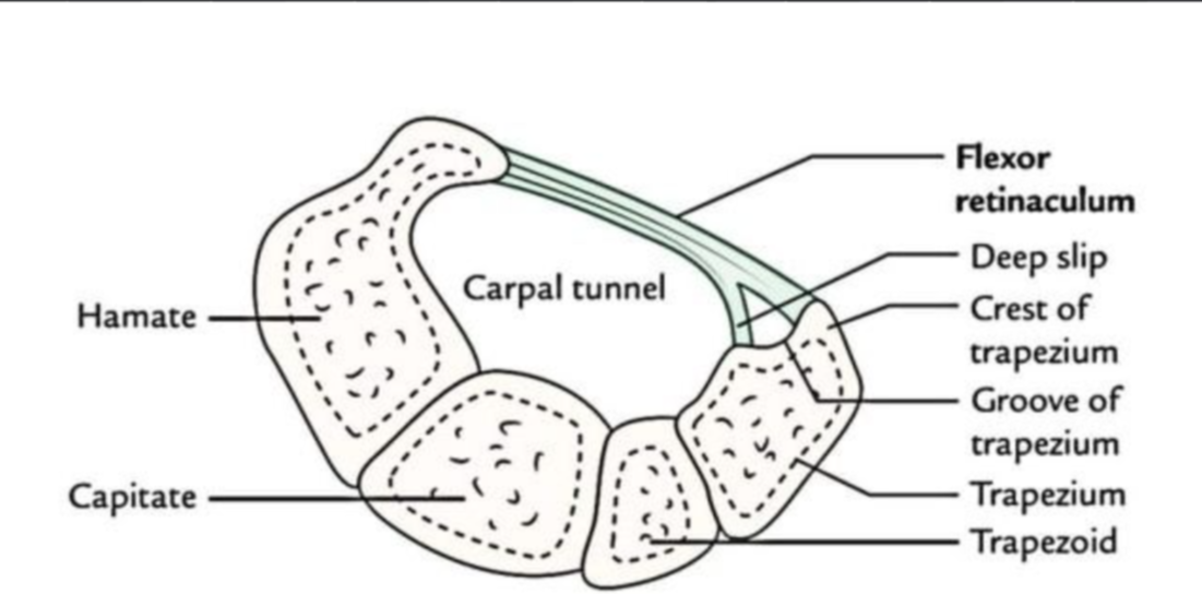
1. Carpal Bones
The carpal bones form the wrist and are arranged in two rows:
Proximal row: Scaphoid, Lunate, Triquetrum, Pisiform.
Distal row: Trapezium, Trapezoid, Capitate, Hamate.
These bones articulate with the radius and ulna (proximal forearm bones) to form the wrist joint, allowing for a wide range of movement in the hand.
2. Carpal Tunnel
The carpal tunnel is a passage formed by the carpal bones and the transverse carpal ligament. It serves as a conduit for several tendons and the median nerve to pass from the forearm to the hand.
Boundaries:
Anterior boundary: The flexor retinaculum (also known as the transverse carpal ligament), a strong fibrous band that stretches across the wrist.
Posterior boundary: The carpal bones, which form the floor and walls of the tunnel.
Structures Passing Through the Carpal Tunnel:
Tendons of the Flexor Muscles (flexors of the fingers and thumb):
Flexor pollicis longus (tendon for the thumb).
Flexor digitorum superficialis (tendon for fingers 2–5).
Flexor digitorum profundus (tendon for fingers 2–5).
Median Nerve:
The median nerve enters the hand through the carpal tunnel, innervating muscles such as the thenar muscles (for thumb movements) and the lateral lumbricals (for flexing the proximal phalanges).
The carpal tunnel is vital for movement and sensory function in the hand, particularly in fine motor skills like typing, grasping, and pinching.
Clinical Significance of the Anterior Carpal Region and Carpal Tunnel
The carpal tunnel is clinically significant due to its association with carpal tunnel syndrome and its involvement in various hand and wrist injuries.
1. Carpal Tunnel Syndrome (CTS)
Definition: Carpal tunnel syndrome is a condition that arises when the median nerve becomes compressed or irritated as it passes through the carpal tunnel. This compression can lead to symptoms such as pain, numbness, tingling, and weakness in the thumb, index finger, and middle finger.
Causes:
Repetitive wrist movements (e.g., typing, assembly line work).
Wrist injury or fracture.
Inflammation (from conditions such as rheumatoid arthritis or tenosynovitis).
Pregnancy (due to hormonal changes leading to fluid retention).
Diabetes (which can cause nerve damage).
Symptoms:
Numbness and tingling in the thumb, index, and middle fingers.
Weakness in the thumb, making it difficult to grip objects.
Pain in the wrist and hand, often worse at night.
Diagnosis:
Tinel's sign (tapping over the median nerve leads to tingling sensations in the fingers).
Phalen's maneuver (flexing the wrist for a prolonged period to elicit symptoms).
Treatment:
Conservative measures such as wrist splints, anti-inflammatory medications, or steroid injections.
Severe cases may require surgical release of the transverse carpal ligament to relieve the pressure on the median nerve.
2. Other Clinical Conditions
Tendinitis and Tenosynovitis:
Inflammation of the tendons passing through the carpal tunnel, such as flexor tendinitis (inflammation of the tendons of the flexor digitorum superficialis and flexor digitorum profundus) can lead to swelling in the carpal tunnel, increasing pressure on the median nerve.
Wrist Fractures:
Fractures of the distal radius or the scaphoid can damage the structures of the carpal tunnel, leading to compression of the median nerve and symptoms similar to carpal tunnel syndrome.
Ganglion Cysts:
Ganglion cysts can form on the anterior aspect of the wrist and may put pressure on the carpal tunnel. These fluid-filled sacs can compress the median nerve and cause symptoms similar to carpal tunnel syndrome.
Blood Supply and Nerve Supply to the Anterior Carpal Region
1. Blood Supply
The anterior carpal region receives its blood supply from branches of the radial artery and the ulnar artery.
Ulnar artery: Supplies the palmar branch, which contributes to the superficial palmar arch.
Radial artery: Contributes to the deep palmar arch.
2. Nerve Supply
The median nerve is the most significant nerve passing through the carpal tunnel, providing motor and sensory innervation to the thenar muscles and the lateral lumbricals.
The ulnar nerve and radial nerve also provide innervation to structures in the hand, but they do not pass through the carpal tunnel.
Summary
The anterior carpal region and the carpal tunnel are crucial for the movement and function of the hand, allowing tendons and the median nerve to pass from the forearm into the hand. The carpal tunnel is a confined space bounded by the flexor retinaculum and the carpal bones, and it contains tendons of the flexor muscles and the median nerve. Carpal tunnel syndrome is a common condition that results from compression of the median nerve, leading to pain, numbness, and weakness in the hand. Other conditions such as tendinitis, ganglion cysts, and wrist fractures can also affect the structures within the carpal tunnel. Understanding the anatomy of the anterior carpal region and the carpal tunnel is essential for diagnosing and treating these disorders.