The Cardiovascular an Lymphatic Systems
The Circulatory System
The cardiovascular system consists of the blood vessels and the heart.
The blood vessels conduct blood in continuous loops.
The heart is a muscular pump.
Cardiovascular disease is a major killer in the United States.
The lymphatic system functions in the circulatory and immune systems.
The Cardiovascular System
The cardiovascular system is composed of
Blood
Blood vessels
Heart
Parts of the Cardiovascular System:
Veins
Carry blood back to the heart.
Superior vena cava
Carries blood from the upper body back to the heart.
Renal vein
Carries blood from the kidney to the heart.
Radial vein
Carries blood from the hand back to the heart.
Femoral vein
Carries blood from the thigh and inner knee back to the heart.
Jugular veins
Carry blood from head to the heart.
Pulmonary veins
Carry oxygenated blood from the lungs to the heart.
Inferior vena cava
Carries blood from the lower body back to the heart.
Iliac vein
Carries blood from the pelvic organs and abdominal wall back to the heart.
Arteries
Carry blood away from heart.
Carotid arteries
Deliver blood to the head and the brain.
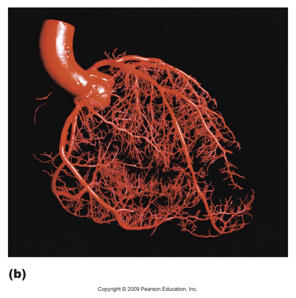
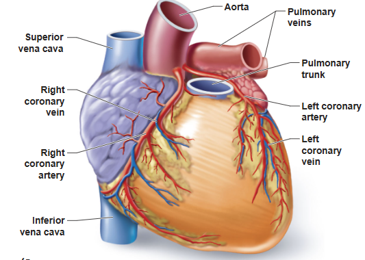
Coronary arteries
Deliver blood to the heart muscle cells.
Iliac artery
Delivers blood to pelvic organs and abdominal wall.
Aerta
Delivers blood to the body tissues.
Pulmonary arteries
Deliver oxygen-poor blood to the lungs.
Renal artery
Delivers blood to the kidney.
Radial artery
Delivers blood to the hand.
Femoral artery
Delivers blood to thigh and inner knee.
The Blood Vessels Conduct Blood in Continuous Loops
Blood passes through the following loop of vessels moving away from the heart.
Arteries
Arterioles
Capillaries
Venules
Veins
Blood returns to the heart from the veins.

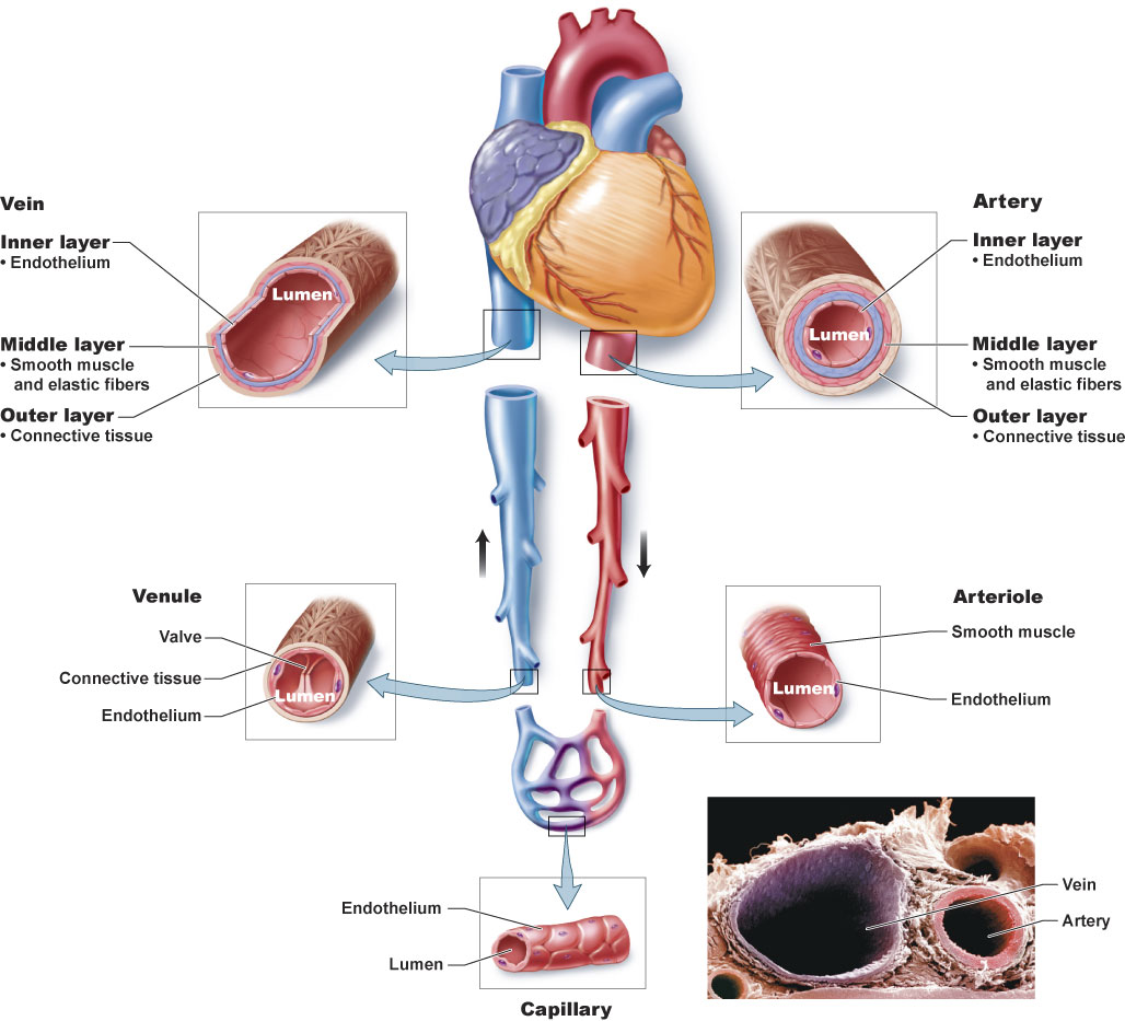
Blood Vessels
The hollow interior of all blood vessels is called the lumen.
Arteries
Thick, muscular vessels that carry blood away from the heart.
Are able to withstand high blood pressure.
The elasticity of the arteries maintains pressure on the blood between heartbeats to keep it flowing through the vessels.
As the heart pumps blood into the arteries, they expand such that one is able to feel a pulse.
The pulse rate is the same as the heart rate.
Vasoconstriction
When muscle contracts and the diameter of the lumen narrows, reducing blood flow.
Vasodilation
When muscle relaxes and the diameter of the lumen increases, increasing blood blow.
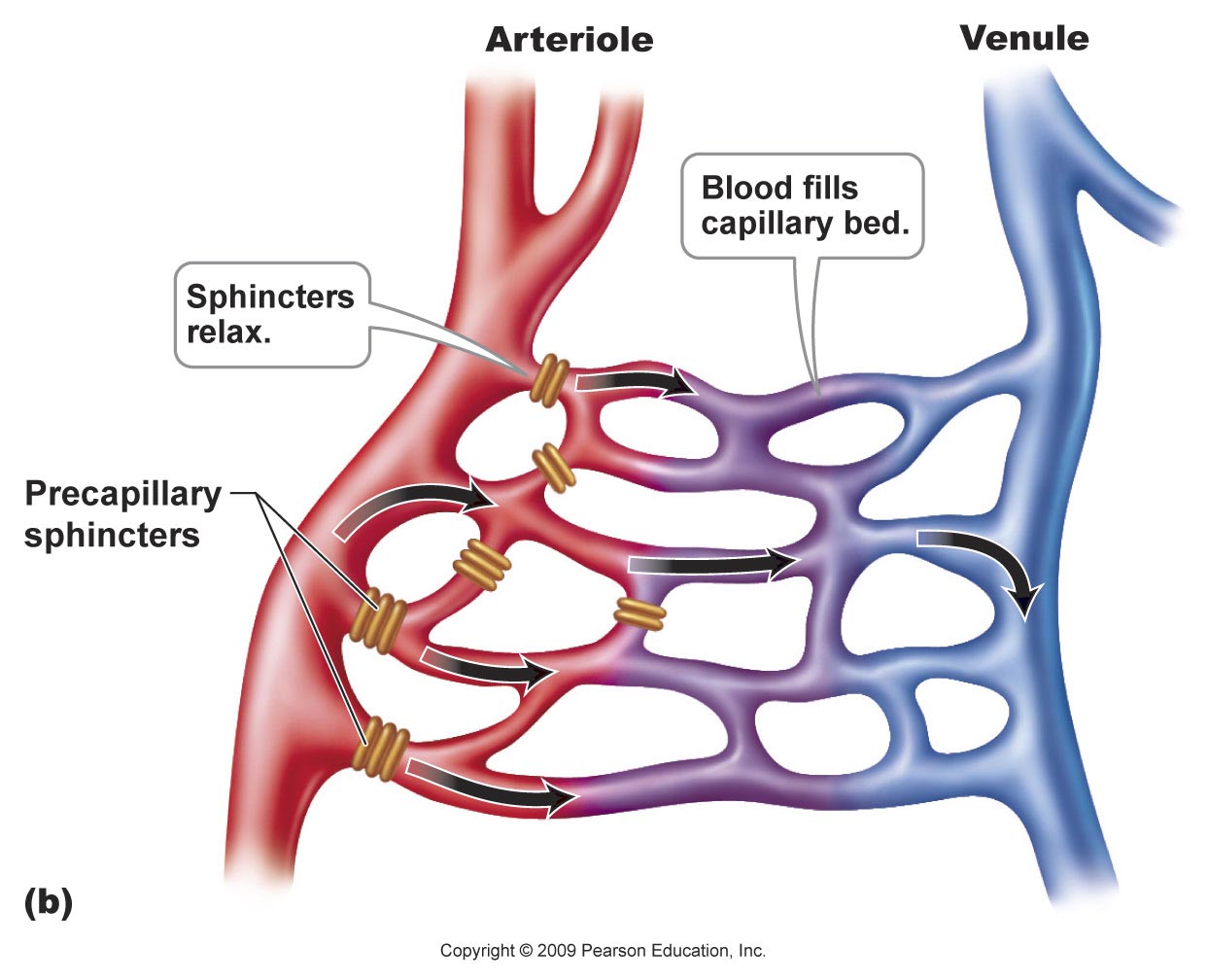
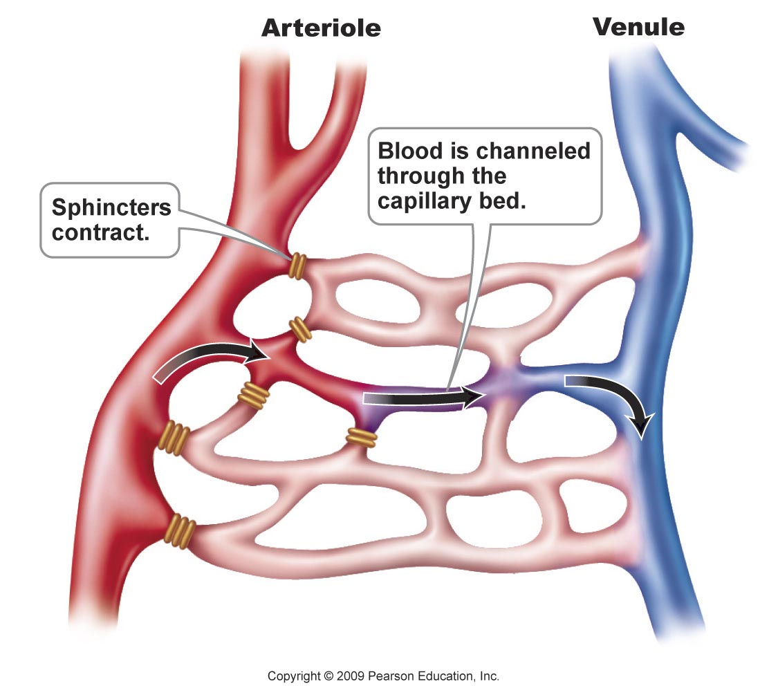
Arterioles are the prime controllers of blood pressure.
Arterioles serve as gatekeepers to the capillary networks keeping them open or closed.
An aneurysm occurs when the wall of an artery is weakened and swells.
The primary risk is that it will burst, causing blood loss.
If it does not burst it can form life-threatening clots.
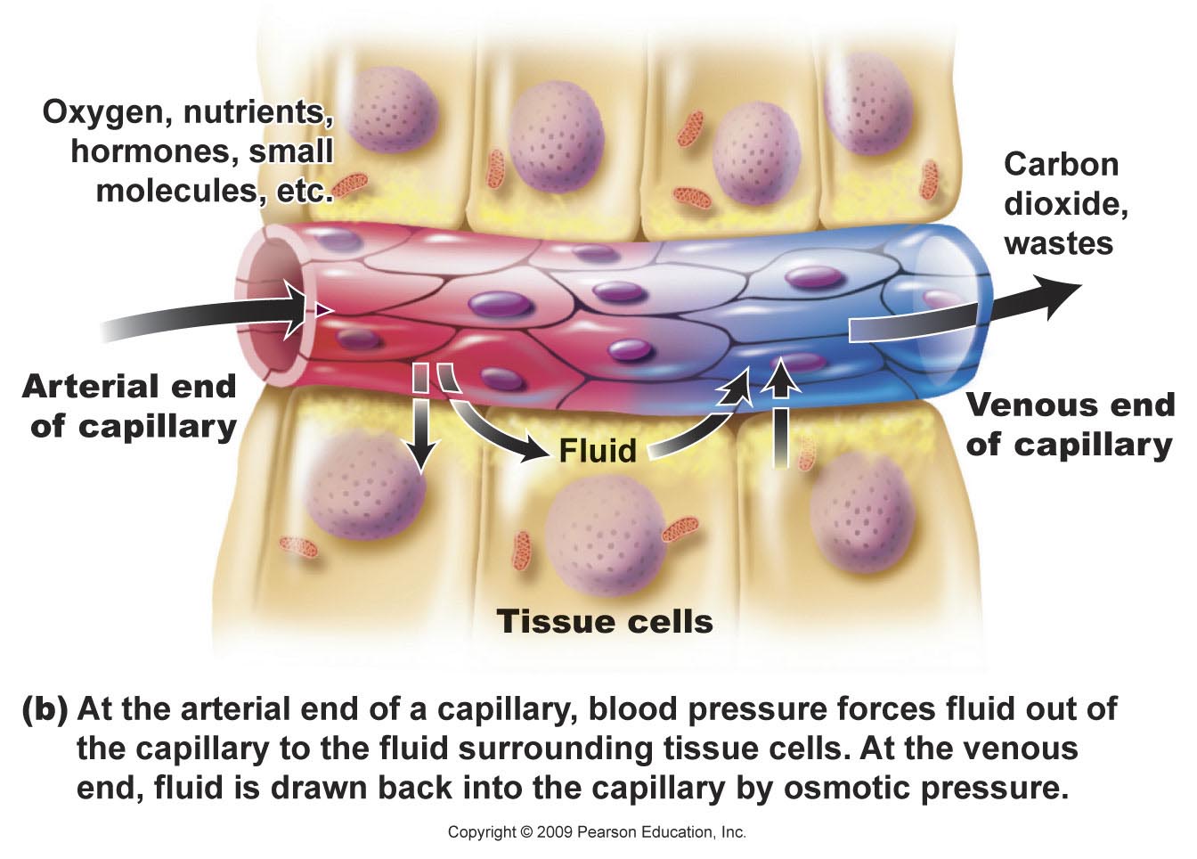
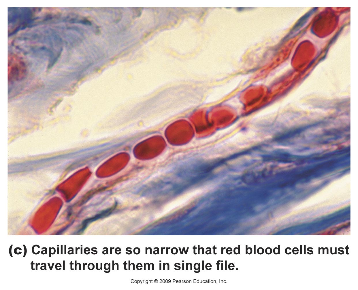
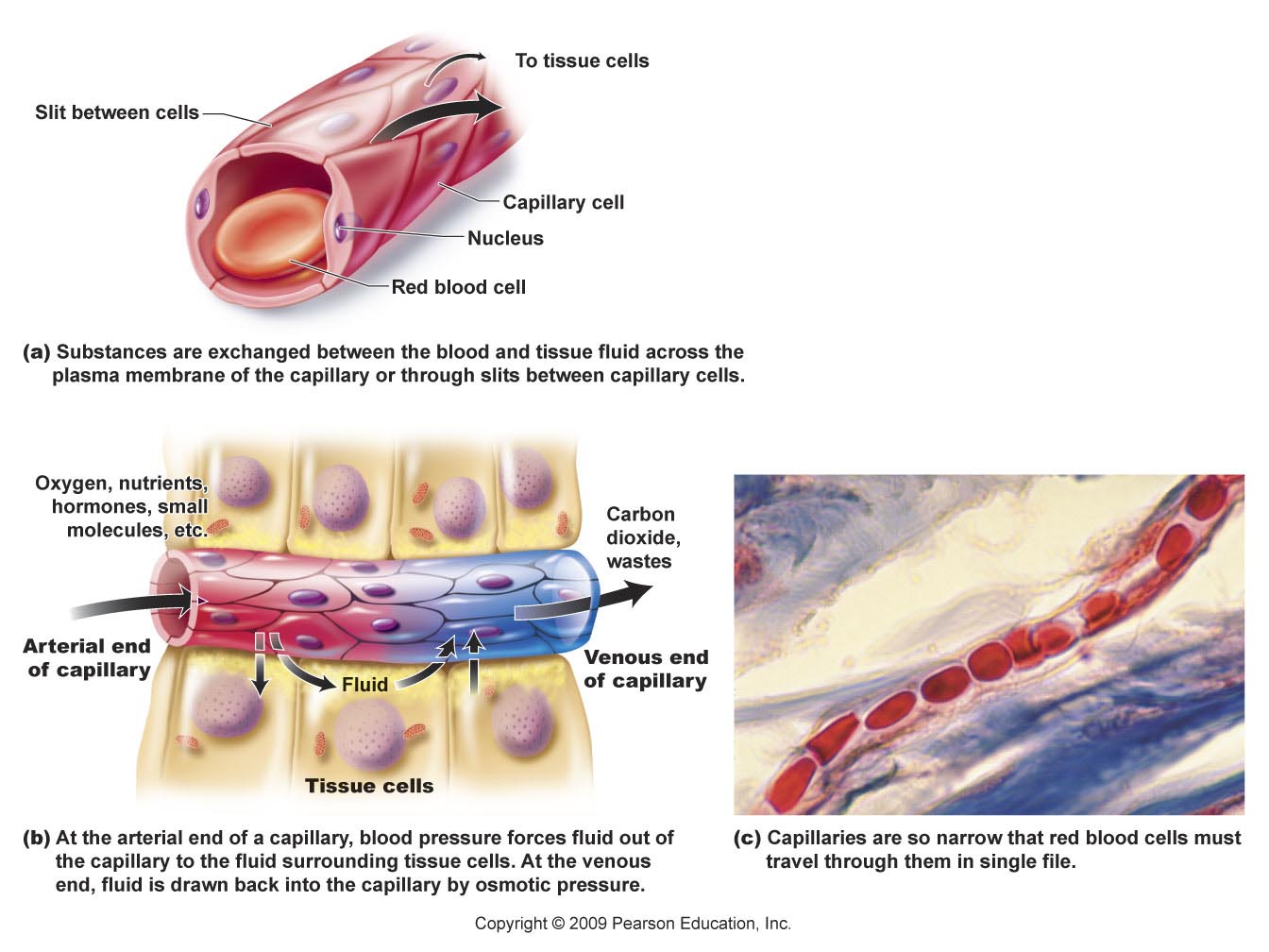
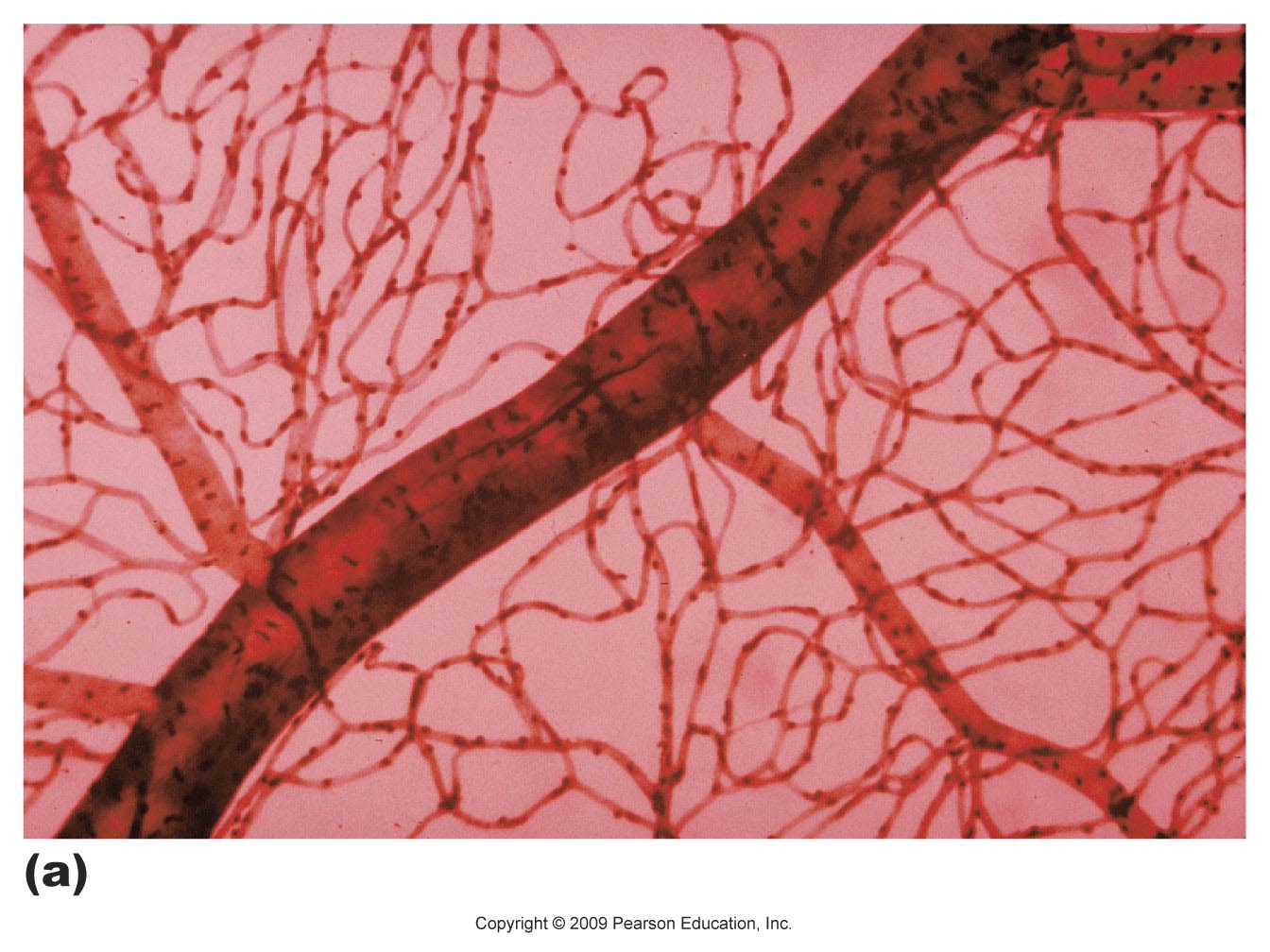
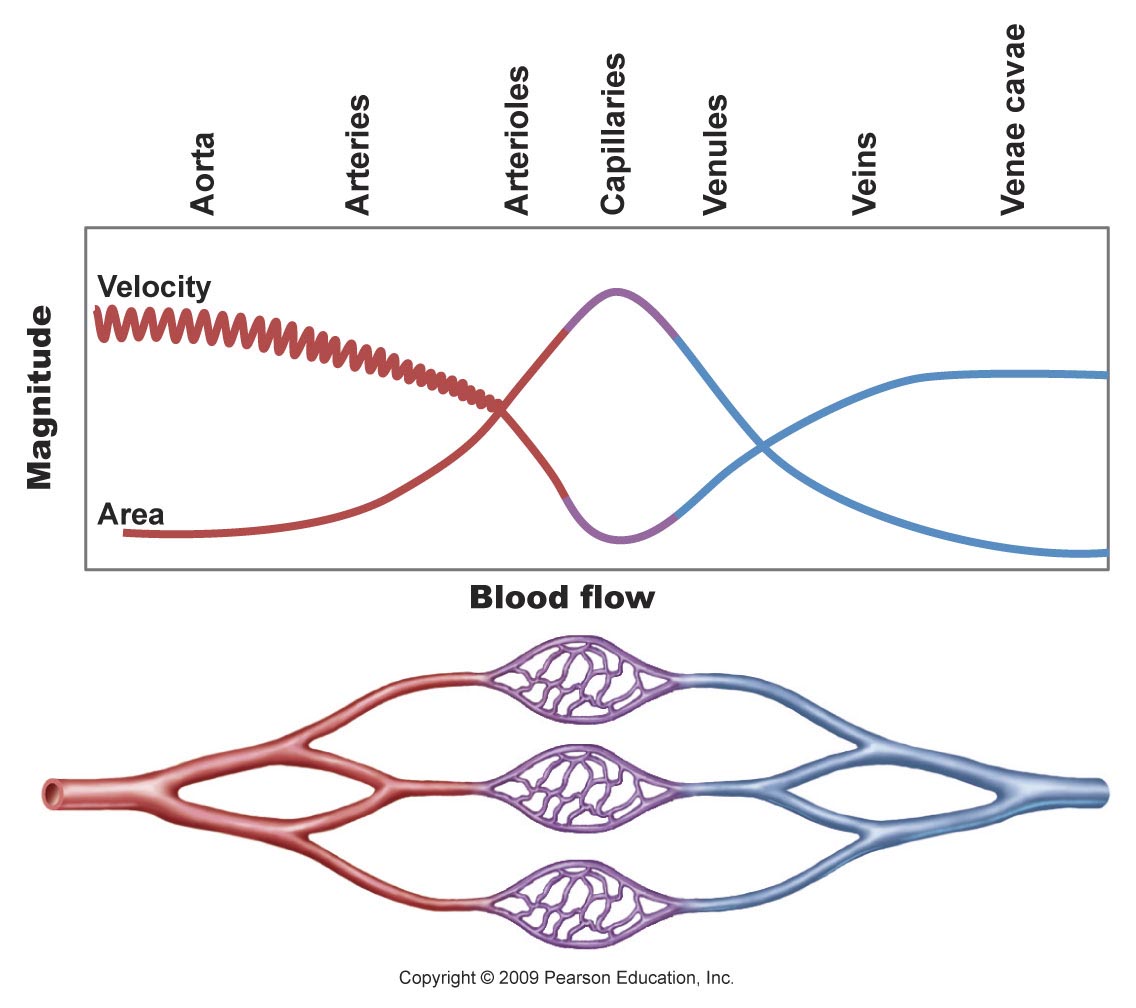
Capillaries have walls that are one cell thick and connect arterioles and venules.
Capillaries form branching networks that allow for the exchange of materials between the blood and tissues.
Blood flows more slowly due to the large surface area.
Provides more time for the exchange of materials.
Capillaries merge to form the smallest kind of vein, a venule.
Veins
Carry blood back to the heart.
Serve as reservoirs for blood volume.
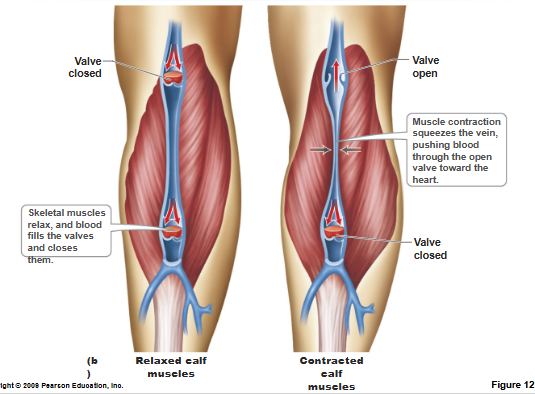
Veins
Blood is moved against gravity toward the heart by:
Contracting skeletal muscles.
Pressure differences caused by the movement of the thoracic cavity during breathing.
Valves.
Prevent blood flowing backwards.
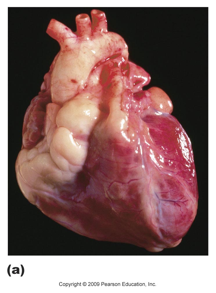
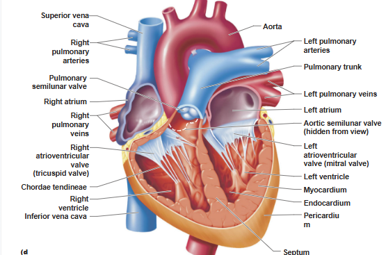
The Heart is a Muscular Pump
The heart is made of cardiac muscle tissue called myocardium.
The interior of the heart is lined by endocardium.
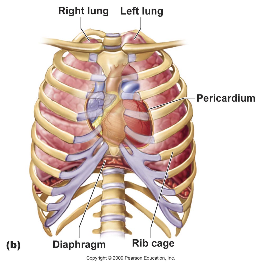
A fibrous sac, the pericardium, encloses the heart and holds the heart in the center of the thoracic cavity.
The two halves of the heart are separated by a septum.
Each half has two chambers.
One smaller and thin-walled atrium.
One larger, more muscular ventricle.
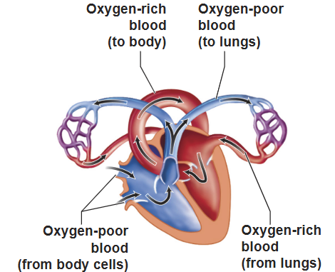
The right side of the heart.
Contains blood rich in carbon dioxide.
Returns from the issues.
Flows out to the lungs.
The left side of the heart.
Contains blood rich in oxygen .
Returns from the lungs.
Flows out to the tissues.
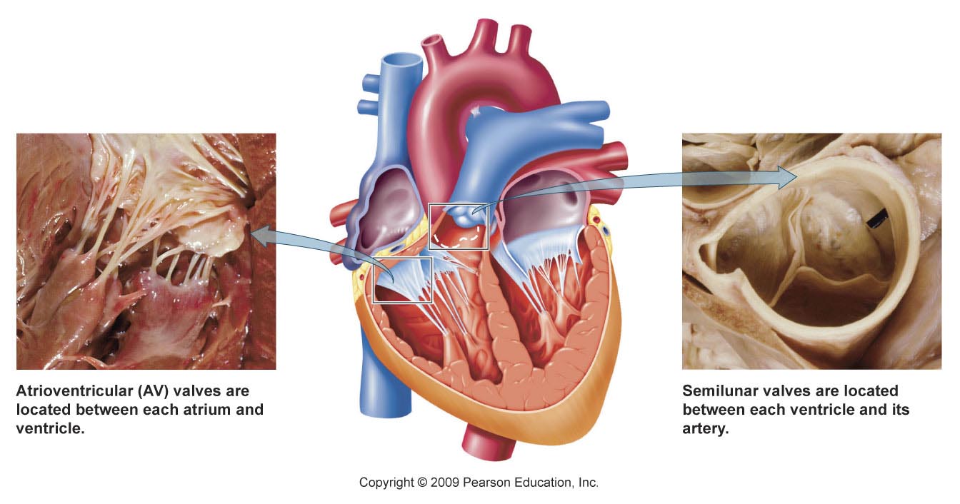
Valves
Atrioventricular (AV) valves
Separate the atria from ventricles
Semilunar valves
Separate the ventricles from the exit vessels.
Keep blood from flowing backwards.
Give rise to the typical “lub-dup” sounds of the heartbeat.
The AV valve on the right
Called the tricuspid valve
Has three flaps
The AV valve on the left.
Called the bicuspid or mitral valve
Has two flaps
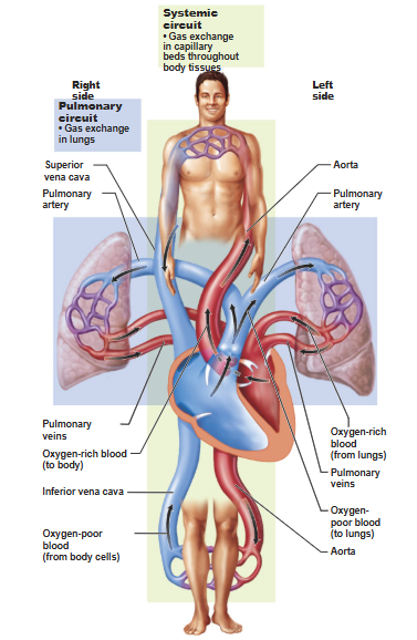
Pulmonary Circuit
The right side of the heart pumps blood to and from the lungs.
Systemic Circuit
The left side of the heart pumps blood to and from the tissues.
The heart muscle is nourished by coronary circulation.
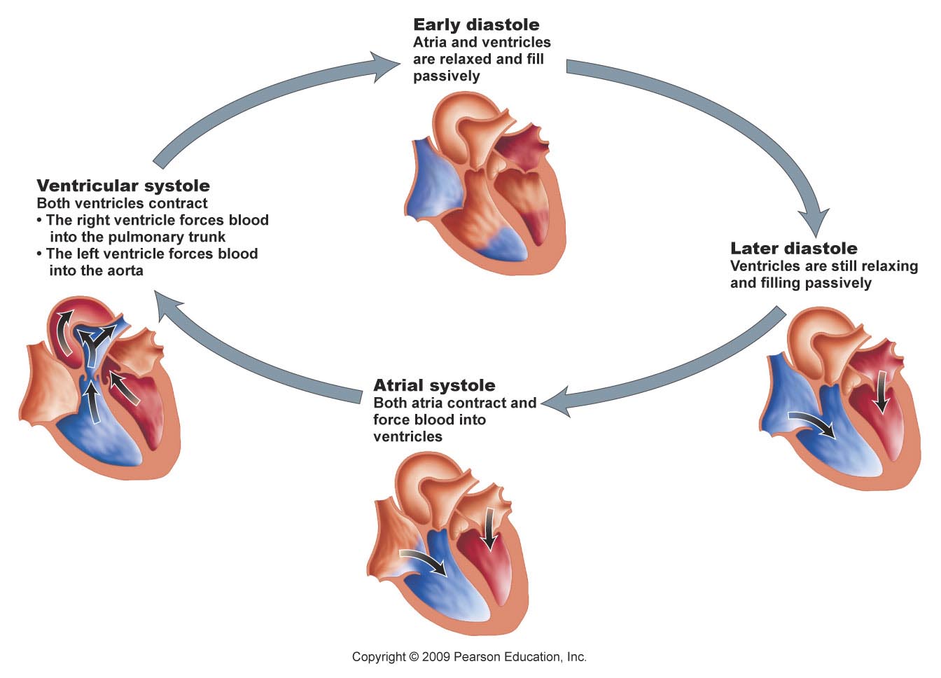
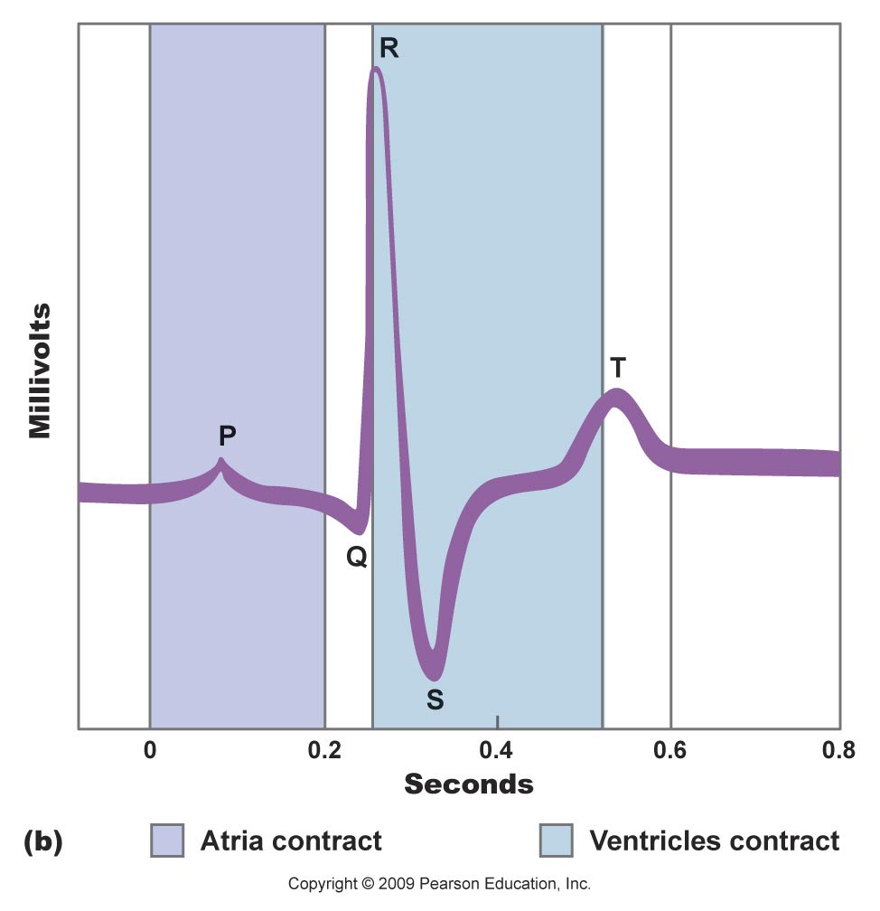
The cardiac cycle
Contraction of the atria.
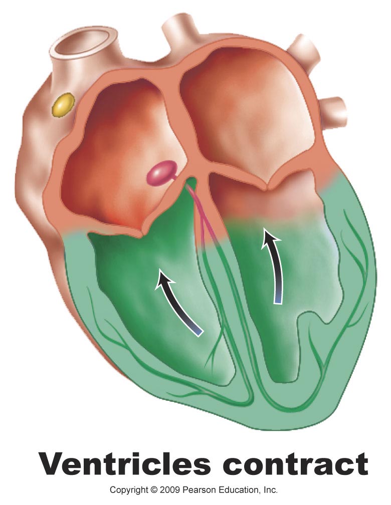
Followed by contraction of the ventricles.
Followed by a rest when neither chamber is contracting.
Contraction is called systole.
Relaxation is called diastole.
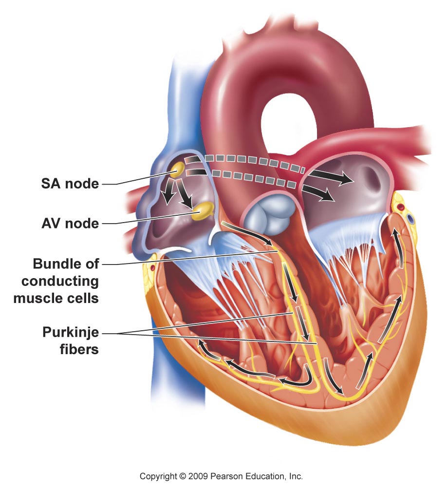
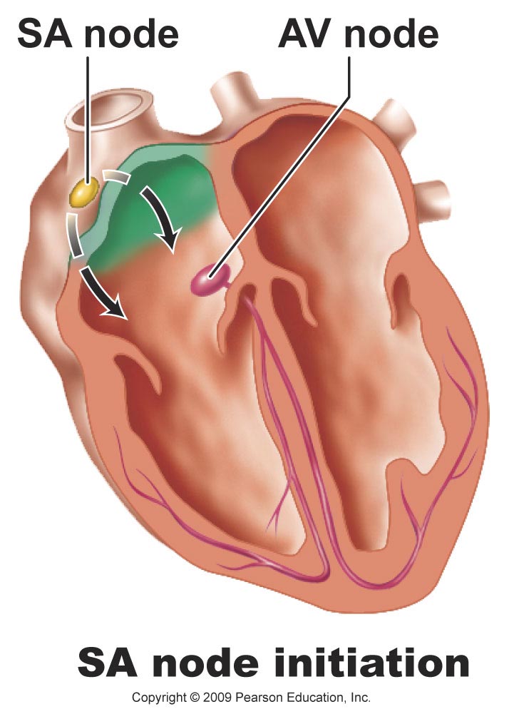
The sinoatrial (SA) node
Generates an electrical signal that sets the tempo.
Called the pacemaker.
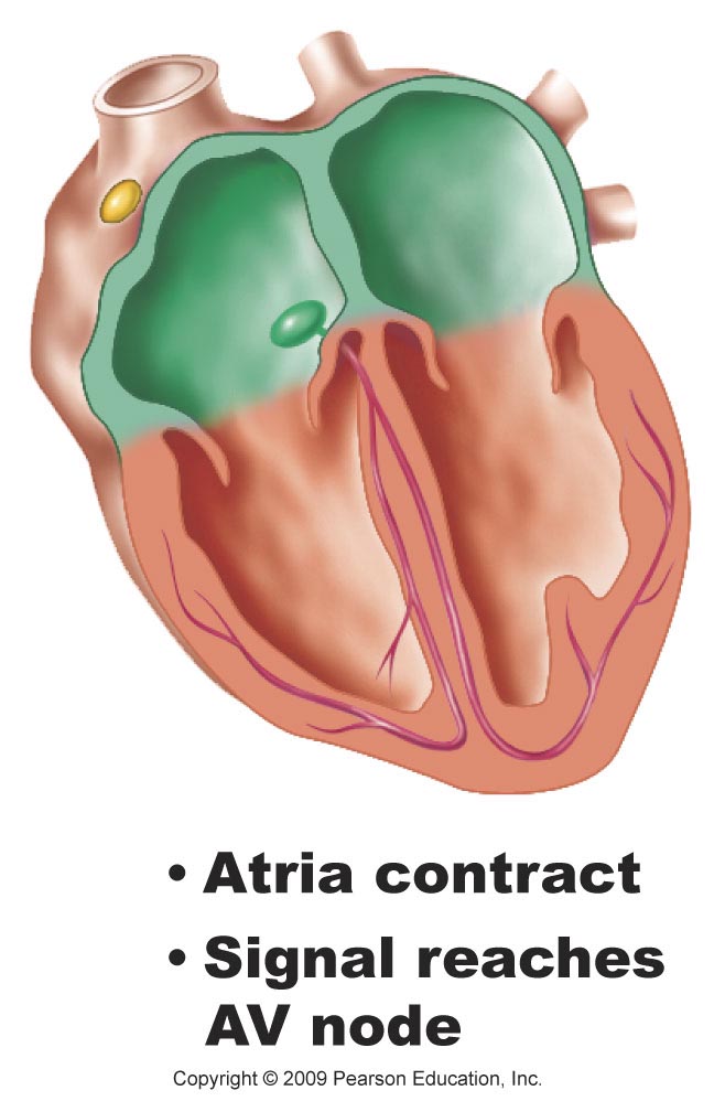
The SA node
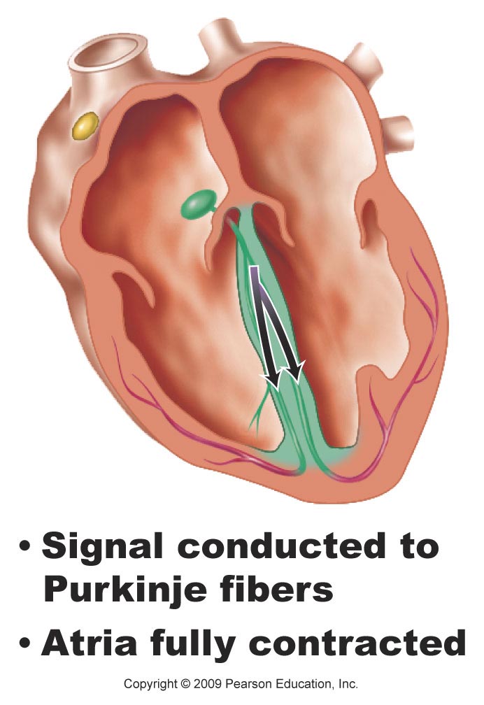
Causes contraction of the atria and sends a signal to the atrioventricular (AV) node, which relays information to the atrioventricular bundle and out through the Purkinje fibers, causing the ventricles to contract.
A combination of nervous and endocrine signals control the strength and rate of contraction of the heart.
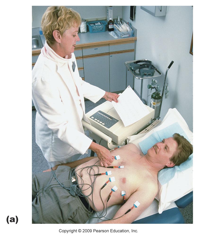
An electrocardiogram (ECG/EKG)
Recording of the electrical events associated with the heartbeat.
A powerful diagnostic tool.
Abnormal patterns can indicate heart problems.
A typical ECG/EKG consists of three distinguishable deflection waves.
P wave
QRS wave
T wave
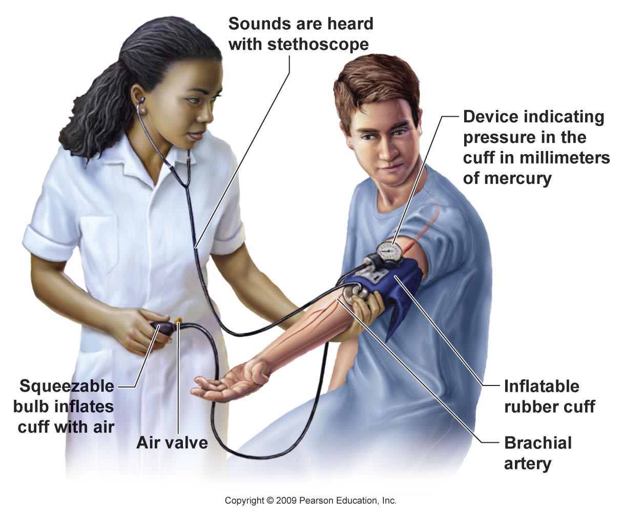
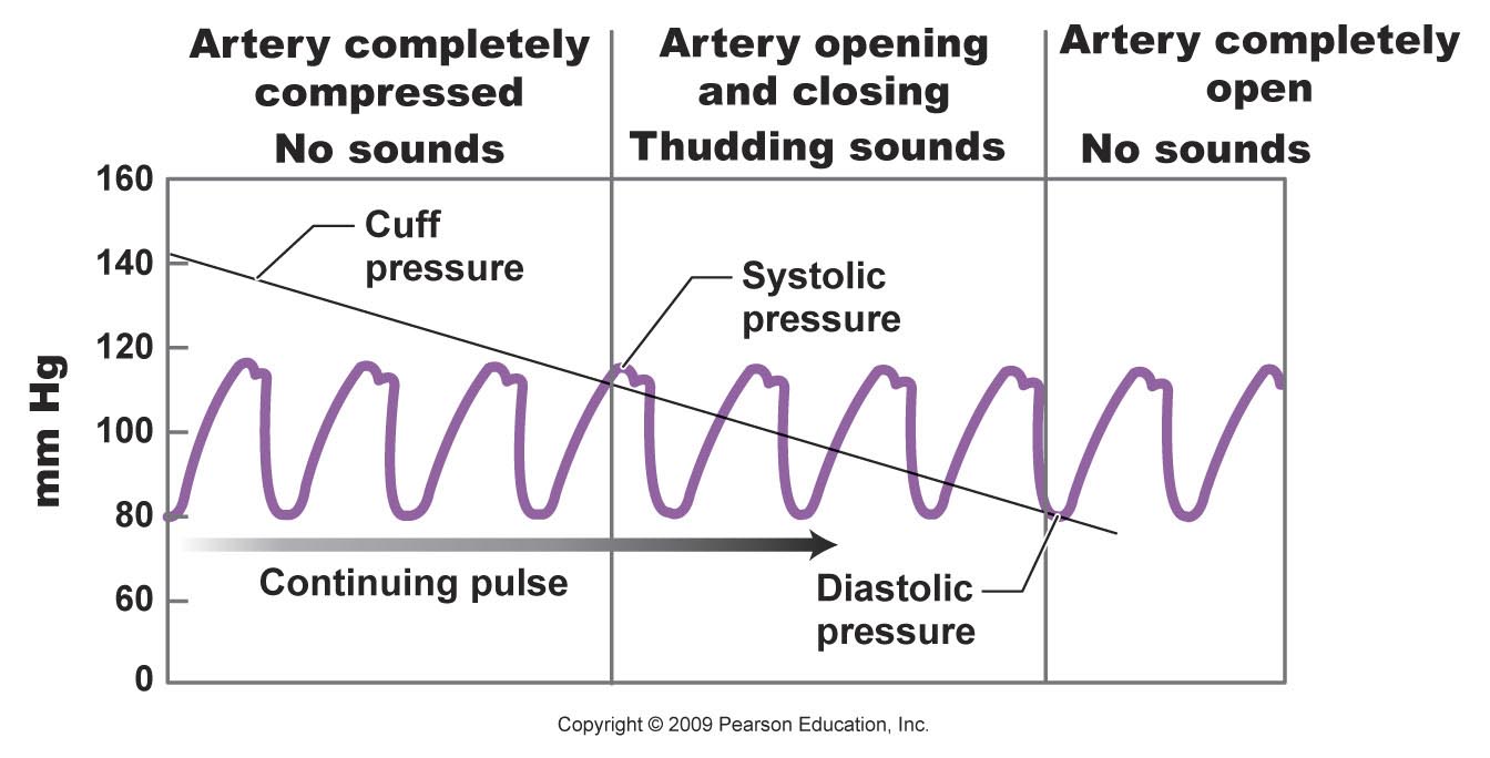
Blood pressure
Highest (systolic) when the ventricles contract, sending blood into the arteries.
Lowest (diastolic) when the heart relaxes between beats.
Cardiovascular Disease Is a Major Killer in the United States
Sphygmomanometer
Measures blood pressure.
Can provide early identification of hypertension, or high blood pressure, the silent killer.
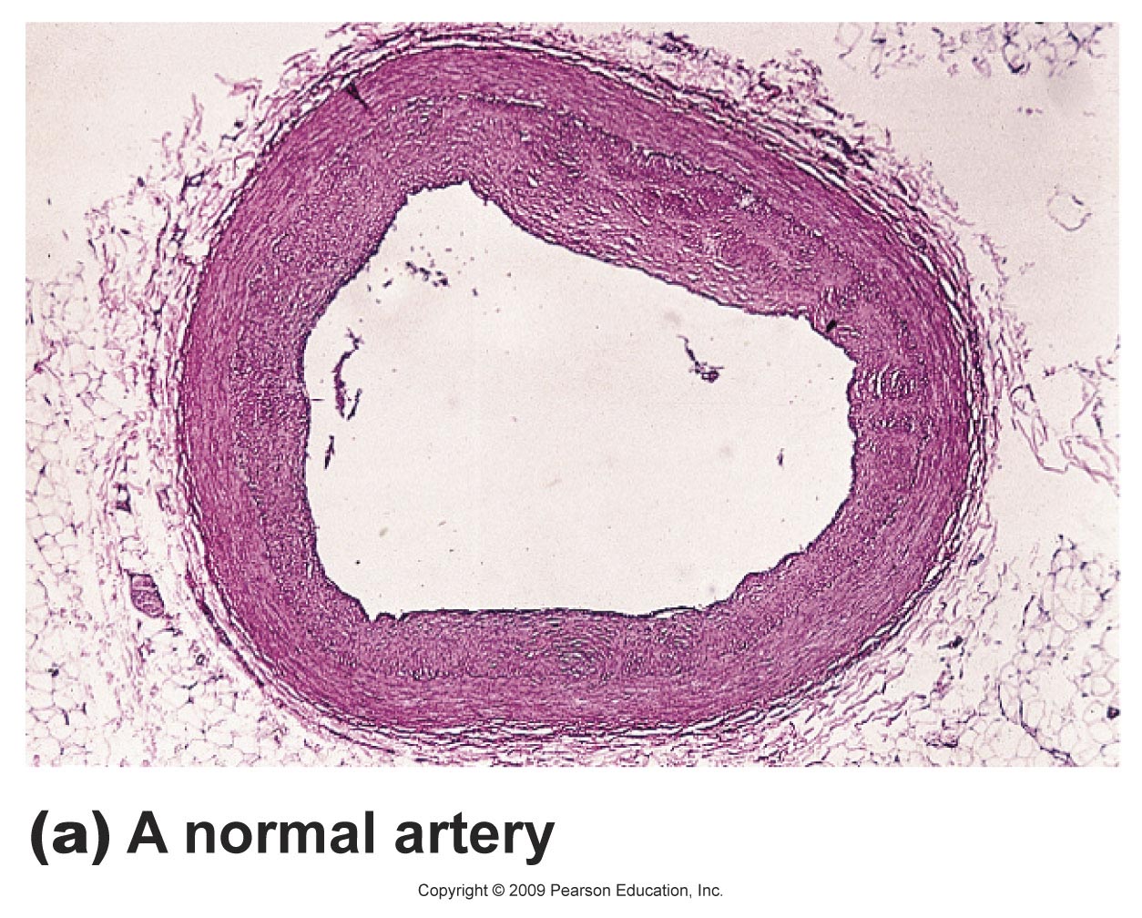
Cardiovascular Disease
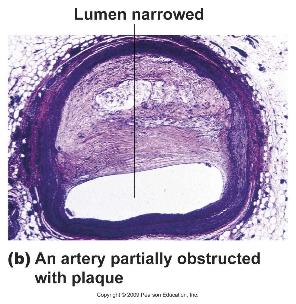
Atheroscloerosis
A narrowing of the arteries due to fatty deposits and thickening of the wall.
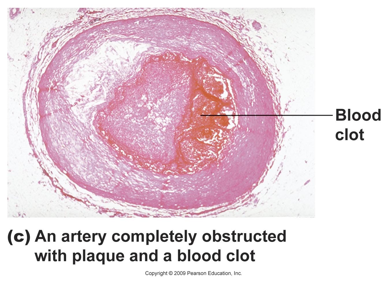
Can lead to heart attack or stroke.
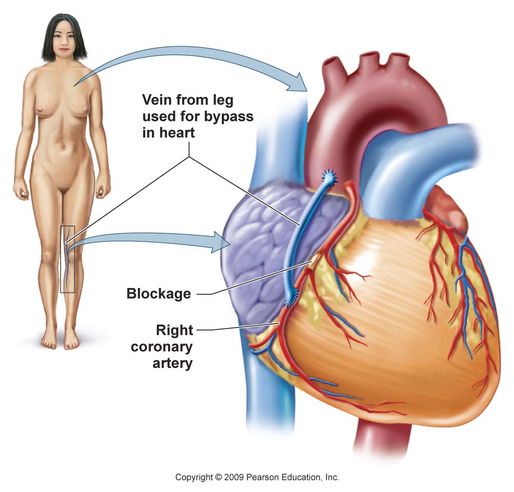
When this occurs in the arteries of the heart muscle, it is called coronary artery disease.
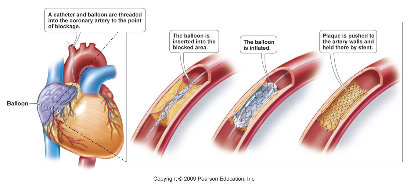
Angiography
Can show coronary artery blockage, which can then be treated with medicines or surgical operations such as angioplasty or coronary bypass surgery.
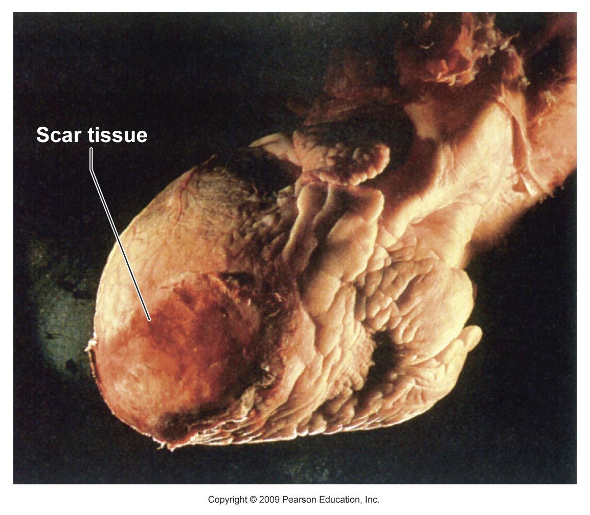
Heart muscle dies because an insufficient blood supply during a heart attack (myocardial infarction) and is gradually replaced by scar tissue.
Scar tissue cannot contract, so part of the heart permanently loses it pumping ability.
Heart failure
Condition in which the heart becomes an inefficient pump.
Leads to shortness of breath, fatigue, and fluid accumulation.
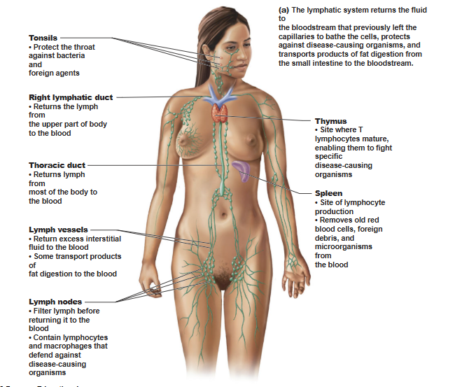
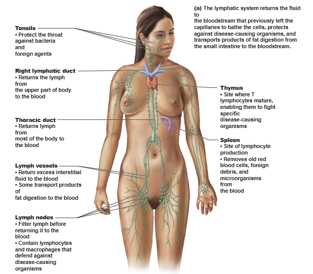
The Lymphatic System
Lymphatic system functions.
Return interstitial fluid to the blood stream.
Transport products of fat digestion.
Defend the body against disease causing organisms and abnormal cells.
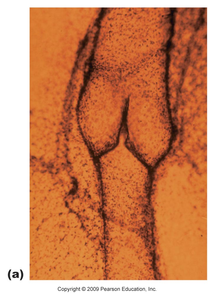
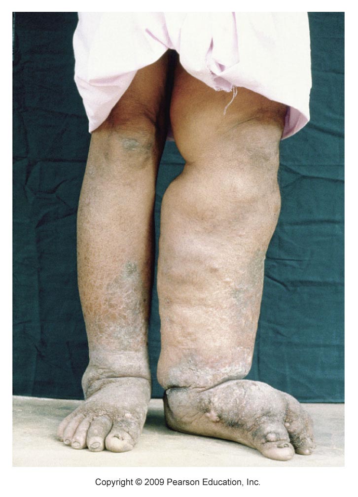
Elephantiasis
A condition in which parasites block the passage of lymphatic fluid returning to blood.
Results in massive swelling, darkening, and thickening of the skin in the affected area.
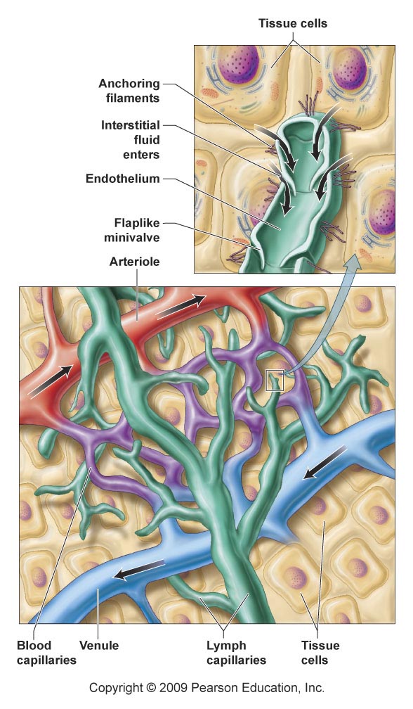
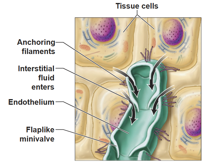
Lymph
Interstitial fluid that builds up around the cells.
Enters the lymph capillaries, then passes through a series of vessels and is returned to the circulatory system.
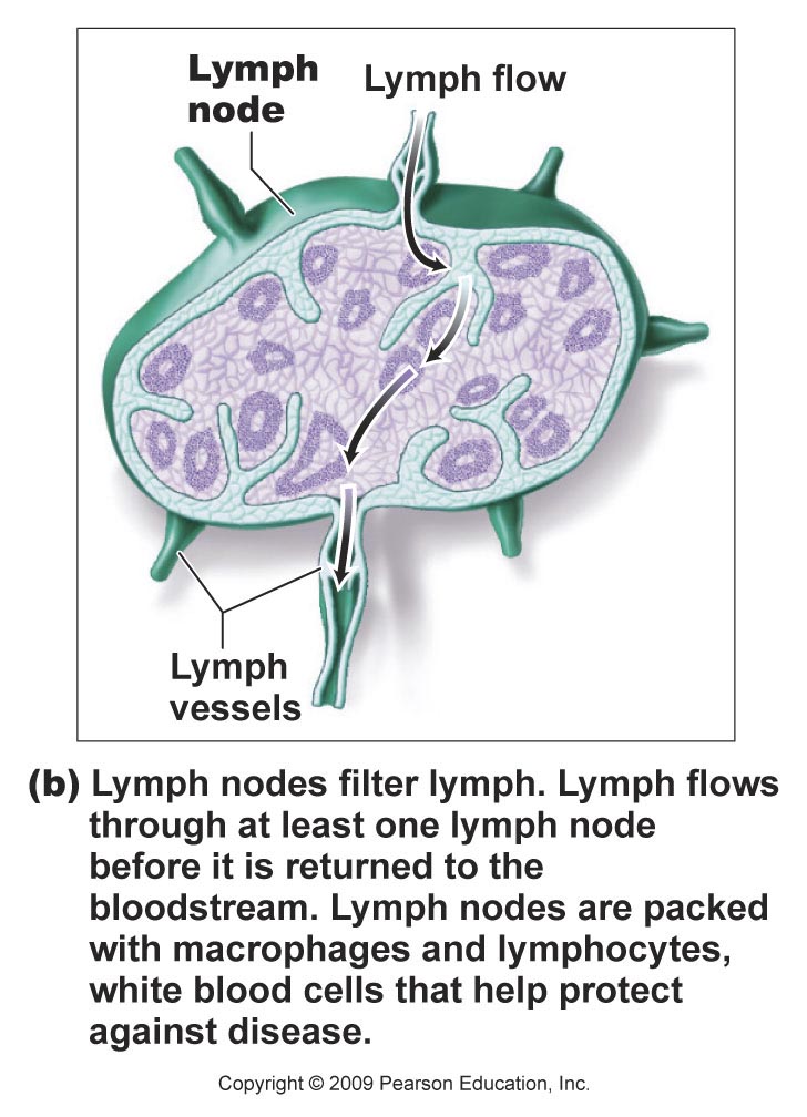
Lymph nodes
Bean-shaped structures.
Filter lymph.
Contain macrophages and lymphocytes that actively defend against disease-causing organisms.
Lymphoid organs include
Tonsils
Thymus gland
Spleen
Peyer’s patches