saiyans
The life process of respiration
Human Respiratory System
I. Human Respiratory System
Purpose
-Your external body surface is dry and impermeable to gases. Lungs provide a thin, moist internal surface for the exchange of gases.
-Oxygen is required for cellular respiration and carbon dioxide, a waste gas, needs to be removed from the body.
-Gases are transported throughout the body by hemoglobin in the red blood cells.
Types of Respiration
External - Exchange of air between lungs and atmosphere
Internal- Gas exchange at the cellular level
bb
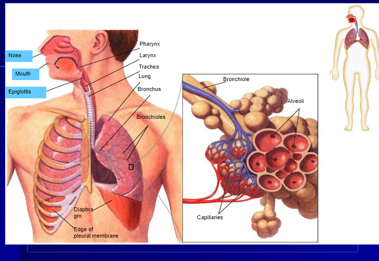
B. Structures
1. Nose: Air enters the body through the nostrils.
a. Nostrils contain hairs that trap dirt and foreign particles from entering the body.
b. Walls of the nasal cavity are lined with mucus which also traps dirt and moistens the air.
c. Large number of capillaries near the surface of the nostrils warm the air as it enters the body.
2. Pharynx and Larynx
a. Air enters the Pharynx (throat) from the nasal cavity.
b. The air then passes into the Larynx (voice box)
- composed of cartilage.
- vocal cords: pairs of membranes stretched across the larynx; their vibration creates sound.
4. Bronchi and Bronchioles
a. Bronchi: 2 cartilage ringed tubes that branch off the trachea
-Lined with cilia
-Entrance way to the lungs
b. Bronchioles: branch off the bronchi
-Divide and become smaller, thinner with less cartilage
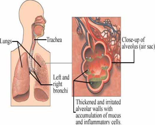
5. Alveoli
a. Tiny air chambers at the end of the bronchioles.
b. Walls are 1 cell thick and moist from mucus.
c. Surrounded by capillaries.
d. Through the alveoli walls, the exchange of oxygen and carbon dioxide takes place.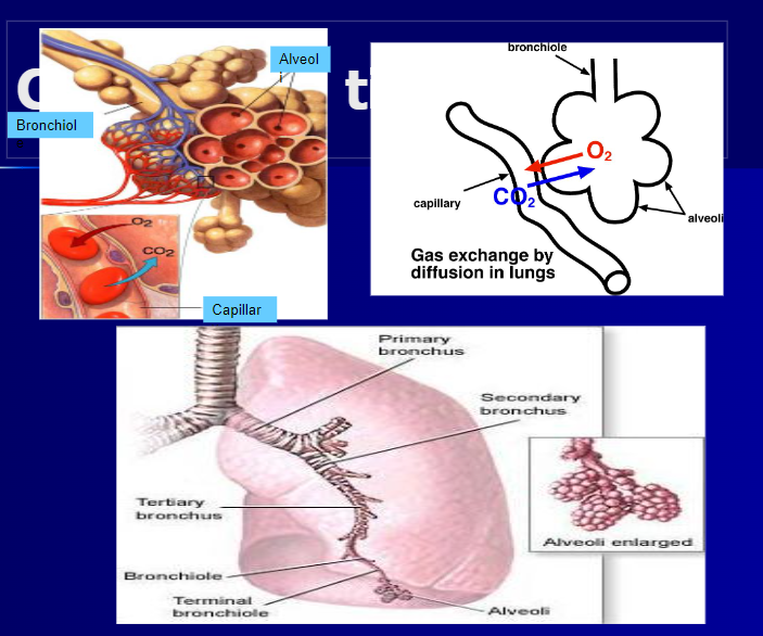 II. Breathing
A. Definition: the physical process by which air is moved into and out of the lungs.
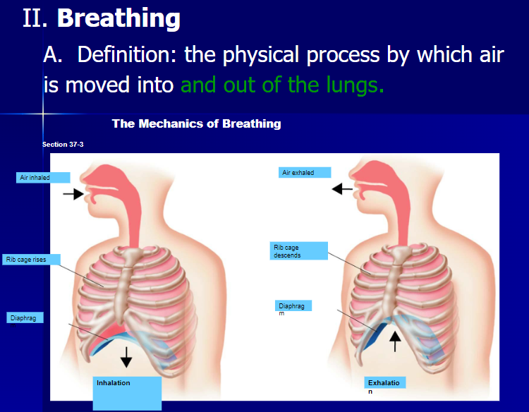 1. Inhalation draws air into the lungs.
a. Active phase of breathing.
b. Ribs are pulled up and out, while the diaphragm is pulled downward.
-The chest cavity becomes larger.
This causes pressure within the chest cavity to decrease which brings air into the lungs forcing them to open.
2. Exhalation allows air out of the lungs.
a. Passive phase of breathing
b. Diaphragm relaxes and moves up. Rib muscles relax causing the ribs to drop. The chest cavity becomes smaller which increases the pressure inside; this will force air out of the lungs and into the environment.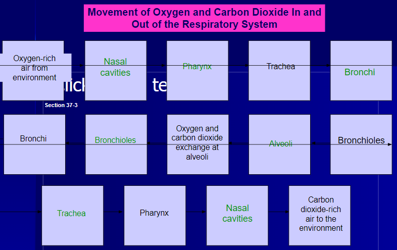 III. Malfunctions of the Respiratory System
A. Emphysema:
1. Caused by smoking.
2. Particles from cigarette smoke accumulate on the alveoli walls causing inelastic scar tissue to form.
3. This decreases the working area of the respiratory surface.
- Lungs lose their elasticity.
4. Characterized by shortness of breath, difficulty exhaling, and decreased lung capacity.
B. Lung Cancer
1. Disease in which tumors (masses of tissue) form in the lungs as a result of irregular and uncontrolled cell growth.
2. Linked to smoking.
C. Asthma
1. Severe allergic reaction in which the contraction of the bronchioles makes breathing difficult.
D. Bronchitis
1. Inflammation of the lining of the bronchial tubes.
2. Passageways to alveoli become swollen and clogged with mucus.
3. Marked by severe coughing and difficulty breathing.
E. Pneumonia
1.
Circulatory System
The Circulatory System is responsible for transporting materials throughout the entire body.
It transports nutrients, water, and oxygen to your billions of body cells and carries away wastes such as carbon dioxide that body cells produce.
It is an amazing highway that travels through your entire body connecting all your body cells.
Components
- Heart
- Blood
- Vessels
- Arteries
- Veins
- Capillaries
Which gases are transported to and from the body’s cells by the blood flowing in the circulatory system?
Oxygen is the gas needed for respiration and is transported to the body’s cells.
Carbon Dioxide is a waste gas released from transportation and into the atmosphere
Two types of blood
Oxygen-rich blood
- Dark blood traveling to the body cells. High oxygen content; Low carbon dioxide content
Oxygen-poor blood
- Bright blood traveling away from the body cells. Low oxygen content; High carbon dioxide content
These two types of blood cannot mix
Heart - organ at the center of the circulatory system, pumps blood around the body
- inside is divided into two sections so two types of blood do not mix
Heart walls:
Epicardium - Outermost layer, fat to cushion heart
Myocardium - middle layer, primarily cardiac muscle
Endocardium - Innermost layer, thin and smooth, stretches as the heart pumps
Myocardium - Muscle of the heart, strong and thick, can conduct electricity like nerves, composed of spontaneously contracting cardiac muscle fibers, It’s blood supply comes from the coronary arteries
The right and left sides of the heart are separated by a septum or wall.
The septum prevents the mixing of oxygen-rich and oxygen-poor blood.
On each side of the septum are two chambers.
The upper chamber (which receives blood) is the atrium.
The lower chamber (which pumps blood out of the heart) is the ventricle.
Four chambers
Two Atria
Upper chambers
Left and right
Separated by interatrial septum
Two Ventricles
Lower chambers
Left and right
Separated by interventricular septum
Atria - collects blood that enters the heart
Ventricles - pump blood out of the heart
The valves between the atria and ventricles are connected to the inner walls of the heart by tough tendons.
The tendons allow the valves to close and hold the valve flaps in place. They prevent the valves from flipping up and turning inside out
Valve - acts like a door that only opens in one direction
If the door is held by someone at a fixed point, only the arm moves as the door opens and closes. When the door is closed the arm is fully extended, so the door can only be opened in one direction.
In the heart, the valve's tendons are like the arm holding the door.
One end of each tendon is fixed to the wall of the heart so the valve can only open in one direction.
Superior Vena Cava (upper body), Inferior Vena Cava(lower body) - Right atrium - tricuspid valve - right ventricle - pulmonary valve -pulmonary artery - lungs (gas exchange) - pulmonary veins - left atrium - mitral valve - left ventricle - aortic valve - aorta - to the body - cycle repeats
The heart can pump blood because it is made of muscle.
Muscle tissue works by contracting (squeezing) and relaxing
All the parts of the heart on either side, work together in a repeated sequence.
The two atria contract and relax; then the two ventricles contract and relax.
This is how blood moves through the heart and is pumped to the lungs and the body. One complete sequence of contraction and relaxation is called a heartbeat.
Vessels:
Arteries: Carry blood away from the heart.
Capillaries: Link arterioles to veins.
Veins: Carry blood towards the heart
Arteries
-Large vessels
-Carry blood from the heart to tissues of the body.
-Carry oxygen-rich blood, with the exception of pulmonary arteries.
-Thick walls need to withstand pressure produced when the heart pushes blood into them.
Capillaries
-Smallest blood vessels
-Walls are only one cell thick and very narrow.
-Important for bringing nutrients and oxygen to tissues and absorbing CO2 and other waste products.
Veins
Once blood has passed through the capillary systems it must be returned to the heart. Done by veins
Walls contain connective tissue and smooth muscle.
The largest veins contain one-way valves that keep blood flowing toward the heart.
Many are found near skeletal muscles. When muscles contract, blood is forced through veins.
Blood Pressure
The heart produces pressure
The force of blood on the wall of the arteries is known as blood pressure.
Blood pressure decreases as the heart relaxes, but the rest of the circulatory system is still under pressure.
When blood pressure is taken, the cuff is wrapped around the upper portion of the arm and pumped with air until blood flow in the artery is blocked.
As the pressure in the cuff is relaxed, 2 numbers are recorded.
- Systolic pressure- the first number taken, is the force felt in the arteries when the ventricles contract.
- Diastolic pressure- the second number taken, is the force of the blood on the arteries when the ventricles relax.
Blood
What percent of your body is blood? 8%
How much blood do we contain? On average 4-6 liters. We contain about a pint of blood for every 15 pounds of body weight
Composition of Blood:
What percent of your blood is cellular? 45%
What percent of your blood is plasma? 55%
Composed of plasma and blood cells
Types of Cells are:
- Red Blood Cells
- White Blood Cells
- Platelets
Plasma
- Straw colored
- 90% water
- 10% dissolved gases, salts, nutrients, enzymes, hormones, wastes, and proteins.
Plasma proteins
3 Types: Albumins, globulins and fibrinogen.
Albumins and Globulins- transport substances such as fatty acids, hormones and vitamins. Fibrinogen- Responsible for blood’s ability to clot
Red Blood Cells
- Most numerous type
- Transport oxygen
- Get color from hemoglobin
- Disk-shaped
- Made in red bone marrow
- Circulate for 120 days
White Blood Cells
- Guard against infection, fight parasites, and attack bacteria
- The number of WBCs increases when the body is fighting
- Lymphocytes produce antibodies that fight pathogens and remember them
Platelets
- Aid the body in clotting
- Small fragments
- Stick to edges of broken blood cells and secrete clotting factor to help form clots.
Blood has 3 main functions
- transport
- protect
- temperature