BIOB34 Module 5
Terminology:
P50: Partial pressure at which pigment is 50% saturated
Coagulogen: a protein that coagulates to trap bacteria
Hypoxic Ventilatory Response (HVR): Behavioural response to lower Po2
Osmotic Pressure: pressure needed to offset movement of pure solute across semipermeable membrane
Hydrostatic pressure: pressure fluid physically exerts on surroundings (due to pull of gravity, pressure of blood on vessel wall)
Antidiuretic hormone (ADH): peptide hormone
Module 5 - Respiration
Diversity of Digestive Systems
- Direct absorption for sugars, amino acids, etc.
- Endocytosis (cellular phenomenon) for larger food particles
- Size is important; if it is small enough, it is able to get its nutrients through direct absorption
Diversity of Respiratory Systems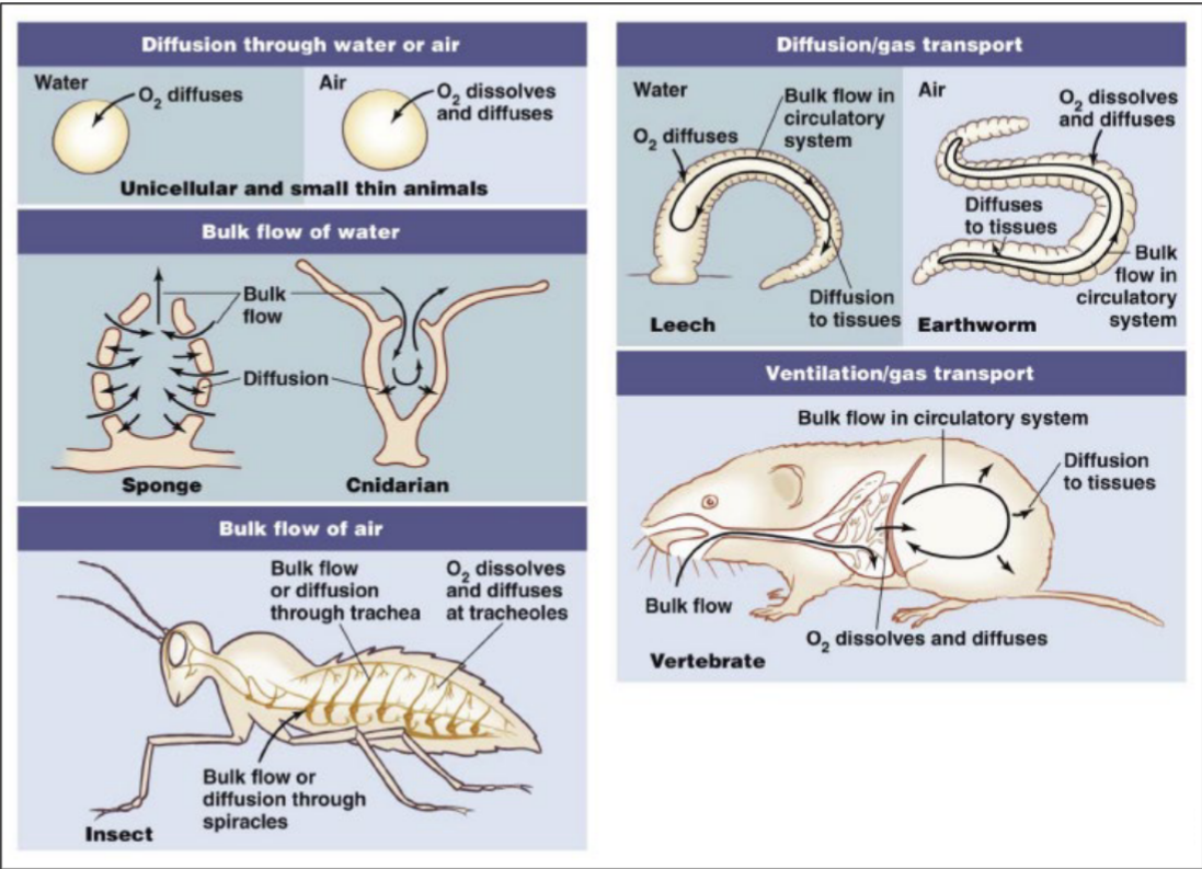
- If small, diffusion is sufficient
- If larger, need for both conductive/diffusive and convective (bulk flow) steps
- Step where gases must diffuse across a membrane
- Involves movement of fluid
Respiratory Systems can interface with circulatory system
- Lungs or gills designed with lots of surface area for exchange
- Respiratory system has great surface area where the diffusion occurs
- Exchange is with a delivery fluid (blood) and circulation system
Closed (e.g. Mammalian) circulatory system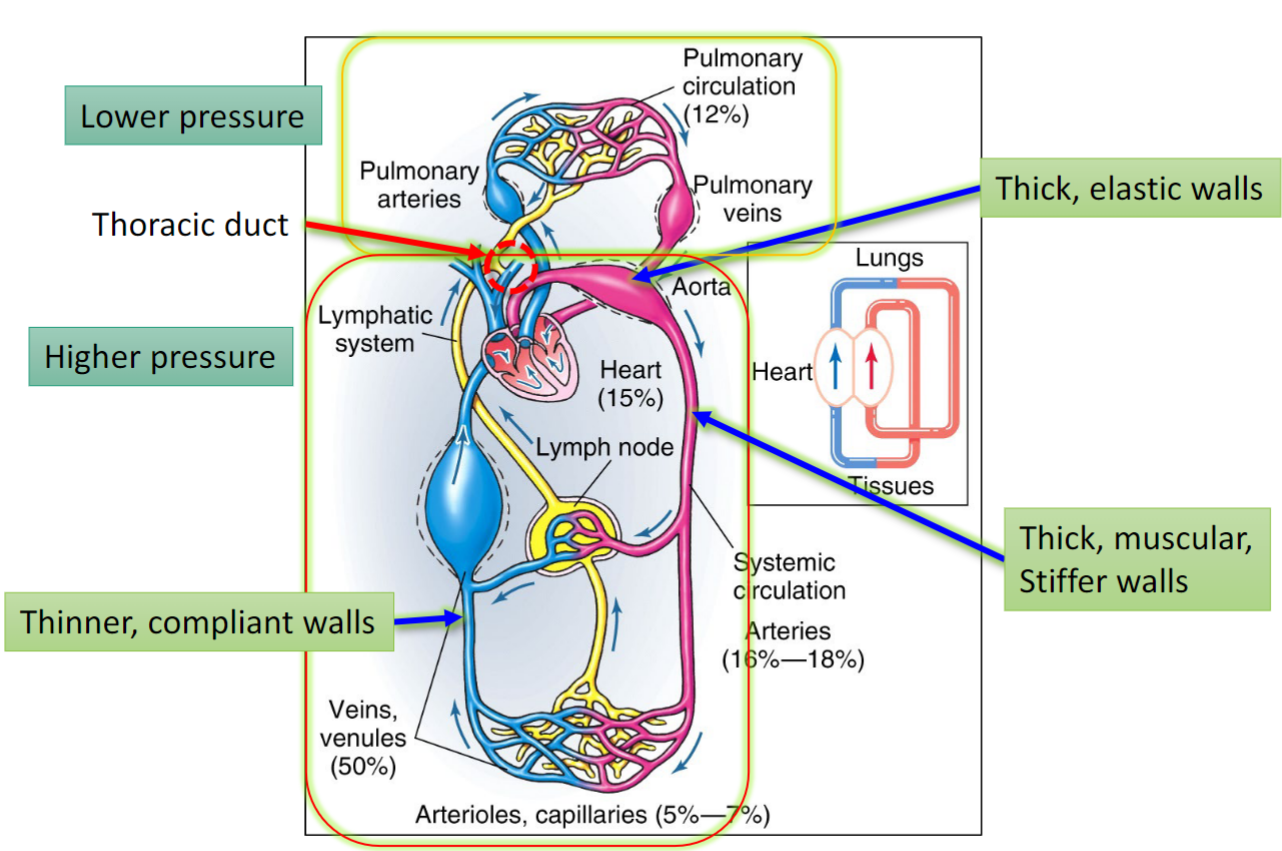
- Closed meaning that the blood is contained within the vascular network
- Some plasma leaks out where the capillary beds are very dense; where the vessels are smallest
- Fluids leak from vessels out into extracellular fluid
- Rely on lymphatic system to drain fluid which can be directed & dumped back into circulation close to the hearts
- Blood pumped by heart → pulmonary circulation (deoxygenated blood, arteries) & interphase with alveolar sacs & can diffusively pick up oxygen + diffusively unload CO2 ; pulmonary venous circulation returns blood to the heart, now red & oxygenated → pumped through the left ventricle → systemic circulation
- 4 chambered heart; pump with an atria & ventricle on the right side directing flow to the lungs + atria & ventricle on left side that pumps to systemic circulation
- Lower pressure (at top of diagram); shorter trip to pulmonary circulation
- Thick elastic walls:
- Needs greater force/pressure to meet the demands necessary
- Connective tissue
- Acts as a dampener; moderating force of blood pressure
- Thoracic duct: where lymph system is interfacing with the vascular system
- Can feed back into circulation
- Lymphatic system important for immune system
Open (e.g. insect) circulatory system and tracheal system
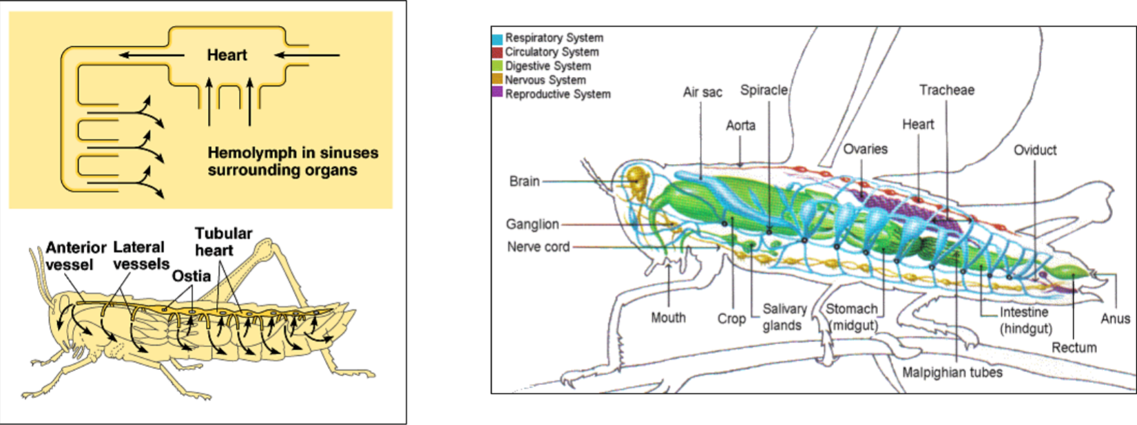
- Open; hemolymph pumped by a muscular heart, goes out through arteries, then fluid gets dumped out into general extracellular fluid that bathes the tissue of the animal
- Blind openings close to the heart where extracellular fluid can leak back into the heart and be pumped again
- Insects; separate system for respiratory gases; respiratory and circulatory system are more distinct in their operation compared to mammalian system
- Tracheal system requires sphericals, openings on the side of the insect → Alveoli spread throughout the body
- “Pumping”; convective movement of air in & out of sphericals
- Aqueous environment in tracheae at most terminal portions; need fluid lining since oxygen in the air needs to dissolve into the fluid layer which can then diffuse into alveoli into pulmonary capillaries & into red blood cells
- Tissues themselves can be diffusing CO2 outwards
Circulatory Fluid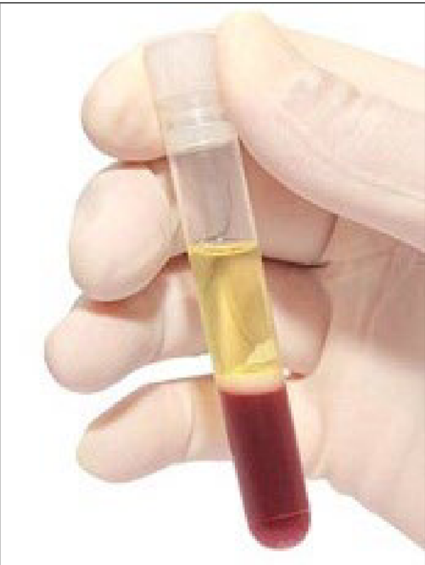
Vertebrates:
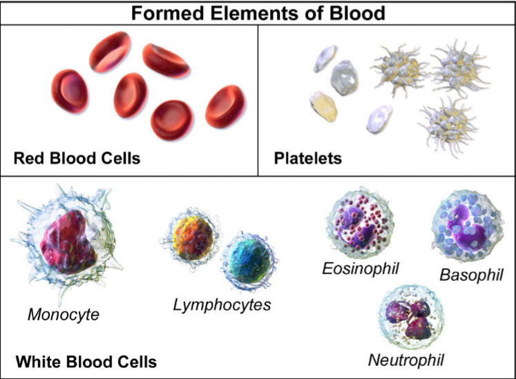
- RBCs (red blood cells)- where resp. Pigments are
- Also carry other nutrients, hormones, etc.
- Fractionated - spin blood in centrifuge
- Plasma layer; aqueous stuff (dissolved nutrients) - yellow
- Buffy coat; platelets at top and white blood cells + other - white
- RBCs - concentrated blood cells
Invertebrates:
- In many insects, doesn’t provide O2/CO2 delivery role
- Tracheal system does
- (IF PRESENT) Resp. pigments in plasma (not contained in specialized pigment containing blood fells; floating freely)
- E.g. some crustaceans, other arthropods, some worms
- Also carries other nutrients, hormones, etc.
- Contains hemocytes
- Role in immune response like WBCs (white blood cells)
Red blood cells: no nucleus, may be different in classes of vertebrates (e.g., birds; nucleated and has mitochondria)
Platelets: crucial, allows injured vessels to be temporarily clotted to avoid losing too much blood and blood pressure
Hemoglobin (Haemoglobin - Hb)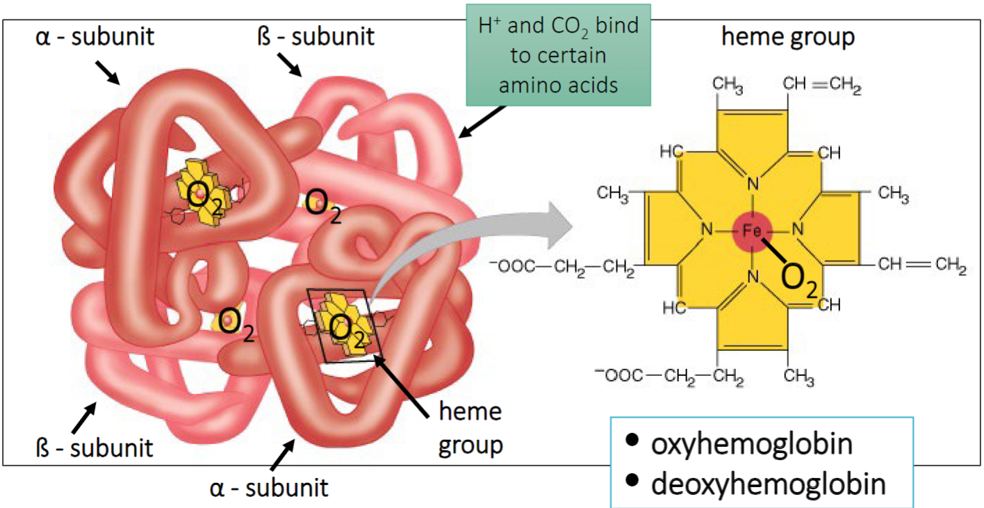
- ≈ 98.5% of the O2 in blood is bound to hemoglobin; Found inside red blood cells
- If hemoglobin is found outside red blood cells, something is wrong
- Red blood cells are bursting
- 1.5% is O2 dissolved in plasma/aqueous environment
- Hemoglobin increases capability of a volume of blood to carry oxygen
- Oxygen can bind with high affinity to heme groups (Fe)
- Proteins subject to modification and regulation (heme units with protein subunits)
- Give system the ability to modify the activity of hemoglobin to push and pull oxygen holding capacity in those proteins when appropriate
- H+ and CO2 bind to certain amino acids; causes conformational changes in the protein (influences its activity → how tightly it does/does not bind oxygen)
- 2 a-subunits and 2 b-subunits
- Tetromeric combination
- Different alleles of a and b-subunits are present in our genomes
- Oxyhemoglobin: O2 bound to most
- Deoxyhemoglobin: most heme groups are free; O2 not bound
O2 delivery via circulatory system (the typical vertebrate example)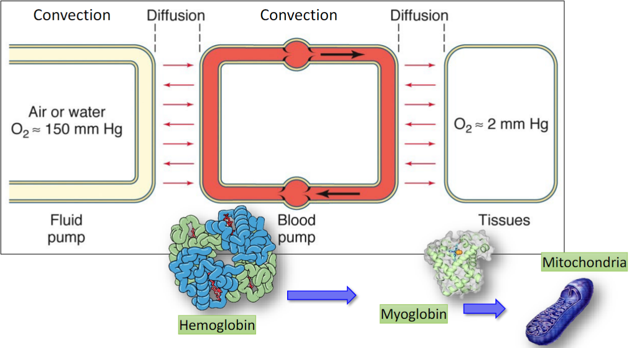
- Tuna; RAM ventilators → must swim forward in order for there to be fluid flowing across their gills
- Pump is done by skellular ? muscle of the animal moving it through stable aqueous environment
- Other fish; gulp water into oral cavity, squeeze cavity pushing water back and across their gills
- Diffusion:
- At least a couple of cell membranes
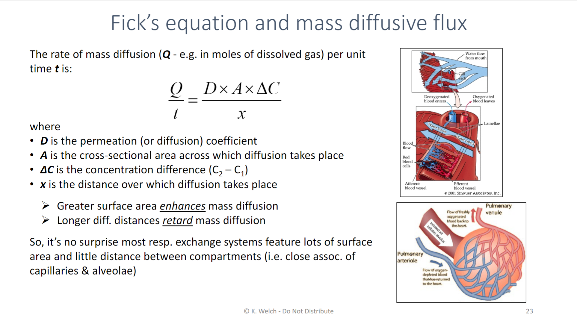
- Try to minimize the diffusional distance; enhances diffusion of oxygen
- Diaphragm causes negative pressure in lumen of lungs; increased pressure gradient to drive air in
- Exhale: muscularly contract and pushes air back out
Diagram:
- Convection + fluid pump: involves bulk flow of fluid
- Convection + blood pump: capillary system and pulmonary circulation
- Muscular pump: by bulk flow or convection moving fluid around
- Second diffusion:
- Oxygen and CO2 must move between the hemoglobin and where the plasma in the blood across membranes (of tissues at least) and into tissue to make its way into the mitochondria
- CO2 produced in mitochondria and elsewhere through various decarboxylation (enzymatic steps)
- In first few moments when muscle cell transitions from rest to exercise, there is a rapid potential increase in rate of O2 consumption at tissues
- Not instantaneously met by all the previous steps
- So, drawing down the pool of oxymyoglobin making deoxymyoglobin; making store of O2 to buffer
- Oxygen and CO2 must move between the hemoglobin and where the plasma in the blood across membranes (of tissues at least) and into tissue to make its way into the mitochondria
- Myoglobin:
- While hemoglobin is a tetrameric protein, myoglobin is a monomeric
- Temporal & spatial buffer for oxygen in the tissue itself
- Found widely distributed in our tissues (cardiac muscle); can reversibly bind oxygen in the heme group that it contains
- “Storage locker” that oxygen temporarily binds to
- Mitochondria:
- Becomes sink for O2 itself; sucking up and using O2
Respiratory pigments
P50: Partial pressure at which pigment is 50% saturated
- Analogous to makalousmentin? Coefficient; concentration of substrates that the enzyme will operate at 50% of its maximum enzymatic activity
- NOT A RATE!
Sigmoidal versus hyperbolic shape; cooperativity!
- P50 for hemoglobin is higher
- P50 for myoglobin is lower value; takes much lower PO2 in the test tube to get 50% of the myoglobin to be loaded with oxygen
- Affinity of myoglobin for O2 is higher/stronger than the affinity of hemoglobin for O2
- Hemoglobin shows up at tissue (has a relatively low PO2); hemoglobin is happy to give up O2, little oxyhemoglobin leftover → at the same moment, myoglobin has a higher affinity for O2 than hemoglobin which encourages the movement of O2 from the circulation of where hemoglobin is into the tissue where myoglobin is
- If you took out respiratory pigments and purified them from samples of blood:
- Put them in a spectrophotometer?
- Can tell or red or how blue the colour, then change O2 level the test tube experiences → how much O2 bound to hemoglobin or myoglobin at various partial pressures of O2
- Put them in a spectrophotometer?
Graph:
- Myoglobin curve; exponential curve → no cooperativity since not multiple subunits
- Hemoglobin curve; sigmoidal shape → can exhibit cooperativity
- Binding of O2 can cause conformational change
- H+ and CO2 binding to amino acid in protein can cause conformational change and impact affinity (impacts P50)
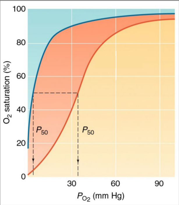
- Conformational change: as more O2 binds to more heme groups, the conformation changes and makes it easier for O2to bind to the remaining unfilled heme groups
- Can work in reverse; O2 going off of heme groups
- (0,0) is a deoxygenated situation
- Curve approaching 100%: saturation effect (O2 saturation approaches 100%); only a number of binding spots left on heme groups
Hemocyanin
- Respiratory pigment found in some Arthropods (Chelicerates)
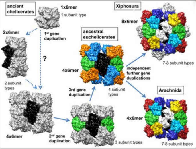
- E.g. horseshoe crabs
- Does NOT contain a heme group
- Copper-containing → copper can reversibly bind O2 the same way that an iron molecule can
- Floats freely in the plasma
- Harvesting horseshoe crab blood to extract “coagulogen”
- Tests human blood samples and others for contamination
- Binds strongly to bacteria
- Many monomer units bound together (into octamers, dodecamers, etc.)
- What do you predict the shape of the hemocyanin-O2 binding curve is?
- Wouldn’t exhibit cooperativity (in ancient form) due to monomer
- Like myoglobin
- Elaborate sigmoidal curve later when multiple subunits
- Wouldn’t exhibit cooperativity (in ancient form) due to monomer
Image:
- As the genes duplicated, evolved into complex multisubunit protein
Other respiratory pigments
- Hemerythrin/myohemerythrin
- Contains iron, but not a heme group
- Found in some marine inverts, single annelid worm genus
- Colourless when deoxygenated, pink-violet when oxygenated
- Chlorocruorin
- Found in plasma of many annelids (especially polychaete worms)
- Composed of multiple monomers
- Cooperativity → sigmoidal curve for O2 binding
- Contains “abnormal” heme group
- Heme group with iron; but heme group is different in its molecular structure
- Incidentally, some Nematodes and Arthropods have Hb floating in plasma (or even inside nerves or muscle fibers - kind of similar to role of myoglobin in us)
- Not surprising to the convergent evolution in the respiratory pigment
Integrating effects on % O2 saturation
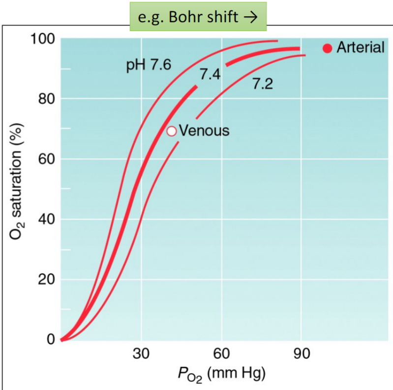
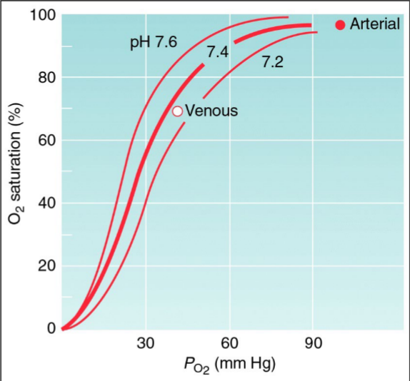
- Effectors of P50 (the following lower affinity = increase in P50):
***If it moves to to the right across the x-axis: lowered affinity
- Increase in temperature
- Impacts weak bonds in proteins and helps destabilize to make it more chemically active
- Active sites of O2 consumption that will correlate with local increased temperature
- More levels (more binding of…)
- 2,3-DPG (diphosphoglycerate); metabolite; substrate has the effect of binding to hemoglobin; the more it binds the lower the affinity
- ATP, GTP (e.g. fishes, hemoglobin in fishes are sensitive to ATP and GTP); red blood cells are nucleated and contain mitochondria (the relative O2 availability to red blood cell, mitochondria can have an influence on the relative concentration of ATP and GTP that those cells are maintaining over time) If ATP levels go up, it signals a good O2 environment, decline in hemoglobin
- IP5 (birds)
- Decrease in pH - greater amount of protons floating around
- Called Bohr shift; as red blood cells are encountering a local environment that becomes more acidic, it shifts their O2 binding curve a little bit to the right (hemoglobin has less affinity for oxygen; more apt to let it go)
- Increase in CO2 (carbamino-hemoglobin formation: HbCO2)
- Pulmonary circulation: want blood to load up oxygen on hemoglobin
- Want affinity to be relatively strong; pulmonary circulation red blood cells flowing closely by alveoli sacs and able to diffusively pick up O2; also site releasing CO2 (diffuse from blood to alveolar sacs)
- Blood at lungs losing CO2 which decreases concentration of dissolved CO2 and decreases carbamino-hemoglobin formation, pH will increase and temperature of microenvironment of pulmonary circulation might lower creating a higher affinity
- CO2 itself can bind to hemoglobin → carbamino-hemoglobin
- Effects by pH
- Effects by binding itself
- Both of the above leading to a decrease in affinity and an increase in P50 in the local area where there is an increase in CO2 (through its effect in lowering pH and binding of CO2 in higher concentrations to hemoglobin)
- Pulmonary circulation: want blood to load up oxygen on hemoglobin
- Increase in temperature
- Where there are high levels of O2 consumption, tend to see a decline in pH; encourages hemoglobin to let go of O2 at most demanding tissues
- When blood reaches active tissues, it can be accompanied by a pH change; as muscle becomes active initially before O2 is delivered and before blood flow can be regulated to those tissues, there is a temporary increase in anaerobic glycolysis to buffer over time the ATP levels in the tissues (with anaerobic glycolysis comes some acidification → due to CO2 that cells are generating naturally combines with water forming carbonic acid which then dissociates into bicarbonate and a proton)
- Blood sample carries certain volume percent of O2 and percent of saturation at Hb with O2
- Anemia; too little red blood cell volume in their blood
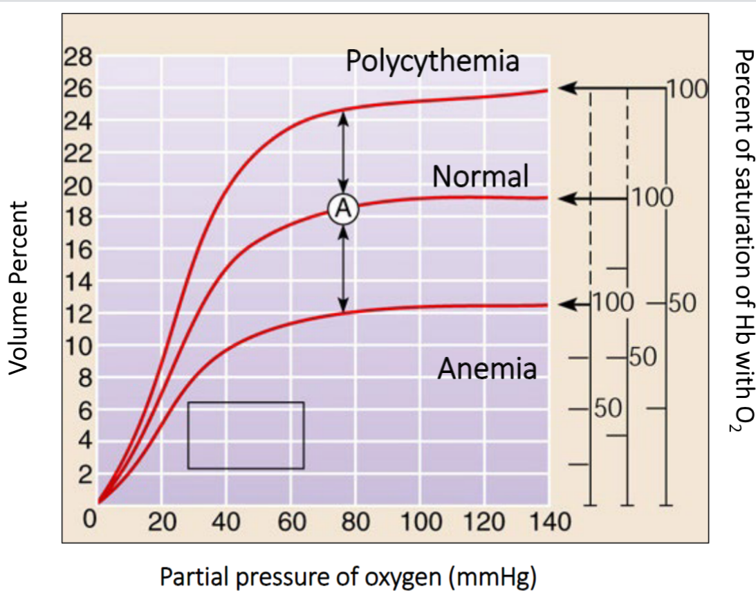
- Not getting enough iron
- Total O2 carrying capacity of the blood sample would be lower
- Fewer hemoglobin molecules per unit volume of blood to carry O2 around
- Polycythemia; can be adaptive or maladaptive, increase red blood cell production
- Increases the absolute carrying capacity of a given volume of blood
- By packing more cells into a given volume of blood, the blood will be thicker and more viscous (makes it harder to pump and could lead to a greater risk of blood clot formation)
- P50 for the curves; might be containing hemoglobin that is equally high affinity for O2
- In all 3 conditions shown in the graph, the O2-hemoglobin saturation reaches 100% of its capacity but the total O2 content differs
CO2 and pH (and H2O) are related: HCO3- + H+
- Almost all of the CO2 floating in blood is in the form of HCO3-
- Good thing; bicarbonate is a good buffer of pH (helps keep pH relatively stable)
- Bohr shift is subtle because even at tissues that pump large amounts of CO2; a lot of them are relatively changing into HCO3- and that is moderating pH (flexible in ability to absorb extra protons and release the, when needed)
- Most of CO2 is in the form of bicarbonate and is being carried around dissolved in the plasma
- CO2 + H2O ⇆ H2CO3 ⇆ H+ + HCO3-
- Carbonic acid happy to become bicarbonate since weak acid
- This process is not enzymatically catalyzed
- Carbonic acid happy to become bicarbonate since weak acid
Graph:
- Snapshot of what the curves would look like if took a sample of blood and exposed them to different partial pressures of CO2
- Wouldn’t expect to see sigmoidal curves since it’s not a respiratory pigment based phenomenon
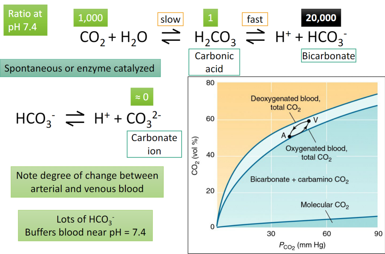
- Difference between oxygenated and deoxygenated blood matters; CO2 can bind to hemoglobin and when it does it reduces the affinity of hemoglobin for O2, oxygenated hemoglobin is more likely to kick CO2 off and lose the carbamino part
- Venous blood; returning has picked up CO2 from tissues where it delivered CO2, PCO2 higher
- Arterial blood; more oxygenated
- Note degree of change between arterial and venous blood
The Challenge of elevation
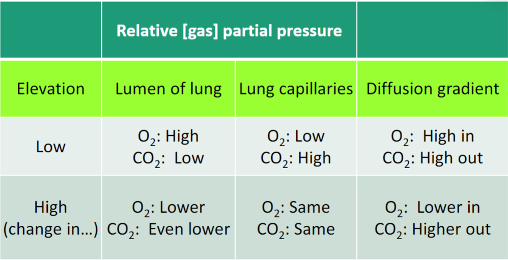
- Partial pressure of all gases (including O2 and CO2) are lower at higher elevations
- This has different effects on two diffusion gradients
- Change in CO2 stores makes it way through systemic circulation
- If blood makes way from pulmonary circulation to hungry tissues; while blood took up O2 easier at pulmonary surfaces, its holding onto it a little more tighter in systemic circulation
- Changed one portion; reduced gradient for O2 at higher elevation
- Makes it harder for pulmonary circulation to pick up oxygen to meet needs of the tissues
- Difference in CO2 from lumen to capillaries creates gradient for CO2 to move out
- Enhancing CO2 diffusion gradient; easier to exhale CO2
- Problem since CO2 and pH are linked and ability for CO2 to bind to hemoglobin can be problem
Acclimatization to high elevation
- Behavioural response to lower PO2: hyperventilation
- Called the “Hypoxic Ventilatory Response” (HVR)
- Feel out of breath; ventilating more strongly to offset
- Initial response; increase breathing rate
- Called the “Hypoxic Ventilatory Response” (HVR)
- Hyperventilation (increased convective flow) enhances O2 and CO2 exchange
- But it’s already easier to lose CO2
- Lowering of CO2 levels in blood leads to slight increase in pH
- Higher affinity for O2 won’t rapidly release O2 optimally at tissue
- Remember Bohr Effect
- Lower P50 for O2 encourages uptake by Hb at lungs, but discourages release at tissues
- After a few days, there is increased release of 2,3-DPG into circulation
- As shown several slides ago, this increases P50 (offsetting effects of pH change)
- Balancing act of what’s good at the lungs vs. what’s good at systemic circulation
- As shown several slides ago, this increases P50 (offsetting effects of pH change)
- Over similar/slightly longer time frame, body responds with increased EPO (hormone)
- This encourages erythropoiesis (formation of red blood cells)
Evolution of mechanics of ventilation
- Tuna:
- Must be swimming forward all the time; only way to ensure that water is moving past their gills where gas exchange with their pulmonary circulation occurs
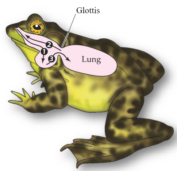
- Fish and amphibians: buccal oscillations (or ram ventilation in some fish - tuna)
- Floor of mouth is lowered; buccal cavity expanded; air flows through the nares (nostrils) into the bottom of the mouth
- Creates negative pressure; pressure gradient for air to move into buccal cavity
- Glottis closed
- Fish: use water and pushes it out through their gills
- Glottis opens; air flows from the lungs across the top of the mouth and out of the nares
- Elastic recovery of energy in torso which pushes whatever air that was in the lungs out; exits out in top of buccal cavity
- Floor of the mouth is raised; buccal cavity is compressed; air is pumped into the lungs; the glottis closes
- Reptiles, birds and mammals: axial musculoskeletal oscillations
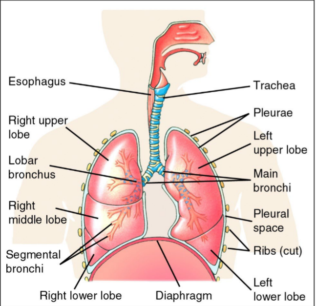
- Inspiration creates negative pressure in lungs because pleural space (between ribs and lungs) cannot expand (fluid filled)
- Lower lobes: when they contract; causes rib cage to expand → enhances inspiration and exhalation
- When the diaphragm contracts, it lowers which pulls on the bottom space defined by ribs → lungs expanding and are deriving low pressure in lungs (gradient for air to move in)
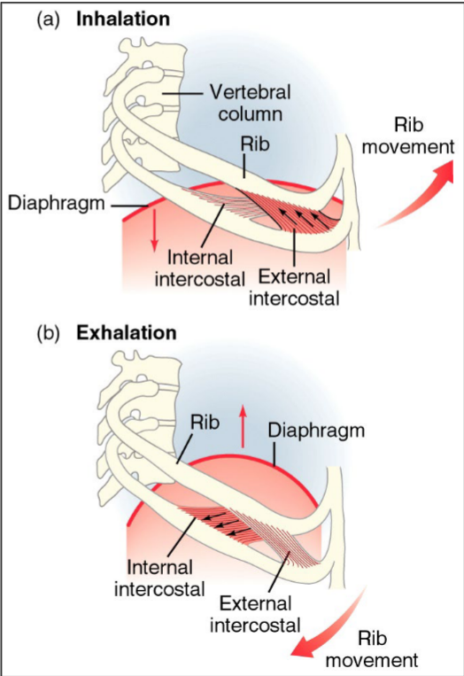
- Diaphragm springs back into place
- Quiet breathing → exhalation is passive (system elasticity)
Quantification of ventilation
- Anatomical dead space: volume of non-respiratory portion; volume of air/gas due to anatomical position
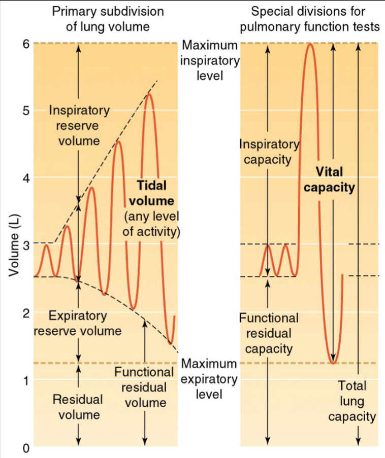
- Still air present in lungs
- Physiological dead space: volume not involved in gas transfer (includes less perfused alveoli)
- Special divisions for pulmonary function tests → clinical; more physiological
- Maximum respiratory level; can hold max 6L in lungs, trachea etc.
- Maximum expiratory level; max. Exhale
Fick’s equation and mass diffusive flux
Hypoxic Pulmonary Vasoconstriction (HPV)
- Circulatory system only works as strongly as it needs
- Arterioles (sensitive to pH, CO2 levels) feeding areas of lung that are hypoxic constrict, reducing blood flow to that area
- Normally, this helps match lung perfusion to pulmonary blood delivery
- Think of physiological dead space
- In hypobaric (lower partial pressure of O2 in environment because of lower total pressure) hypoxia at high elevation, all of pulmonary circulation gets constricted (lung uniformly hypoxic)
- This increased resistance to flow raises pressure, possibly leading to pulmonary edema (leakage of more fluid)
- Tibetans shows blunted HPV response (vessels little less sensitive)
Adaptation to high elevation
- At high elevation, Tibetans tend to show lower increase in hematocrit than lowlanders
- Increase hematocrit means increase in viscosity of blood → harder to pump
Concurrent (model approximates mammals) vs. Countercurrent (e.g. fish)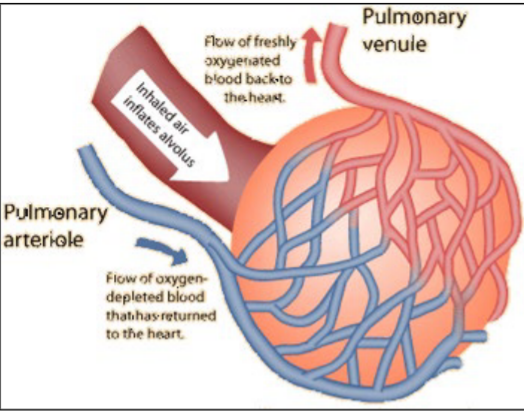
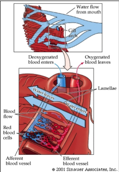
- Difference in architecture has consequences for differences in exchange
Image 1:
- Inhaled air comes into alveolar sac
- Pulmonary circulation from heart (deoxygenated blood; carries more CO2) and exchanges with volume of air
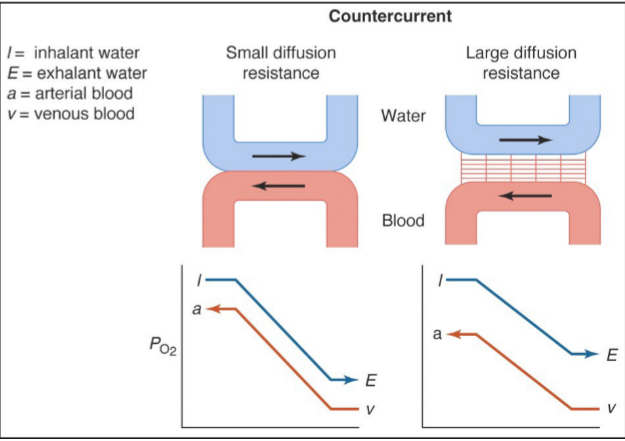
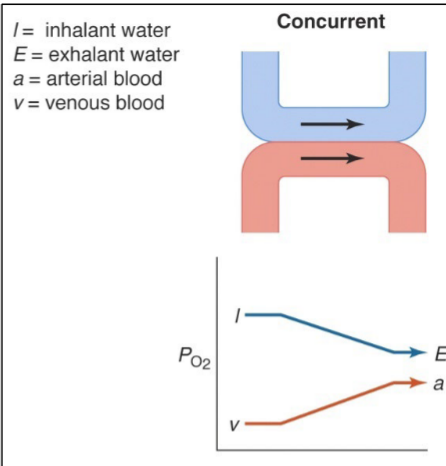
Image 2:
- Moves upstream relative to water flow
- Enhances diffusion of heat
Concurrent: our circulation, reduced diffusion gradient
Countercurrent: gills, more efficient due to water, modest diffusion gradient
Crosscurrent exchange in birds
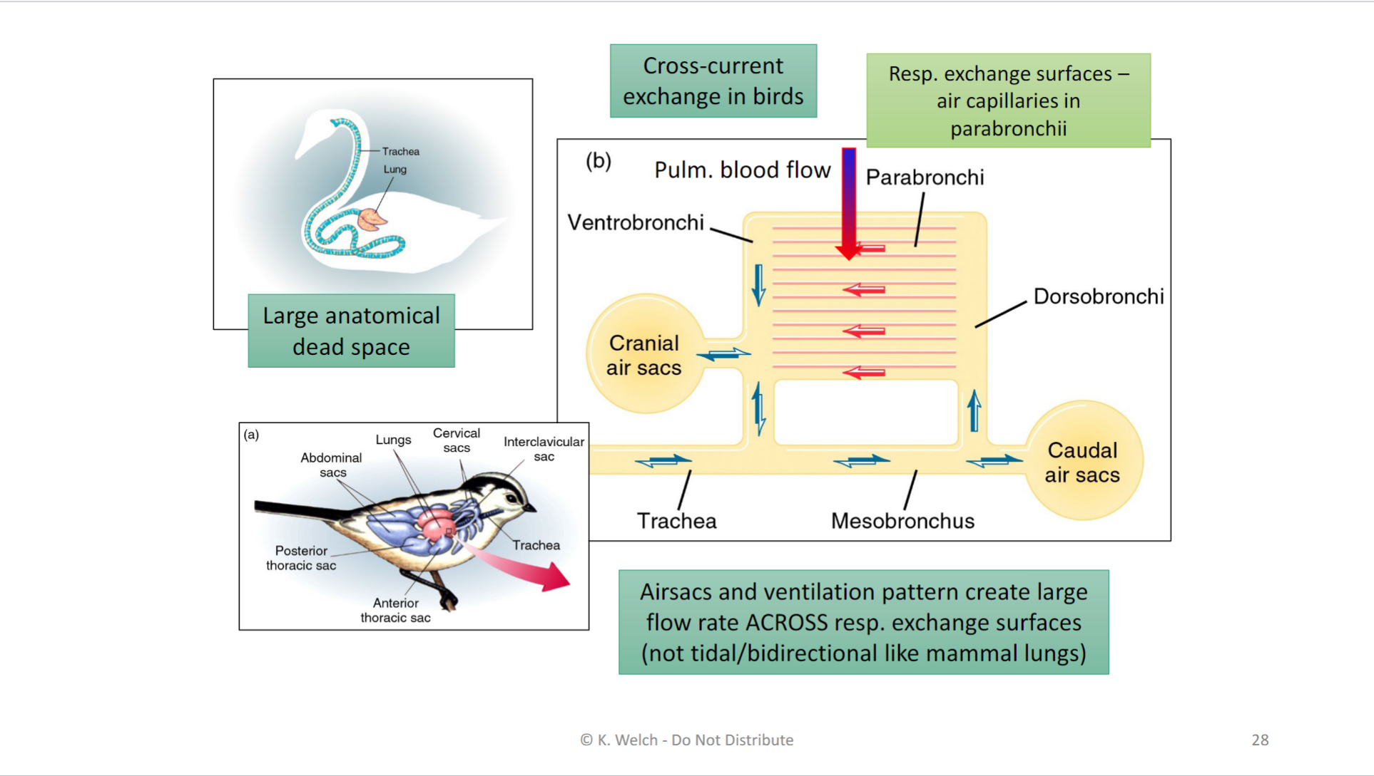
- Caudal air sacs; encourages unidirectional flow
- Enhances rate of blood diffusion