Science 10
Central Nervous System
acts as the body’s control center
coordinates the body’s activities
made up of the brain and spinal cord
Impulses travel through the neurons in your body to reach the brain.
Peripheral Nervous System
made up of all the nerves that carry messages to and from the Central Nervous System
Central Nervous System and Peripheral Nervous System work together to make rapid changes in your body in response to stimuli
2 parts of the Peripheral Nervous System
Somatic Nervous System
Autonomic Nervous System
Somatic Nervous System
relay information between the skin, skeletal muscles, and Central Nervous System
you consciously control this pathway by deciding whether or not to move a muscle (except reflexes)
Responsible for carrying motor and sensory information
Is made up of nerves that connect to the skin, sensory organs, and skeletal muscle movements
Processes sensory information from external stimuli (hearing, touch, and sight)
Autonomic Nervous System
relays information from the Central Nervous System to the organs
Involuntary: you cannot consciously control these
Sympathetic Nervous System
controls in times of stress, such as the fight or flight response
Parasympathetic Nervous System
controls the body in times of rest
Neurons
the basic unit of structure and function in the Nervous System
cells that conduct impulses
made up of dendrites, cell body, and axons
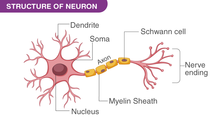
Dendrites
receive impulses from many other axons
branch-like extensions which the receiver impulses and carry them toward the cell body
Axons
single extension of the neuron that carries impulses away from the cell body
branches out an ending to send impulses to many different neurons
Cell Body
also called the soma, is the spherical part of the neuron that contains the nucleus
connects to the dendrites, which bring information to the neuron, and the axon, which sends info to other neurons
when information is received from another neuron, the dendrites pass the signal to this
Types of Neurons:
Sensory Neurons
carry impulses from inside and outside the body to the brain and spinal cord
Interneurons
found within the brain and spinal cord, process incoming impulses and pass them onto motor neurons
Motor Neurons
carry impulses away from the brain and spinal cord
Six Main Sections of the Brain
Cerebrum
Cerebellum
Diencephalon
Midbrain
Pons
Medulla Oblongata
Cerebrum
controls memory, intelligence, muscles, conscious activities
divided into 4 lobes which are:
Frontal Lobe - reasoning and thought
Temporal Lobe - processes auditory information from the ears
Parietal Lobe - integrates sensory information
Occipital Lobe - processing visual information from the eyes
Cerebellum
controls balance, posture, coordination, memory of physical skills
when injured, movements become jerky
Diencephalon
Is found between the cerebrum and the midbrain
Two Structures:
Thalamus - directs sensory impulses to the cerebrum
Hypothalamus - controls and regulates temperature, appetite, water balance, sleep, and blood vessel constriction and dilation
Also controls emotions
Brainstem
Made up of medulla oblongata, pons, ad midbrain
controls involuntary activities such as breathing
Also called the reptilian brain
Cerebral Cortex
Wrinkled countless folds and groove
Medulla Oblongata
involuntary activities such as heart rate and breathing
The lowest part of the midbrain
Regulates heart and blood vessel function
Center for respiration
Pons
located below the midbrain (in the brainstem)
Responsible for certain reflex actions such as chewing, tasting, and saliva production
Regulates
Heart and blood vessel function
Digestion
Swallowing
Coughing
Sneezing
Blood pressure
Spinal Cord
The link between the brain and the nerves in the rest of the body
Divided into 4 regions:
Cervical
Thoracic
Lumbar
Spinal nerves
Afferent Spinal Nerves
Carries information from the body to the brain
Efferent Spinal Nerves
Carries information from the brain to the body
Afferent Sensory Neurons and Efferent Motor Nerves
Allows communication to happen between the nerves in the body in the central nervous system
Afferent Sensory Neurons
Conducting inward
Takes information from the nerves to the central nervous system
Efferent Motor Neurons
Conducting outward
Takes information from the central nervous system to the muscle nerves throughout the body
Nervous System
Coordinates all activities of the body
Responds and adapts to change
Spinal Cord
The link between the brain and the nerves in the rest of the body
4 Regions:
Cervical
Thoracic
Lumbar
Spinal nerves
Endocrine System
Collection of glands that produce hormones, that regulate metabolism, growth and development, tissue function, sexual function, reproduction, sleep
Includes endocrine glands and hormones
Hormones
Produces notable effect
Chemicals secreted by endocrine glands
Chemical messenger which targets a specific group of cells
Exocrine glands
Release their secretions into ducts or tubes
Secrete sweat, tears, saliva, milk, and digestive
Liver - bile released into the gall bladder > duct > small intestine
Pancreas - releases pancreatic juice into the small intestine via duct
Endocrine glands
Are called ductless glands
Releases hormones directly into the bloodstream
Each hormone acts only on a certain kind of tissue called its target tissue
Parts of the Endocrine System:
Pituitary
Thyroid
Parathyroid
Adrenal
Pancreas
Thymus
Testes and Ovaries
Pituitary gland
At the base of the brain and is no bigger than a pea, located behind the bridge of the nose
Often called the master gland
Stimulates growth and controls function of other glands
Hormones released are oxytocin, vasopressin, growth hormone, ACTH (adrenocorticotropic), prolactin
Round organ, the size of a pea (1cm in diameter)
Secretes different hormones
Growth
Blood Pressure
Regulation of Pregnancy, etc.
Oversecretion of growth hormones - Gigantism
Undersecretion of growth hormones - Dwarfism
Thyroid
Stimulating hormone ~ Stimulates thyroid
Follicle-stimulating hormone ~ regulates puberty
Luteinizing hormone ~ stimulates the production of estrogen & testosterone
Bowtie size
In the neck
Release hormone thyroxin
Regulates metabolism
Oversecretion - weight loss and nervousness
Under secretion - cretinism (mental retardation, small size)
Iodine deficiency in your diet results in goiter
Parathyroid
Control metabolism of calcium
Necessary for normal nerve and muscle function, blood clotting, healthy bones, and teeth
Back of thyroid gland
Releases hormone (Parathyromone)
Under secretion - results in nerve disorder, brittle bones, and blood clotting
Adrenal Glands (Kidney Hat)
Located at the top of each kidney
Release cortisone and adrenaline
Cortisone - regulates carbohydrate, protein, and fat metabolism
Adrenaline - raises blood sugar levels, increases heartbeat, and breathing rates
Under secretion - results in the inability to deal with stress
Islet of Langerhans (Pancreas)
Located in the pancreas
Secretes hormones - insulin and glucagon
Insulin - stimulates glucose uptake by cells
Glucagon - promotes the conversion of glycogen (animal-based carbohydrates) to glucose
Under secretion - results in high blood sugar which leads to diabetes
Diabetes Type 1
Found in children and young adults; the body does not make enough insulin
Frequent urination, unusual thirst, extreme hunger, unusual weight loss, extreme fatigue, and irritability (symptoms)
Diabetes Type 2
The body does not produce enough insulin
Any type 1 symptoms, frequent infections, blurred vision, cuts/bruises that are slow to help, tingling/numbness of hand (symptoms)
Feedback Mechanism
Endocrine System
Input > Process > Output
Positive Feedback
Controls events that can be out of control and do not require continuous adjustment
Rarely used to maintain homeostasis negative feedback
Act like a thermostat in a home
Often used to maintain homeostasis
Testicle
Pair of sperm-producing organs that maintain the health of the male reproductive system
Testes are known as gonads
Their female counterpart is the ovaries
Also, have the distinction of being an endocrine gland because it testosterone - a hormone that is vital to the normal development of male physical characteristics
Located at the lower abdomen
Hormones secreted: androgen, testosterone
Slow
Endocrine System
Long Term
Rapid
Nervous System
Short Term
2 Types of Pituitary Gland:
Anterior - inside
Posterior - outside
Pineal Gland
Mainly connects the endocrine and nervous system
Hypothalamus
Controls the pituitary gland by producing chemical
Parathyromone
Hormones released by the parathyroid
Thymus
Enables the body to produce antibodies
Under secretion - inability to deal with stress
In adulthood, testosterone maintains libido, muscle strength, and bone density
Disorders of the testes are caused by too little testosterone production
Ovary
Girl sex organ
Primary girl/woman reproductive organs
Are a pair of ova-producing organs (produce egg cells) that maintain the health of the female reproductive system
Produces or secrete hormones, estrogen, and progesterone – that are vital to normal reproductive development and fertility
Ovaries are the female gonads
Located in the lower abdomen
Maintain the health of the female reproductive system
Primary Organ
Testes
Ovary
Diseases associated with the ovary are ovary cancer, dysmenorrhea, and myoma.
Female Reproductive System
Vulva
the external female reproductive organs
External Female Structures
Mons Pubis.
Labia Majora
Labia Minora.
Clitoris.
Vestibule.
Perineum
Mons Pubis
rounded, soft fullness of subcutaneous fatty tissue, prominence over the symphysis pubis that forms the anterior border of the external reproductive organs.
is covered with varying amounts of pubic hair.
Labia
Rich in nerve endings and blood vessels (similar…)
Protects internal organs against pathogens
Functions in sexual arousal
Labia Majora
is two rounded, fleshy folds of tissue that extend from the mons pubis to the perineum.
protects the labia minora, urinary meatus, and vaginal introitus.
Labia Minora
located between the labia majora, are narrow.
lateral and anterior aspects are usually pigmented.
inner surfaces are similar to the vaginal mucosa, pink, and mois.
rich vascularity.
Clitoris.
the term comes from a Greek word meaning key.
erectile organ.
it’s rich in vascular, highly sensitive to temperature, touch, and pressure sensation
small knob of tissue above & in front of the vaginal opening
rich supply of nerve endings & blood vessels
important in sexual arousal
• Similar in sensitivity & number of nerve endings to the head of the penis
Vestibule
is an oval-shaped area formed between the labia minora, clitoris, and fourchette?
contains the external urethral meatus, vaginal introitus, and Bartholins glands.
Perineum
is the most posterior part of the external female reproductive organs.
extends from the fourchette anteriorly to the anus posteriorly.
composed of fibrous and muscular tissues that support pelvic structures.
Internal Female Structures
Vagina
Uterus
Fallopian tubes
Ovaries
Fallopian tubes
The two tubes extended from the corner of the uterus to the ovary.
runs in the upper free border of the broad ligament.
length 8 to 14 cm average 10 cm
divided into 4 parts.
Fimbriae are fingerlike processes, one of these is longer than the other and adherent to the ovary.
The fimbriae become swollen and almost erectile at ovulation.
Functions:
Gamete transport (ovum pickup, ovum transport, sperm transport).
Final maturation of gamete post ovulates oocyte maturation, sperm capacitation.
Fluid environment for early embryonic development.
Transport of fertilized and unfertilized ovum to the uterus.
Ovaries
oval solid structure, 1.5 cm in thickness, 2.5 cm in width, and 3.5 cm in length respectively. Each weighs about 4–8 gm.
is located on each side of the uterus, below and behind the uterine tubes
Functions of the ovary
Secrete estrogen & progesterone.
Production of ova
Uterus
is a hollow, pear-shaped muscular organ.
measures about 7.5 X 5 X 2.5 cm and weighs about 50 – 60 gm.
normal position is anteverted (rotated forward and slightly anti-flexed (flexed forward)
divided into three parts
Cervix
a part of the uterus
lowermost position of the uterus “neck”.
length is about 2.5 t0 3 cm.
the os is the opening in the cervix that runs between the uterus and vagina.
the upper part of the cervix is marked by the internal os and the lower cervix is marked by the external os.
Vaginal Opening
Hymen
Located just inside the vaginal opening
Thin tissue stretching across the opening
No known function; not always present
Some females may be born w/o; usually has several holes
Allows passage of menstrual flow
1st-time w/intercourse – female may experience pain & bleeding, NOT ALWAYS true with all females!
Tissue is very flexible & may stay intact during intercourse!
b/c it has openings, sperm released at the vaginal opening can swim into the vagina and up to the ovum
Can get pregnant & still have the hymen intact!
Vagina
An elastic fibro-muscular tube and membranous tissue about 8 to 10 cm long or 4 - 5” long
Lying between the bladder anteriorly and the rectum posteriorly
Connects the uterus above with the vestibule below
“Birth canal”
Function
To allow discharge of the menstrual flow
As the female organs of coitus
To allow passage of the fetus from the uterus