Chapter 14: The Brain and Cranial Nerves (Anatomy & Physiology) – Practice Flashcards
14.1 Overview of the Brain
the brain is the central processor and director of nervous system receives and filters
consciousness, thought, memory, learning, emotions, language, all reside in the brain
is the only known organ to be aware of itself
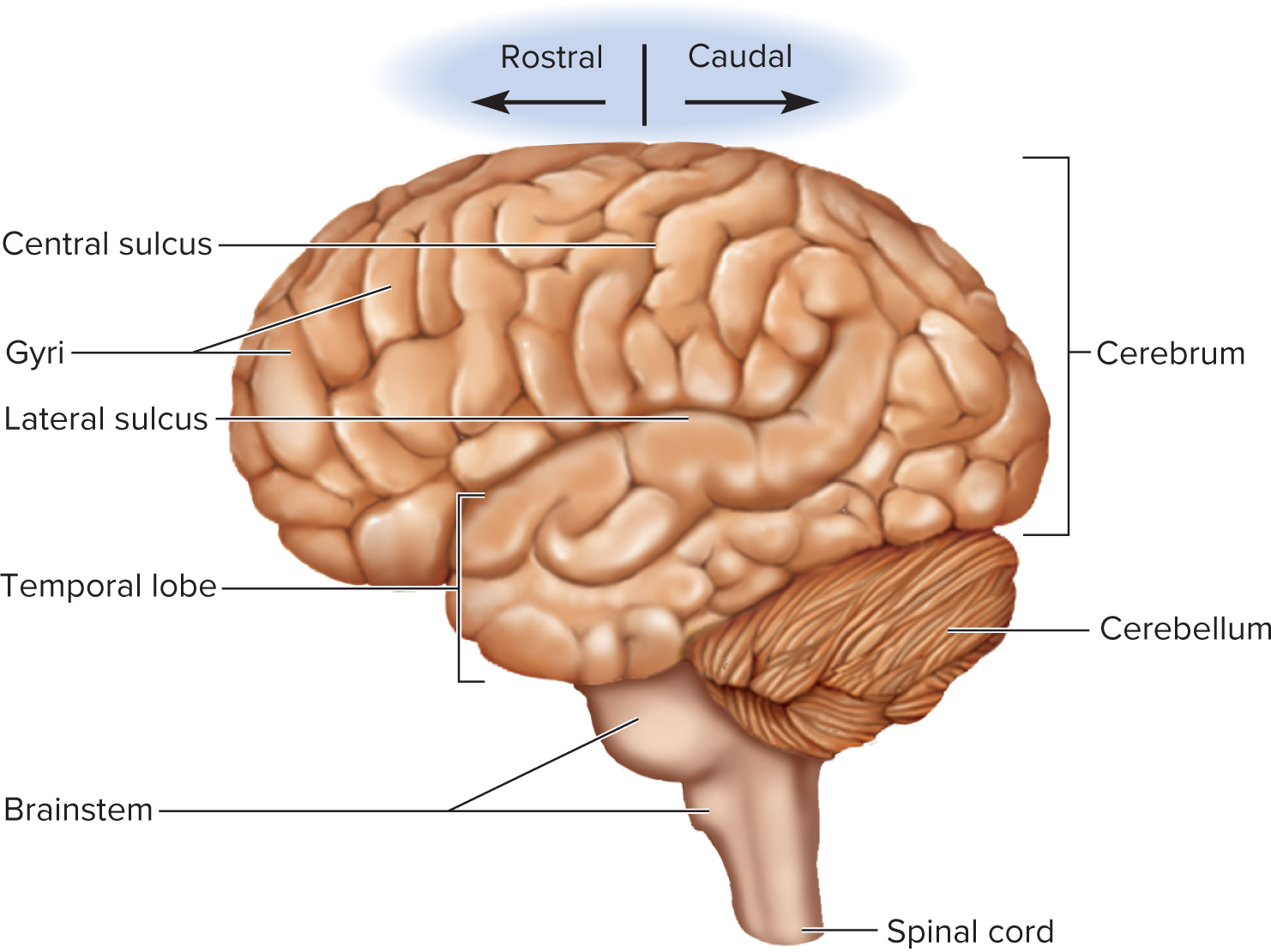
Directional Terms
Rostral — toward forehead
Caudal — toward spinal cord

Major Portions
Forebrain
Cerebellum
Brainstem
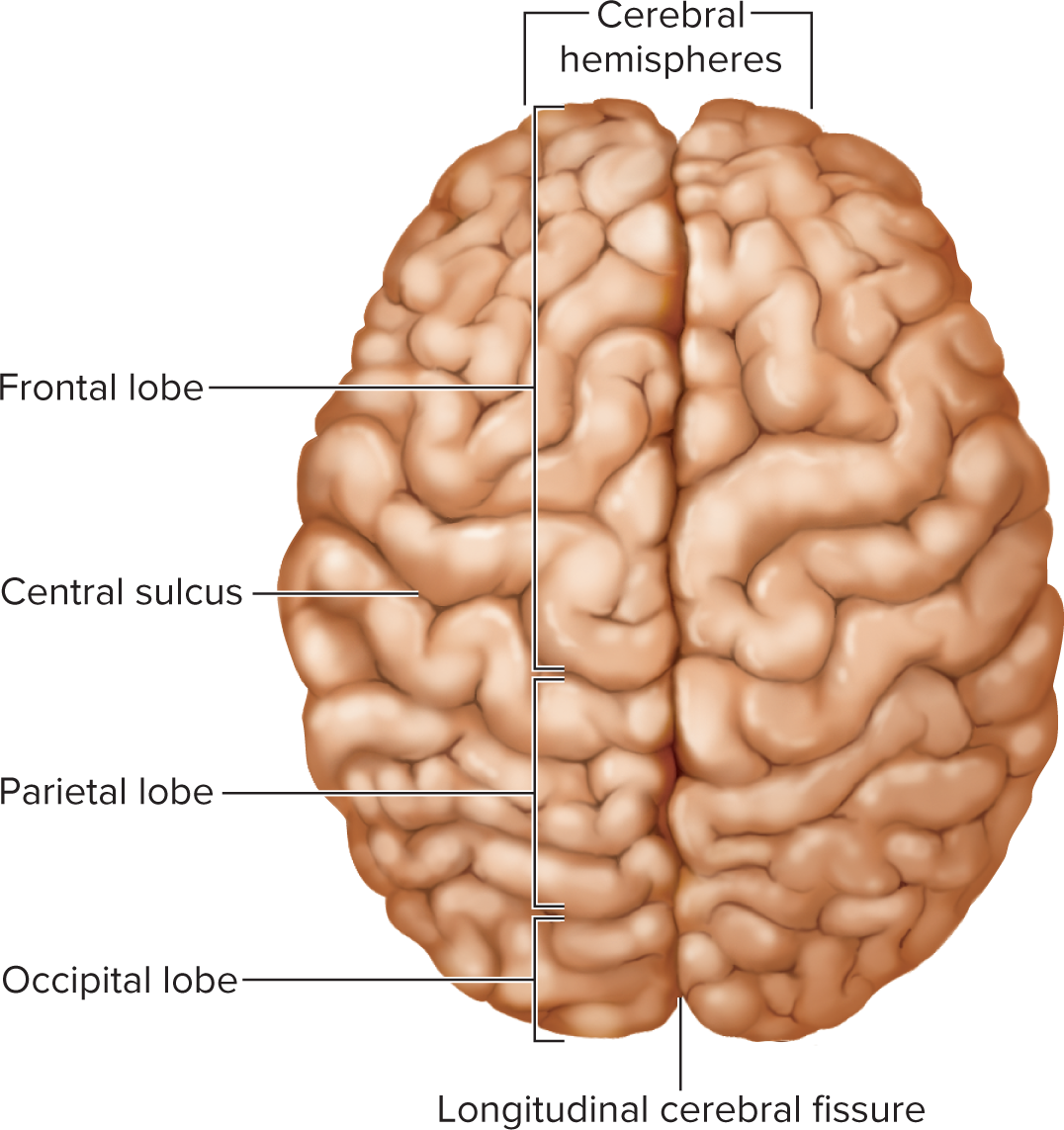
Cerebrum (largest part; 83\% of volume)
Cerebral hemispheres (pair of half-globes)
Gyri (thick folds) on cerebrum surface
Sulci (shallow grooves)
Longitudinal cerebral fissure (separates hemispheres)
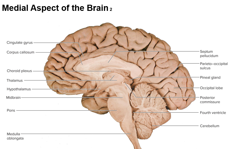
Corpus callosum (thick commissural bundle at fissure base)
Cerebellum (second-largest) 10% of volume
Sits in posterior cranial fossa
has half of the brains neurons with densely packed nerves, enabling precise coordination and control of movement, as well as the integration of sensory information.
Separated from cerebrum by transverse cerebral fissure
Surface folds = folia
Brainstem = remainder (midbrain, pons, medulla oblongata)
diencephalon inside the brain
includes the thalamus, epithalamus, hypothalamus
Midbrain: Located between the forebrain and hindbrain, it plays a crucial role in vision, hearing, and eye movement.
pons: The pons is a bridge connecting different parts of the nervous system, particularly the brain and spinal cord, and it is involved in regulating sleep, respiration, swallowing, and facial expressions.
medulla oblongata: The medulla oblongata is responsible for regulating vital functions such as heart rate, blood pressure, and respiration, making it essential for maintaining homeostasis.
Cranial Nerves: There are twelve pairs of cranial nerves that emerge directly from the brain and are primarily responsible for motor and sensory functions of the head and neck.
Midbrain: The midbrain, situated above the pons, plays a crucial role in processing visual and auditory information and helps in the coordination of motor functions.
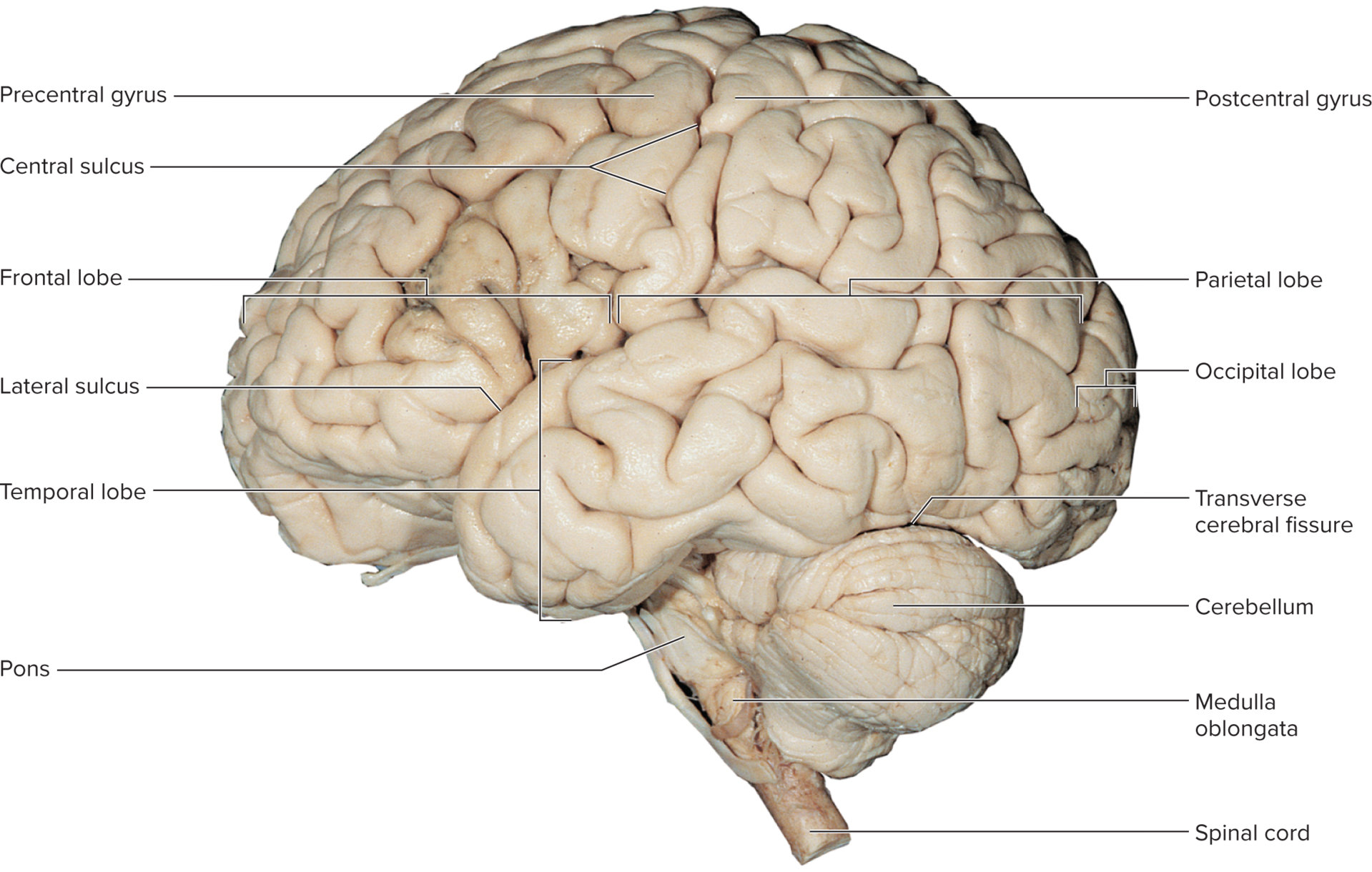
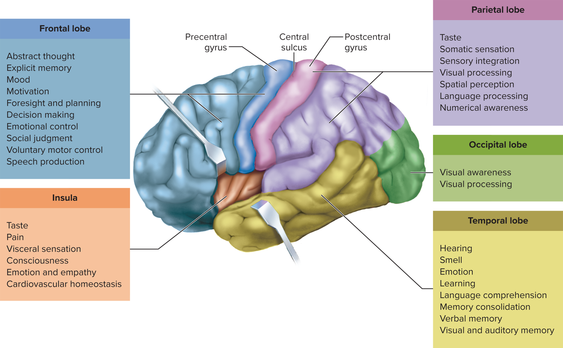
Central sulcus: The central sulcus is a prominent groove in the cerebral cortex that separates the frontal lobe from the parietal lobe and is critical in delineating areas responsible for motor and sensory functions.
Lateral sulcus: The lateral sulcus, also known as the Sylvian fissure, is a significant groove that separates the temporal lobe from the frontal and parietal lobes, playing an essential role in language processing and auditory perception.
precentral gyrus: (motor strip) is prominent fold of cortex located on the frontal lobe rostral to the central sulcus, and it is primarily involved in the planning and execution of voluntary movements.
The postcentral gyrus, located in the parietal lobe, is positioned directly posterior to the central sulcus and is crucial for processing somatosensory information, including temperature, pain, and touch sensations. Is coronal to central sulcus

Gray vs. White Matter
Gray Matter
Neuron cell bodies, dendrites, synapses
cortex (cerebrum & cerebellum)-Surface layers
nuclei-Deeper masses of gray matter, surrounded by white matter
White Matter
composed of tracts-bundles of nerve fibers (axons)
Myelinated axon tracts
Lies deep to cortical gray; connects brain regions & spinal cord
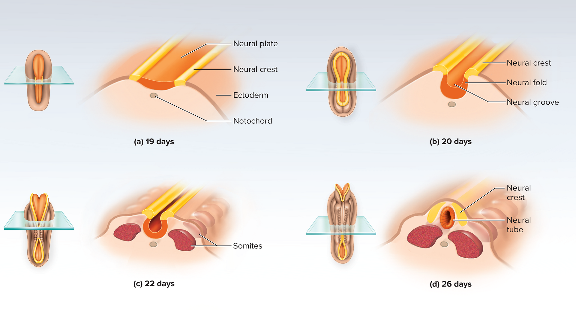
Embryonic Development of CNS
ectoderm: the outermost embryonic primary germ layer layer, crucial for developing the nervous system.
Neurulation (first 3 weeks)
Neural plate → neural groove with neural folds → fusion forms hollow neural tube by day 26
Tube lumen becomes future ventricles & central canal
Neural crest forms PNS, meninges (inner 2 layers), pigment cells, etc.
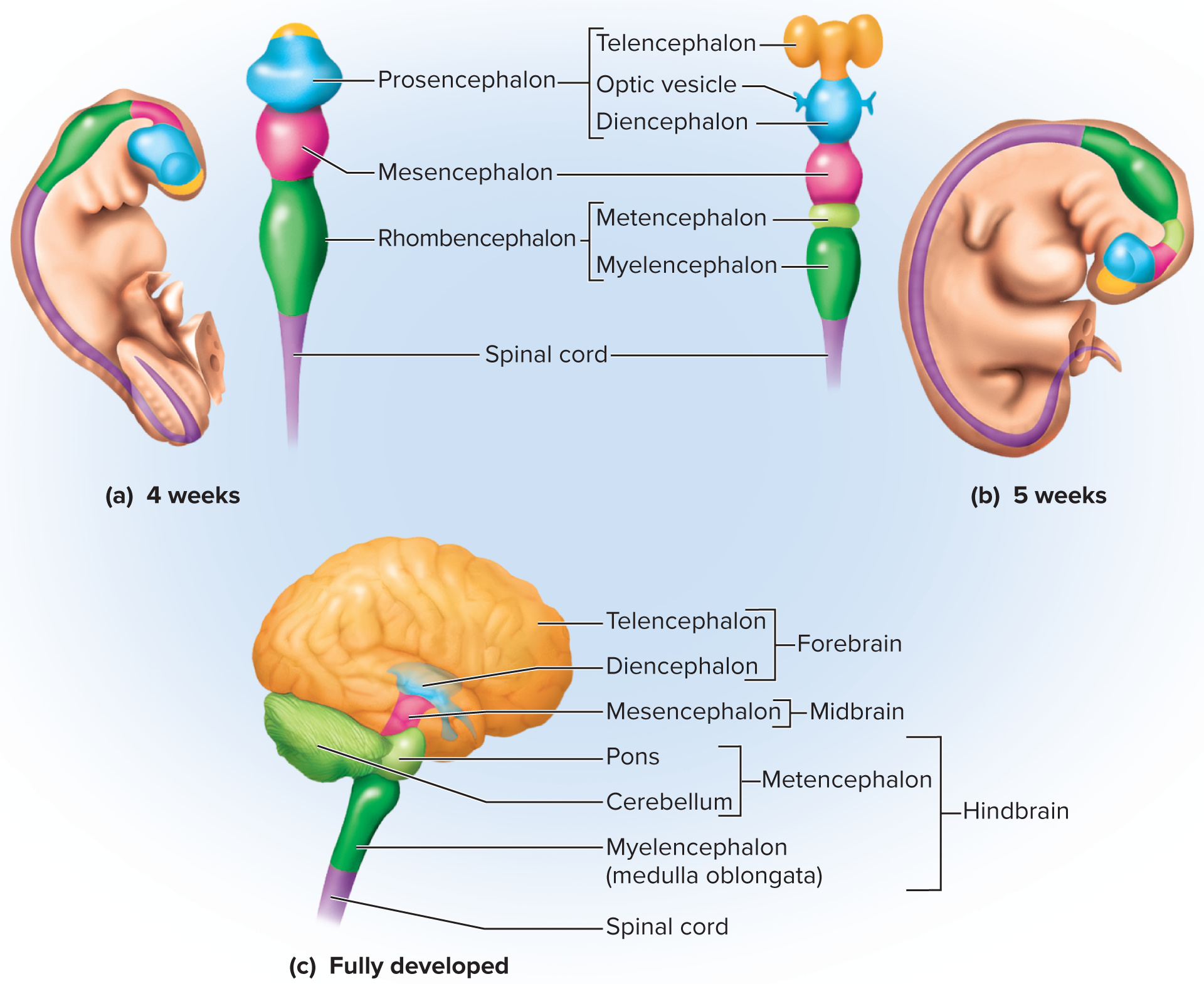
Primary brain vesicles (week 4):
Prosencephalon (forebrain)
Mesencephalon (midbrain)
Rhombencephalon (hindbrain)
Secondary vesicles (week 5):
Telencephalon & diencephalon (from forebrain)
Mesencephalon remains
Metencephalon & myelencephalon (from hindbrain)
14.2 Meninges, Ventricles, CSF & Blood Supply
Meninges
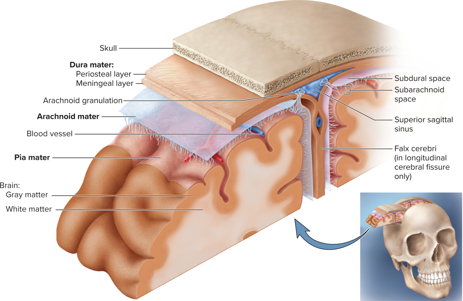
Three membranes (external → internal): dura mater, arachnoid mater, pia mater
Cranial dura mater
Periosteal layer (outer) = cranial periosteum
Meningeal layer (inner) → continues into vertebral canal and forms spinal dural sheath around spinal cord
Dural sinuses- spaces located between periosteal and meningeal layers separate
collect venous blood
Superior sagittal sinus (midline)-just under calvaria along median line
Drains blood from the lateral aspects of the cerebral hemispheres into the confluence of sinuses.
Transverse sinus (horiz. posterior)-runs horizontally rom rear of head toward each ear
Dural folds:
falx cerebri: separates the two cerebral hemispheres
tentorium cerebelli: separates cerebrum from cerebellum
falx cerebelli: separates right and left halves of cerebellum
Subdural space: in between dura and arachnoid
subarachnoid space: space between the arachnoid and pia mater filled with cerebrospinal fluid
Arachnoid mater
Transparent; subarachnoid space with CSF & vessels
Pia mater
Thin, follows gyri & vessels or arteries as they penetrate into cerebrum
Meningitis
Inflammation of meninges (usually pia & arachnoid) from viral/bacterial infection; peaks 3 mo – 2 yrs; diagnosed via lumbar puncture
Pia mater and arachnoid are most often affected
Can cause swelling of the brain, enlargement of the
ventricles, and hemorrhageMay progress to coma, then death within hours of onset
Diagnosed by examining CSF obtained by lumbar
puncture (spinal tap)
Ventricular System & CSF
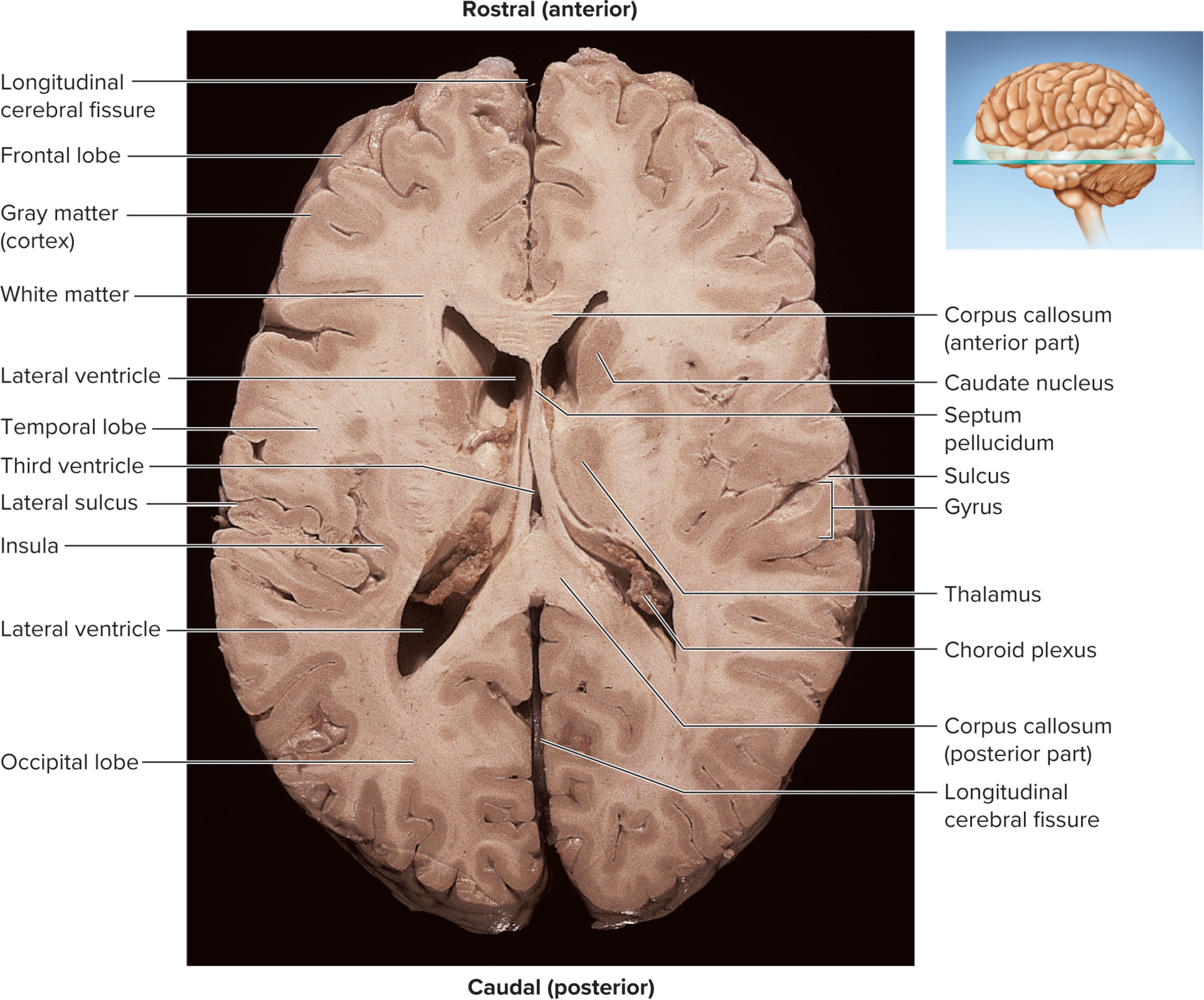
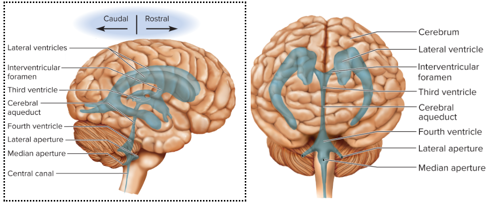
Brain has 4 CSF-filled spaces within the brain called ventricles, which include the two lateral ventricles, the third ventricle, and the fourth ventricle.
ventricles (internal CSF reservoirs)-four internal, fluid-filled chambers of brain
two Lateral ventricles (pair) connected by third ventricle
Third ventricle: narrow medial space beneath corpus callosum (midline, under corpus callosum)
Connected to lateral ventricle by interventricular foramen
Fourth ventricle: small triangular chamber between pons and cerebellum (between pons & cerebellum)
Connections: interventricular foramina → cerebral aqueduct → central canal
interventricular foramen—pore that connects lateral ventricles to third ventricle
Cerebral aqueduct—tube running through midbrain that connects third ventricle to fourth ventricle
Central canal—tube that connects to fourth ventricle and runs through center of spinal cord
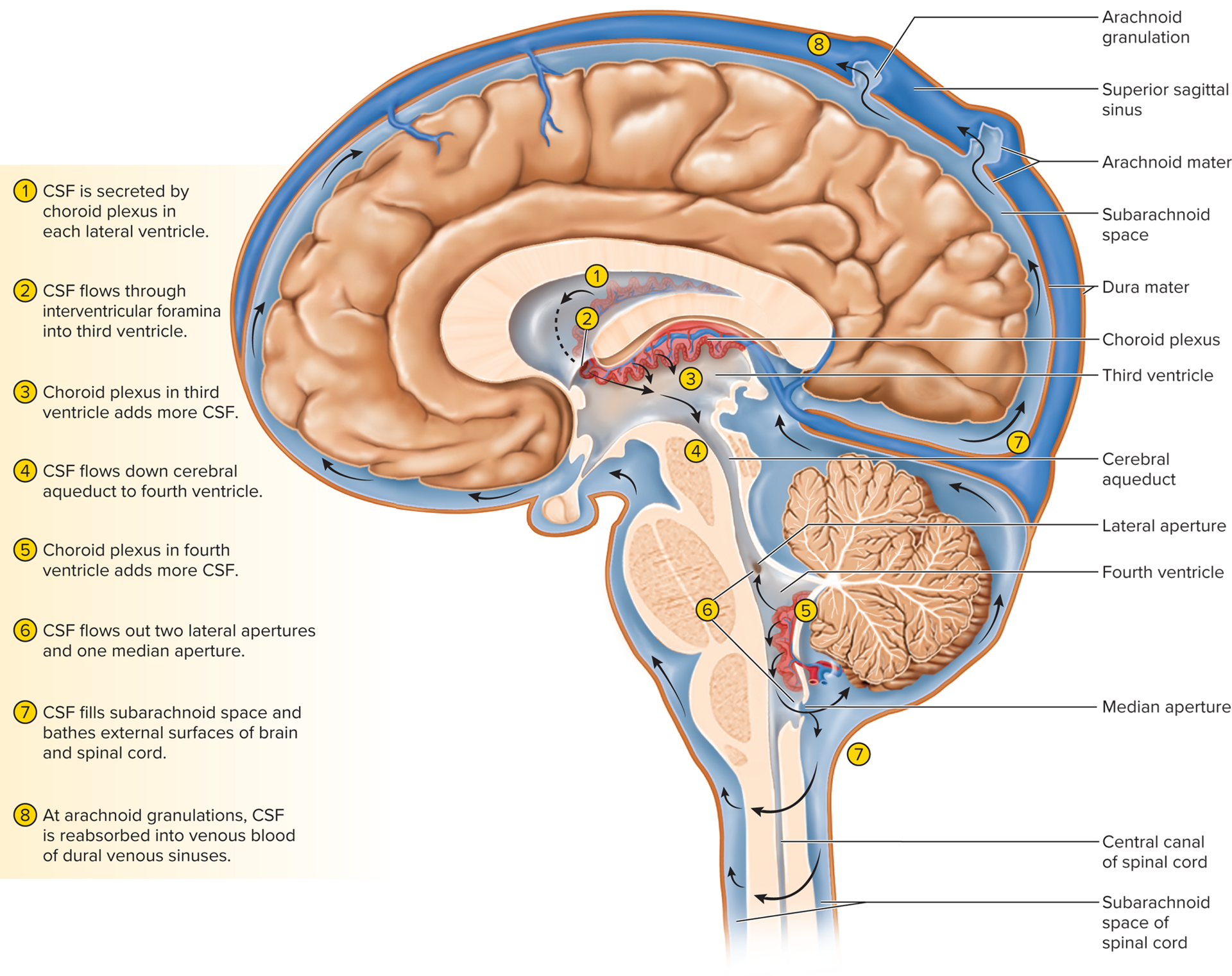
CSF Production
Cerebrospinal fluid (CSF) cushions, nourishes, and protects the CNS
the space between meninges (subarachnoid space) filled with CSF
Production of CSF begins with filtration of blood plasma
through capillaries of the brainChoroid plexus: spongy mass of blood of capillaries in each ventricle that secretes cerebrospinal fluid
Ependymal cells—neuroglia that line ventricles and cover
choroid plexusependymal cells modify the filtrate
Blood plasma filtered at choroid plexus; modified by ependymal cells
Composition: ↑Na^+, Cl^-; ↓K^+, Ca^{2+}, glucose, proteins
Compared to plasma, CSF has more sodium and chloride, less
potassium, calcium, glucose, and very little protein
Flow Path: CSF continuously flows & circulates through ventricles and meninges
Driven by its own pressure, beating of ependymal cilia,
and pulsations of the brain produced by each heartbeatsecreted in Lateral ventricles → through interventricular foramina → into third ventricle (adds CSF) → down cerebral aqueduct → into fourth ventricle (3rd and 4th adds more CSF along the way) → escapes through median & lateral apertures into the → subarachnoid space and spinal cord surface → CSF is reabsorbed by arachnoid granulations → protrude through superior sagittal sinus→ CSF penetrates the walls of the villi and mixes with the blood in the sinus→ allowing for the maintenance of homeostasis within the central nervous system.
All CSF ultimately escapes through three pores that lead
into subarachnoid space of brain and spinal cord surfacemedian aperture
two lateral apetures
CSF is reabsorbed by arachnoid granulations
Cauliflower-shaped extensions of the arachnoid meninx
Protrude through dura mater into superior sagittal sinus
CSF penetrates the walls of the villi and mixes with the blood in the sinus
arachnoid villus reabsorbs CSF into venus blood returned to heart and reestablish pH of blood plasma
Functions
Buoyancy allows brain to be large without being impaired by its size (brain mass ~1.5\,\text{kg} becomes effectively 50\,\text{g})
Protection from blows (though concussion still possible)
Chemical stability (waste removal, pH control)
Blood Supply & Brain Barrier System (BBS)
Brain only contributes = 2\% body weight but receives 15\% of blood cardiac output (≈750\,\text{mL/min})
Neurons have high demand for ATP (therefore oxygen and
glucose) so constant supply of blood critical10\,\text{s} interruption = loss → unconsciousness; 1{-}2\,\text{min} → significant impairment of neural function; 4\,\text{min} → irreversible damage
Barrier Components
Brain barrier system (BBS)—regulates what substances can get from bloodstream into tissue fluid of the brain
Although blood is crucial, it can also contain harmful
agentsBlood-brain barrier (BBB): a selective permeability barrier that prevents certain substances from passing into the brain while allowing essential nutrients to enter.
Highly permeable to water, glucose, and lipid-soluble substances
such as oxygen, carbon dioxide, alcohol, caffeine, nicotine,
anesthetics
• Slightly permeable to sodium, potassium, chloride, waste products
urea and creatinineTwo points of entry must be guarded:
Blood capillaries throughout the brain tissue; guarded by the blood-
brain barrierCapillaries of the choroid plexus; guarded by the blood-CSF barrier
Blood–brain barrier (BBB): protects the brain at the blood
capillaries, consists of tight junctions in cerebral capillaries induced by astrocyte perivascular feetInduce endothelial cells to form impenetrable tight junctions that prevent the passage of potentially harmful substances from the bloodstream into the brain, thereby maintaining the brain's chemical stability.
Blood–CSF barrier: protects the brain at the choroid plexus tight junctions between ependymal cells of choroid plexus
Selective permeability: passes H2O, glucose, O2, CO_2, lipids; limits ions, wastes, pathogens
Circumventricular organs (CVOs)—BBB absent (parts of 3rd/4th ventricles) for homeostatic sensing but allow pathogen entry (e.g., HIV)
Enable the brain to monitor and respond to fluctuations in
blood glucose, pH, osmolarity, and other variables
• CVOs afford a route for invasion by the human immunodeficiency virus (HIV)
Clinical notes
Stroke (CVA): sudden ischemia/hemorrhage; second-leading cause of death; \approx50\% die <1 yr
Hemorrhagic stroke—rupture of a cerebral or subarachnoid blood
vessel
• Ischemic stroke—obstruction of a blood vessel by blood clot or lipid deposit
The Brainstem:
brainstem connects brain to spinal cord, also connecting to cerebrum, thalamus, and cerebellum
3 regions of brainstem
Midbrain
pons
medulla oblongata (is continuous with the spinal cord)
involved in unconscious reflexes and regulations of homeostasis
helps regulate:
blood pressure
heart rate
breathing (pons)
swallowing
coughing/sneezing
relays sensory impulses from spinal cord to thalamus


14.3 Hindbrain & Midbrain
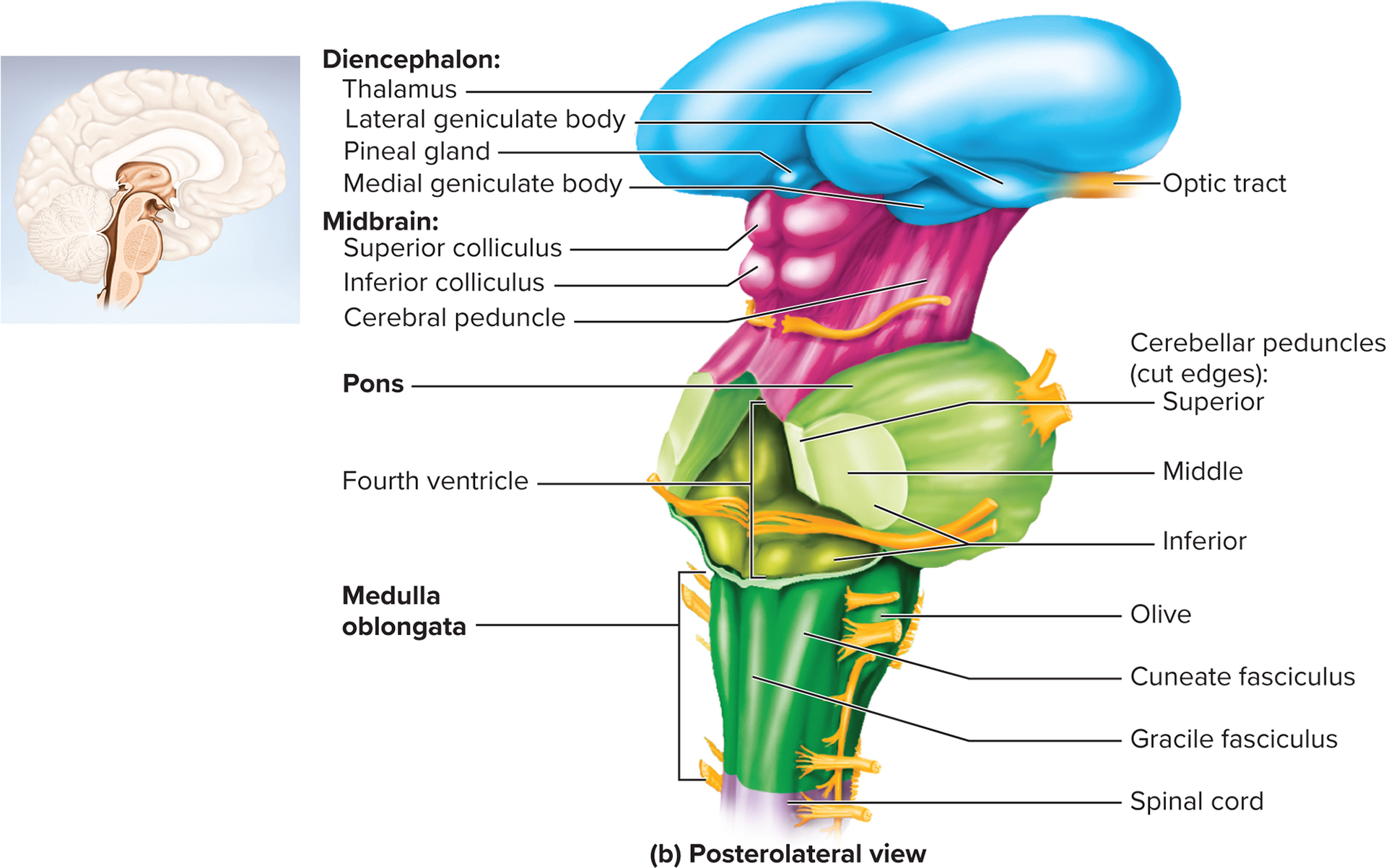
Medulla Oblongata (from myelencephalon)
adult brain region that develops from embryonic myelencephalon
Begins at foramen magnum of skull
• Extends about 3 cm rostrally and ends at a groove just below pons
• Slightly wider than spinal cordLandmarks: pyramids (corticospinal tracts), olives, gracile & cuneate fasciculi
Pyramids—ridges on anterior surface, resemble side-by-
side baseball bats separated by anterior median fissure
four pairs of Cranial nerves begin or end in medulla: VIII (part), IX, X, XII originate/terminate here
Olives—prominent bulges lateral to each pyramid
Gracile and cuneate fasciculi of spinal cord continue as
two pairs of ridges on posterior medulla
Contain sensory fibers; synapse in gracile and cuneate nuclei
Major tracts: medial lemniscus (sensory decussation), corticospinal (motor)
Medial lemniscus—axons of gracile and cuneate nuclei decussate and form ascending (sensory) tract to thalamus
Corticospinal tracts—descending motor tracts in pyramids; carry signals down to skeletal muscles
contains numerous Nuclei: cardiac center, vasomotor center, respiratory centers, inferior olivary
Inferior olivary nucleus—relay center for signals to cerebellum
Reticular formation—loose network of nuclei extending through
entire brainstem; contains cardiac center, vasomotor center, and respiratory centers
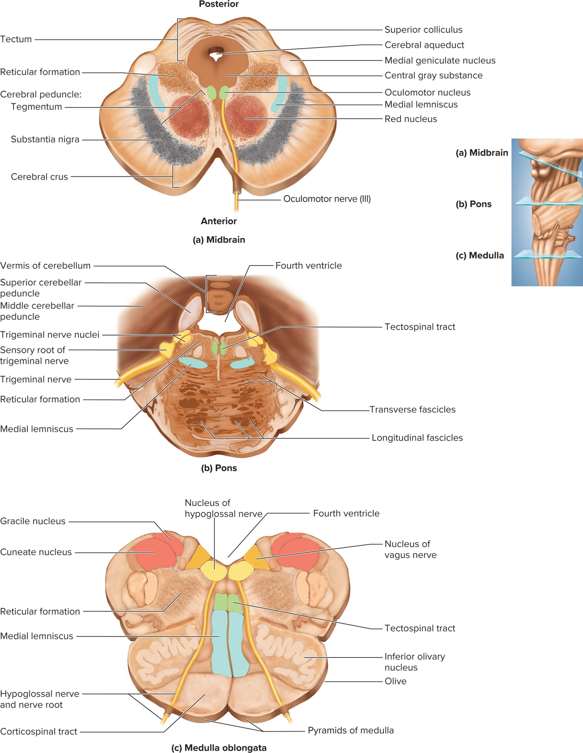
Pons (from metencephalon)
adult brain region that develops from embryonic metencephalon
Anterior bulge 2.5\,\text{cm} long; posterior cerebellar peduncles
Cerebellar peduncles—thick stalks on posterior pons that
connect it (and the midbrain) to the cerebellumCranial nerves V, VI, VII, VIII
Sensory: hearing, equilibrium, taste, facial sensation
Motor: eye & facial movement, chewing, tears, saliva, urination
Reticular formation in pons contains additional nuclei: sleep, respiration, posture
Midbrain (mesencephalon)
brain region that develops from embryonic mesencephalon
Short segment of brainstem that connects hindbrain to
forebrain
Contains cerebral aqueduct, periaqueductal gray substance involved in pain modulation
Motor nuclei of two cranial nerves that control eye
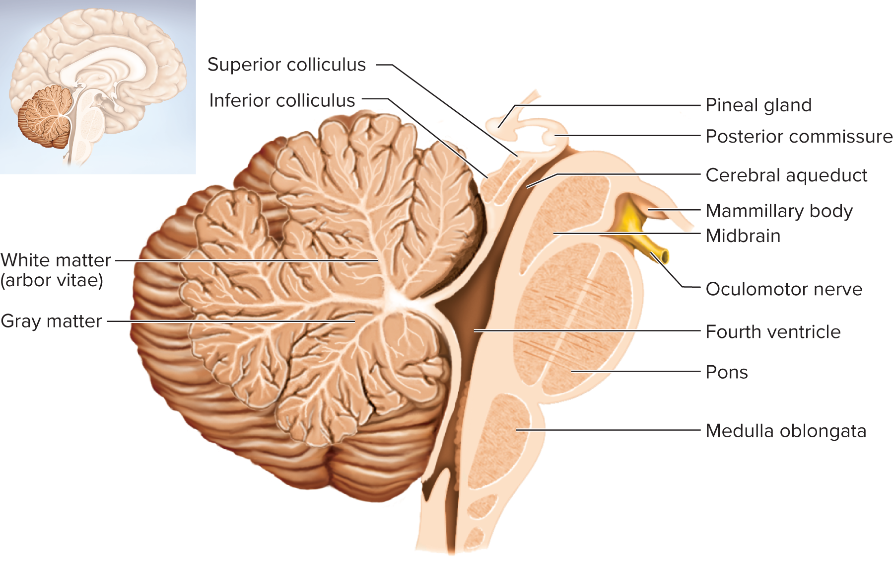
movements: CN III (oculomotor) and CN IV (trochlear)Tectum (roof): four bulges, 2 superior colliculi (visual reflexes), 2 inferior colliculi (auditory relay)
Two superior colliculi—visual attention, tracking moving objects,
and some reflexesTwo inferior colliculi—relays signals from inner ear to thalamus
and other parts of the brain
Cerebral peduncles
are two anterior midbrain stalks that anchor the cerebrum to the brainstem
Each peduncle has three parts: tegmentum, substantia nigra, and cerebral crus
Tegmentum-within cerebral peduncle (dominated by red nucleus → motor relay to cerebellum)
Substantia nigra (dopaminergic inhibitory motor; degeneration → Parkinson)
Nucleus within peduncle; dark nucleus pigmented with melanin
• Motor center that relays inhibitory signals to thalamus and basal
nuclei suppressing unwanted body movement
• Degeneration of neurons leads to tremors of Parkinson’s disease
Cerebral crus (descending corticospinal fibers)
Bundle of nerve fibers that connect cerebrum to pons
Carries corticospinal tracts
Reticular Formation
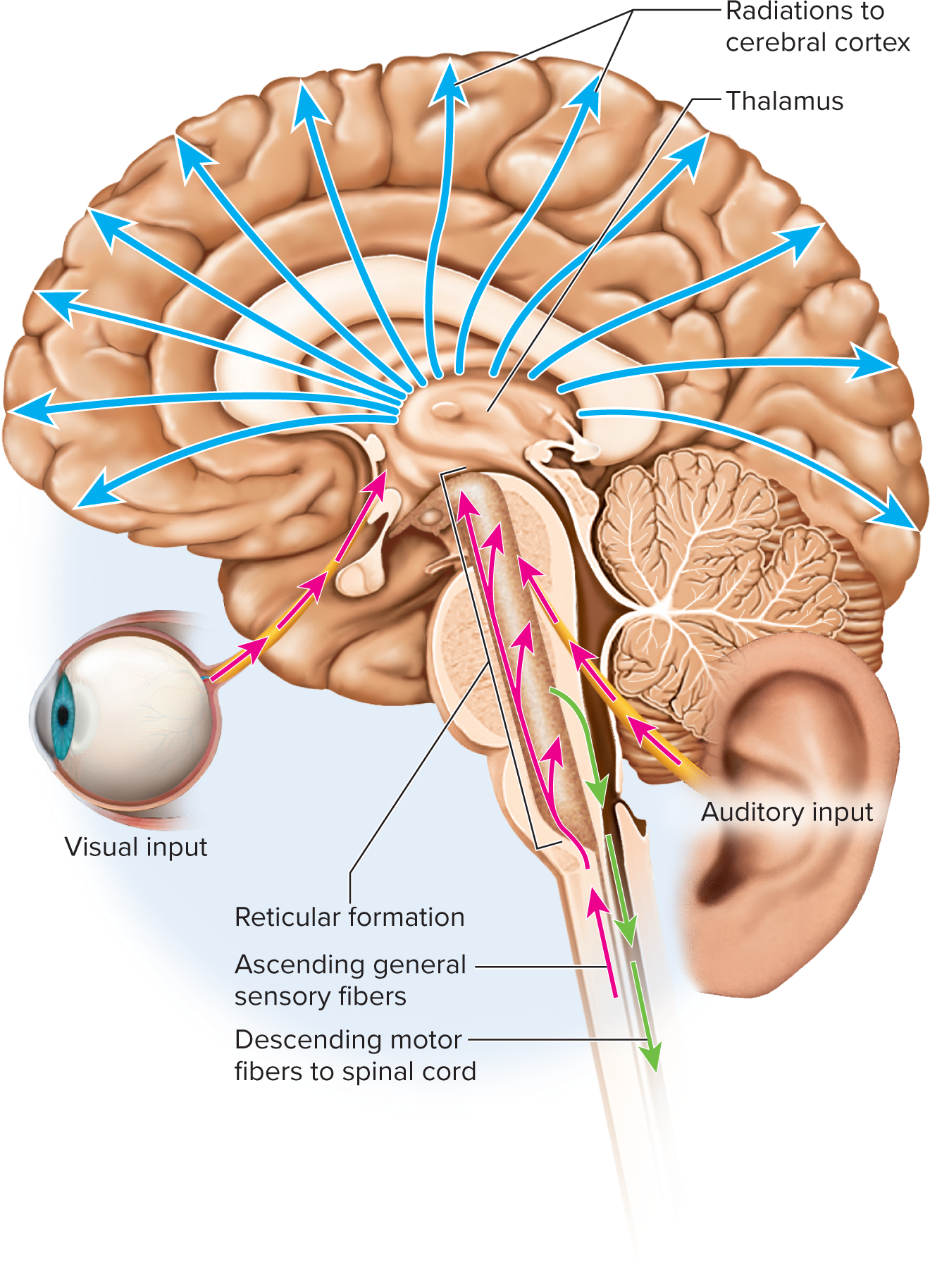
network Web of gray matter through brainstem into spinal cord; >100 nuclei
allows information from eyes and ears to be integrated by rest of brain
sends sensory info to the thalamus, which then relays it to appropriate cortical areas for processing and interpretation.
mediates overall consciousness
Functions:
Somatic motor control (tone, posture, gaze centers, CPGs for breathing/swallow)
Cardiovascular control
Pain modulation (descending analgesic pathways)
Sleep & consciousness (injury → coma)
Habituation/ pa (filters repetitive stimuli)
Cerebellum (metencephalon)
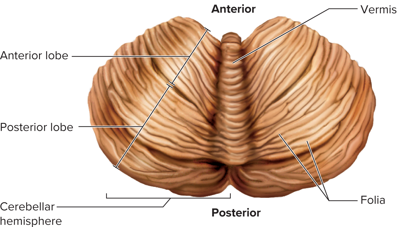
controls balance, posture, and coordination of muscles
>50\% of neurons (granule cells)
Anatomy: hemispheres connected by vermis; cortex (folia), arbor vitae, deep nuclei
Peduncles: inferior (input from medulla/spinal cord), middle (input from cerebrum via pons), superior (output to midbrain/thalamus)
Functions: motor coordination, locomotion, timekeeping, texture, spatial perception, eye movement, language, emotion, planning
14.4 Forebrain
Diencephalon
Connects cerebrum to the rest of the brain: lies between the brainstem and cerebrum
Consists of 3 major structures
Thalamus: Acts as the main relay station for sensory information before it reaches the cerebrum.
Hypothalamus: Regulates vital functions such as temperature, hunger, and thirst, and maintains homeostasis.
Epithalamus: Includes the pineal gland, which secretes melatonin and is involved in regulating the sleep-wake cycle.
Thalamus
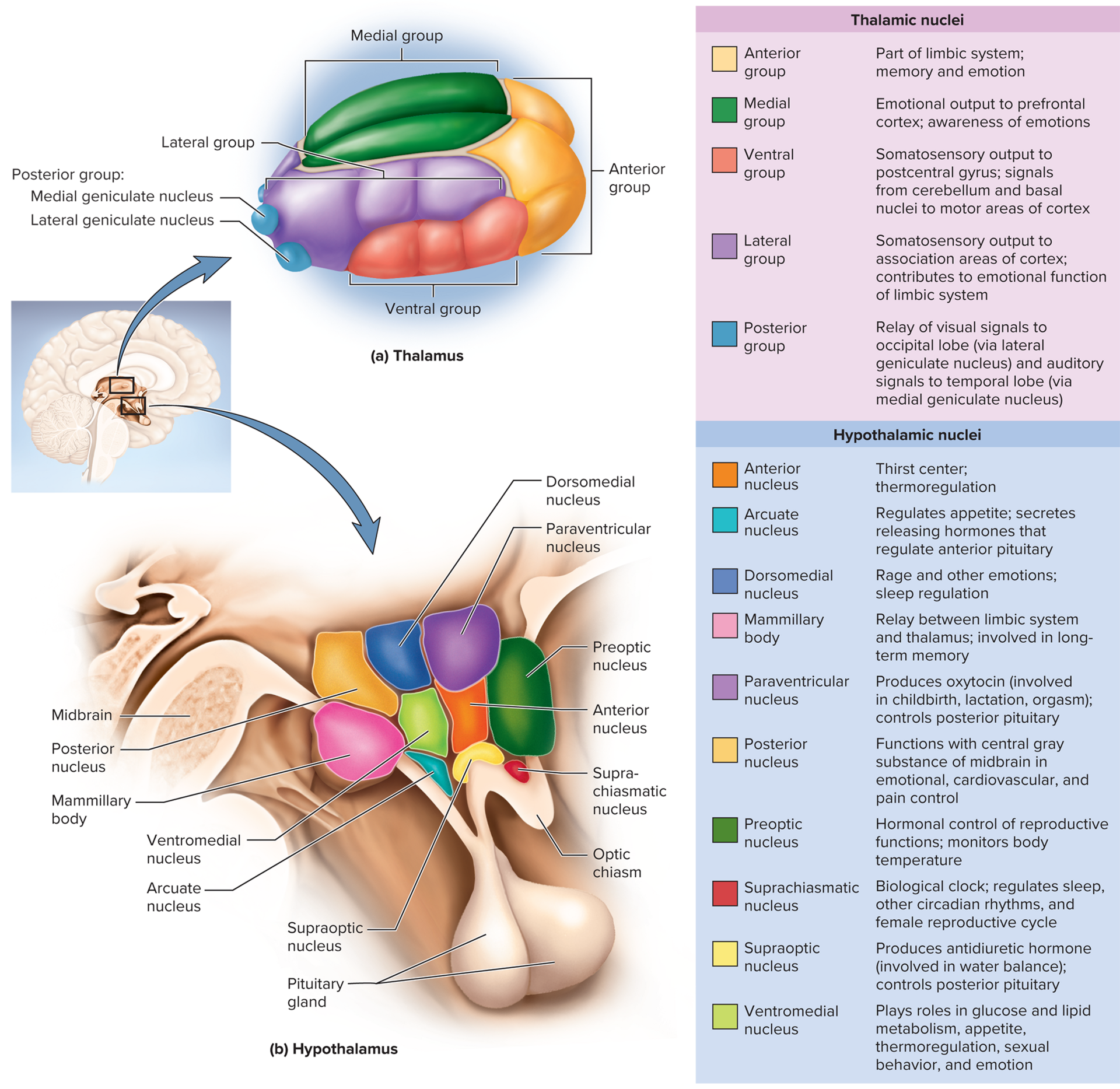
Pair of ovoid masses (~80\% of diencephalon) joined by interthalamic adhesion
\ge23 nuclei (anterior, medial, lateral, ventral, posterior groups)
Gateway to cortex: filters, relays sensory/motor information; role in memory & emotion via limbic links
sensory information: arrives at the thalamus and is relayed to the cerebral cortex
relays motor info between brain regions
involved in memory formation and emotional responses
Hypothalamus
the “unconscious bus driver” that helps regulate autonomic functions such as hunger, thirst, and body temperature, while also influencing emotional behavior and stress responses.
also major integration center for ANS and endocrine system, playing a crucial role in maintaining homeostasis by coordinating the activities of both systems to respond to internal and external stimuli.
Forms walls/floor of third ventricle; extends from optic chiasm to mammillary bodies; attached to pituitary via infundibulum
Nuclei control:
Hormone secretion (anterior & posterior pituitary control)
Autonomic regulation (HR, BP, GI activity)
Thermoregulation (preoptic area)
Hunger & satiety (arcuate, ventromedial)
Water balance & thirst (osmoreceptors; supraoptic \rightarrow ADH)
Sleep/circadian (suprachiasmatic)
Memory relay (mammillary)
Emotion & sexual response
Epithalamus
Regulation of circadian rhythms (pineal gland)
induces sleepiness, promotes repair functions during sleep
may assist in regulating onset of puberty
Pineal gland (melatonin), habenula (limbic relay), thin roof of 3rd ventricle
Cerebrum- the largest part of the brain responsible for voluntary activities, sensory perception, and cognitive processes, including reasoning and problem-solving.
conscious sensory awareness, control of movement, memory and learning, language and speech, emotional responses, intellectual processes
Two hemispheres right and left; connected by corpus callosum, separated by longitudinal fissure
surface shows numerous gyri and sulci↑ 3x surface area to fit in cranial cavity
has sensory areas, motor area, and association areas
sensory area interpret nerve impulses as sensations
primary motor area control voluntary skeletal muscles
association area interrelate sensory and motor areas 75% of brain, responsible for cognition, memoryand higher-level functions such as reasoning and problem-solving, playing a crucial role in integrating and processing complex information.
Lobes & Functions
Frontal: most rostral part voluntary motor, planning, mood, aggression
Parietal: most upper part, after central sulcus, general senses, taste, spatial perception
Occipital: lateral, vision
Temporal: hearing, smell, memory, emotion
Insula: only seen if temporal lobe is cut away language, taste, visceral integration
White Matter Tracts
Projection (vertical cortex ↔ lower centers)
Commissural (corpus callosum, ant/post commissures)
Association (intrahemispheric; long & short)
Cerebral Cortex
2{-}3\,\text{mm} thick; \approx40\% brain mass
Cell types: stellate (local), pyramidal (output)
Neocortex = 90\%, 6 layers (I–VI)
Limbic System (emotion & learning)
Cingulate gyrus, hippocampus, amygdala; interconnected loops; reward/aversion centers
Basal Nuclei (motor control)
Caudate, putamen, globus pallidus
Corpus striatum (caudate + putamen + globus)
Lentiform nucleus (putamen + globus)
14.5 Integrative Functions
Brain Waves (EEG)
Alpha 8{-}13\,\text{Hz} — awake, eyes closed
Beta 14{-}30\,\text{Hz} — mental activity
Theta 4{-}7\,\text{Hz} — drowsy/sleeping adults
Delta <3.5\,\text{Hz} — deep sleep
Sleep
Circadian rhythm (~24 h); stages identifiable on EEG
Stage 1: alpha→mixed
Stage 2: light; sleep spindles
Stage 3: theta + delta; vitals drop
Stage 4: deep; vitals lowest
REM: backtrack to Stage 2; paradoxical (EEG awake-like), dreaming, atonia, penile/clitoral erection
Regulation: SCN (master clock) via orexins, DMN, pineal melatonin; reticular formation; sleep deprivation fatal in animals
Cognition
Distributed association areas (~75\% cortex)
Lesions: parietal → contralateral neglect; temporal → agnosias; frontal → personality change
Memory
Hippocampus: consolidation; teaches cortex long-term memories
Cerebellum: motor skill learning
Amygdala: emotional memory
Amnesia: anterograde (no new), retrograde (no old)
Emotion
Prefrontal cortex: judgement & expression
Amygdala: fear, food, sex, attention; outputs to hypothalamus (autonomic) & prefrontal cortex (behavior)
Sensation
Primary sensory cortices receive; association areas interpret; multimodal areas integrate
Special Senses:
Vision: occipital lobe
Hearing: superior temporal
Equilibrium: cerebellum, brainstem, parietal roof of lat. sulcus
Taste: postcentral gyrus inferior
Smell: medial temporal
General Senses: postcentral gyrus (primary somatosensory); sensory homunculus & somatotopy
Motor Control
Premotor (association) → primary motor cortex (precentral gyrus) → corticospinal tracts (upper & lower motor neurons)
Motor homunculus proportional to motor units, not size
Basal nuclei: movement initiation, patterned behaviors; lesions → dyskinesias
Cerebellum: coordination, posture, learning; lesions → ataxia
Language
Wernicke area: comprehension, formulation of phrases
Broca area: motor program for speech/signing → primary motor cortex
Right hemisphere equivalents handle emotional prosody
Aphasia types: nonfluent (Broca), fluent (Wernicke), etc.
Cerebral Lateralization
Left (categorical): language, analytical, sequential
Right (representational): spatial, holistic, music, pattern
Correlates: 96\% of right-handers left-categorical; lateralization ↑ with age; males > females
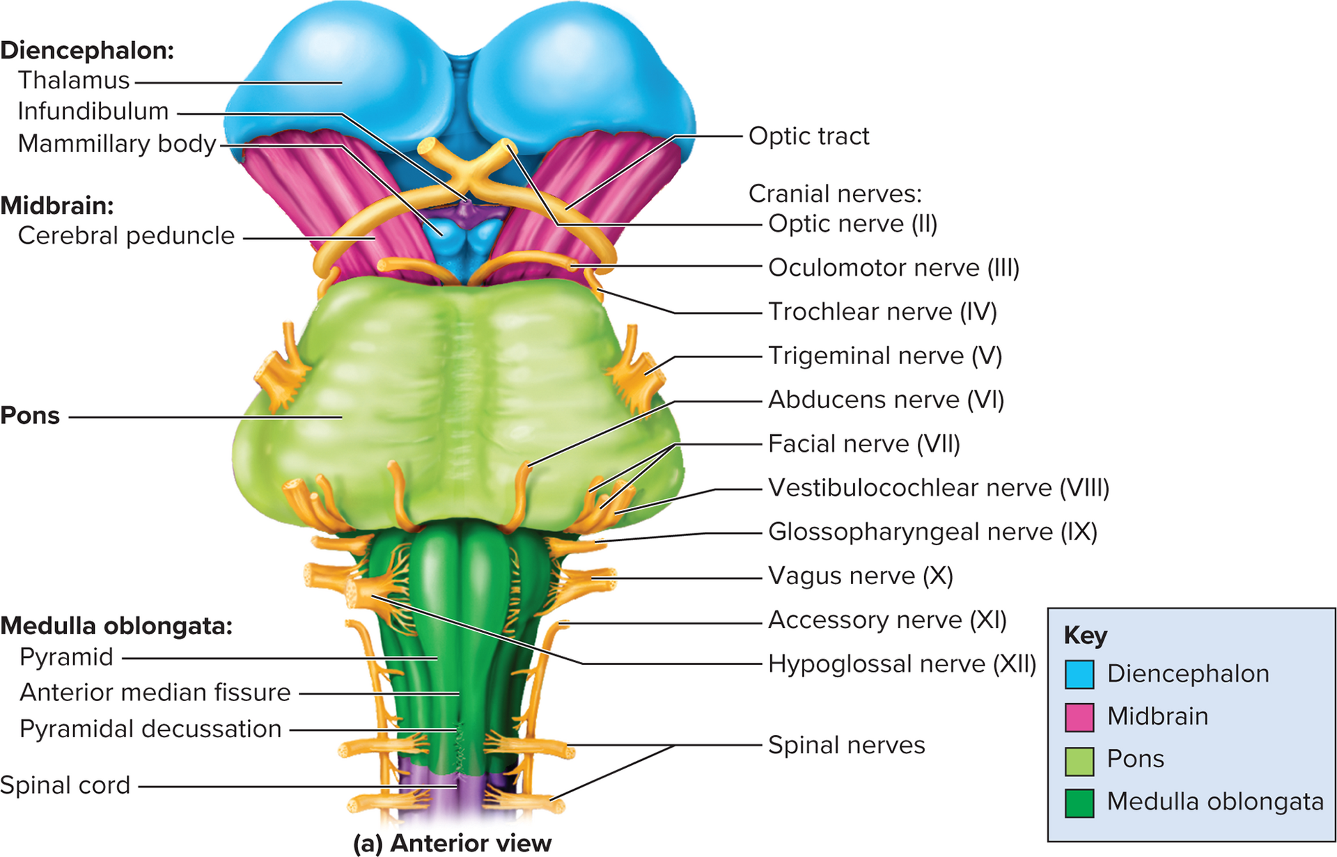
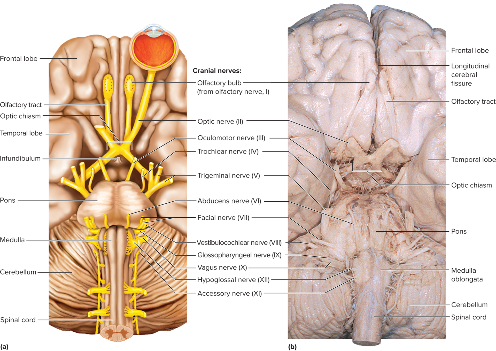
14.6 Cranial Nerves
General Features
12 pairs so 24 total; emerge from brain base via foramina; mostly ipsilateral (exceptions: optic partial decussation; trochlear complete)
Classified: sensory (I, II, VIII), motor (III, IV, VI, XI, XII), mixed (V, VII, IX, X)
most motor fibers of cranial nerves begin in nuclei of the brainstem and lead to the glands and muscles.
sensory fibers: begin in receptors located mainly receptors in the head and neck and lead mainly to the brainstem
sensory fibers for propriception begin in the muscles innervated by motor fibers of the cranial nerves but often travel to the brain in a different nerve from the one that supplies the motor innervation
Most nerves carry fibers between brainstem and ipsilateral receptors and effectors
Sensory nerves CN I and II: carry signals only from outlying sense organs to the brain
Motor nerves CN III, IV, VI, XI, XII carry signals only from the brainstem to outlying muscles and glands
Mixed nerves V, VII, VIII, IX, X carry signals both ways
have sensory functions quite unrelated to their motor functions like facial nerve CN VII, has a sensory role in tatse and a motor role in facial expression
Plexus nerves, such as the brachial plexus, are networks of interwoven nerves from multiple spinal roots that provide motor and sensory innervation to the limbs.
# | Name | Type | Major Functions |
|---|---|---|---|
I | Olfactory | Sensory | Smell via cribriform plate fascicles |
II | Optic | Sensory | Vision from retina |
III | Oculomotor | Motor | Eye movement (4 muscles), lens, pupil |
IV | Trochlear | Motor | Superior oblique muscle (eye) |
V | Trigeminal | Mixed | Face sensation (V1 ophthalmic, V2 maxillary), mastication (V3 mandibular) |
VI | Abducens | Motor | Lateral rectus muscle (eye) |
VII | Facial | Mixed | Facial expression, tears, saliva; taste ant 2/3 tongue |
VIII | Vestibulocochlear | Mostly Sensory | Hearing, equilibrium; motor tune cochlea |
IX | Glossopharyngeal | Mixed | Taste post 1/3 tongue, BP/resp reflexes; salivation, swallow, gag |
X | Vagus | Mixed | Parasympathetic to thoracoabdominal viscera; taste, hunger; speech |
XI | Accessory | Motor | Head, neck, shoulder movement; swallowing |
XII | Hypoglossal | Motor | Tongue movement for speech, food, swallow |
Disorders
Trigeminal neuralgia (tic douloureux): stabbing facial pain (CN V)
Bell palsy: facial paralysis (CN VII)
Imaging Techniques
PET: inject radiolabeled glucose; active areas “light up”
fMRI: measures blood oxygen–dependent signal; maps functional activity