Heart Anatomy
Heart Anatomy
Overview
Anterior Surface: Front of the heart.
Posterior Surface: Back of the heart.
Inferior Surface: Apex (bottom) of the heart.
Superior Surface: Base (pointing towards the right shoulder).
Anterior Structures
Auricles: Fatty pouches on the atria.
Pulmonary Trunk:
Takes deoxygenated blood from the right ventricle to the lungs for oxygenation.
Appears blue in models as it carries deoxygenated blood.
Pulmonary Arteries: Left & Right Pulmonary Artery
Aorta: The Largest Artery coming out of the heart / LV
Aortic Root: Normal Size 2-4 cm/ Connects to the aorta and contains the aortic valve, which opens to allow blood flow from the left ventricle into the aorta.
Ascending Aorta: The Initial part of the aorta.
Aortic Arch: The curved part of the aorta.
Descending Aorta: Thoracic Aorta & Abdominal Aorta
Branches off the Aortic Arch (in order):
Brachiocephalic Artery: Splits into the right subclavian and right common carotid arteries.
Left Common Carotid Artery
Left Subclavian Artery
Right Side Structures
Right Atrium:
Superior Vena Cava: Dumps blood into the right atrium / receives deoxygenated blood from the upper body, including the head, neck, and arms
3 Suppliers of Blood to SVC @ Right Atrium:
Brachiocephalic
Azygos
Coronary Sinus
Azygos Vein: Small veins feeding into the superior vena cava from the posterior side.
Pulmonary Arteries:
Right Pulmonary Artery: Takes deoxygenated blood to the right lung.
Left Pulmonary Artery: Takes deoxygenated blood to the left lung.
Pulmonary Veins:
Carry oxygenated blood back from the lungs to the heart (left atrium).
Left Pulmonary Veins
Right Pulmonary Veins
Coronary Bloodflow
Coronary Arteries:
First branch off the ascending aorta.
Right Coronary Artery
Left Coronary Artery = Left Main
Anterior Aspect
Left Coronary Artery Branches:
Anterior Interventricular Artery:
Also known as the left anterior descending (LAD) artery.
Supplies the anterior walls of both ventricles and the anterior interventricular septum.
Diagonal Arteries Branch off (D1 & D2)
Circumflex Artery:
Supplies the Lateral wall of the heart with blood
Obtuse Marginals Branch off (OM 1 & OM2)
Veins:
Great Cardiac Vein:
Runs alongside the anterior interventricular artery.
Drains blood from the area supplied by the LAD.
Coronary Sinus: Collects deoxygenated venous blood from the heart muscle and drains into the right atrium.
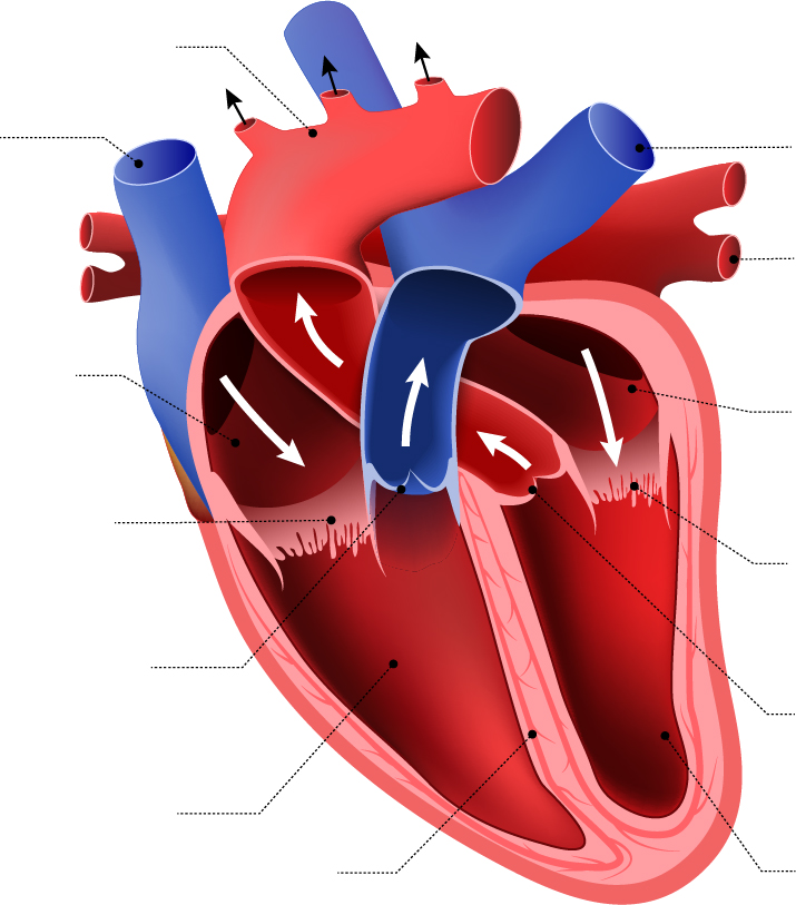
Internal Heart Structures
Atria and Ventricles
Right Atrium
Left Atrium
Right Ventricle
Left Ventricle
Valves → Semilunar & Atrioventricular Valves
ALL valves are held in place by the Chordae tendinae, and are anchored to Myocardium with Papillary Muscles
Tricuspid Valve:
Separates the right atrium from the right ventricle.
Has chordae tendineae (collagen cords) attached to papillary muscles.
Pulmonary Semilunar Valve:
Prevents backflow from the pulmonary trunk into the right ventricle.
Mitral Valve (Bicuspid Valve):
Separates the left atrium from the left ventricle.
Has chordae tendineae attached to papillary muscles.
Aortic Semilunar Valve:
Prevents backflow from the aorta into the left ventricle.
Other Internal Structures
Fossa Ovalis:
Scar tissue in the right atrium.
Remnant of the foramen ovale in fetal circulation, which shunted blood from the right atrium to the left atrium to bypass the pulmonary circuit.
Opening of the Coronary Sinus: Drains deoxygenated blood from the heart into the right atrium.
Ligamentum Arteriosum:
Remnant of the ductus arteriosus in fetal circulation, which shunted blood from the pulmonary trunk to the aorta.
Interventricular Septum:
Separates the two ventricles.
Defects can occur here (e.g., tetralogy of Fallot).
Myocardium:
Muscular layer of the heart.
Made up of cardiac muscle fibers
Endocardium:
Lines the internal chambers of the heart and valves.
Consists of simple squamous epithelial tissue (areolar connective tissue).
Pectinate Muscles:
Muscle ridges in the anterior wall of the right and left atria.
Trabeculae Carneae Muscles:
Irregular muscle fibers in the anterior walls of the ventricles.
Epicardium:
Outer layer of the heart.