ANAPHY Lab ( 2nd reviewer mas mahaba and detailed)
ANAPHY Lab ( 2nd reviewer mas mahaba and detailed)
Components of Skeletal System
Bones
Cartilages
Tendons
Ligaments
Functions of the Skeletal System:
Body support
Organ protection
Body movement
Mineral storage
Blood cell production
Bone Histology
Their characteristics are largely determined by the composition of their extracellular matrix.
The matrix always contain collagen, ground substance, and other organic molecules, as well as water and minerals.
Collagen - is a fibrous protein that provides flexibility but resists pulling or compression.
Proteoglycans - are water tapping proteins that help cartilage to be smooth and resilient.
Bone matrix is 35% organic & 65 % inorganic material by weight.
The organic material is primarily collagen and proteoglycans.
The inorganic material is primarily a calcium phosphate crystal called hydroxyapatite.
Bone Cells
Osteoblasts are responsible for the formation of bone and the repair and remodeling of bone.
Osteoblasts produce collagen and proteoglycans
The formation of new bone by osteoblasts is called ossification.
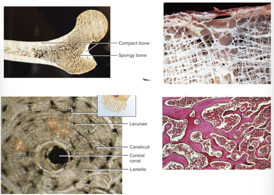
Osteocytes are cells that maintain bone matrix and form from osteoblast after bone matrix has surrounded it.
Osteocytes account for 90-95% of bone cells and are very long lived.
Osteocyte cell bodies are housed within the bone matrix in spaces called lacunae.
Their cell extensions are housed in narrow long spaces called canaliculi.
Osteoclasts are bone-destroying cells.
They contribute to bone repair and remodeling by removing existing bone, called bone reabsorption.
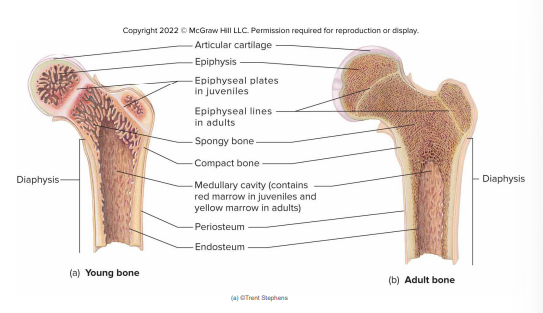
Lamellar bone - Mature bone; it is organized into thin, concentric sheets or layers, called lamellae.
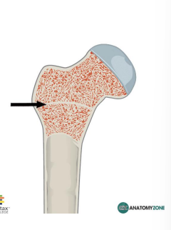
Spongy bone - less bone matrix and more space.
Compact bone - more bone matrix and less space.
Spongy bone - consists of interconnecting rods or plates of bone called trabeculae.
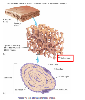
Compact bone - is the solid outer layer surrounding each bone.
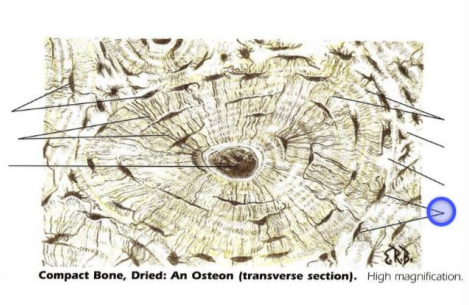
Osteon - the functional unit of compact bone; it is composed of concentric rings of matrix surrounding a central canal.
These are lined with endosteum and cond contains blood vessels, nerves, and loose connective tissue.
Lamellae - are concentric rings of bone matrix which surrounds the central canal.
Lacunae- where osteocytes are located in spaces between the lamellar rings.
STRUCTURE OF LONG BONE 1
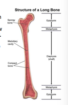
Diaphysis- is the center portion of the bone which is composed of compact bone surrounding a hollow center called the medullary cavity.
Epiphyses- ends of a long bone.
STRUCTURE OF A LONG BONE 2
Articular cartilage - hyaline cartilage that covers the end of a long bone.
Epiphyseal plate - located between the epiphysis and the diaphysis.
Epiphyseal line - the ossification of the epiphyseal plate when the bone stops growing in length.
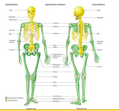
The average adult has 206 bones
Axial Skeleton - consists of the bones of the skull, the auditory ossicles, the hyoid bone, the vertebral column, and the thoracic cage.
Appendicular skeleton - consists of the upper limbs, the lower limbs,and the two girdles.
Girdle - refers to the 2 zones where the limbs are attached to the body. ( pectoral and pelvic girdle _
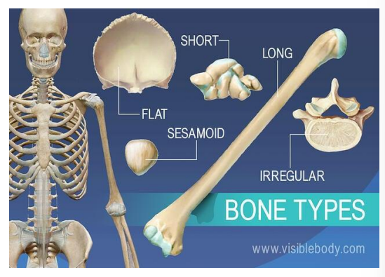
Long - upper and lower limb bones; bones are longer than they are wide
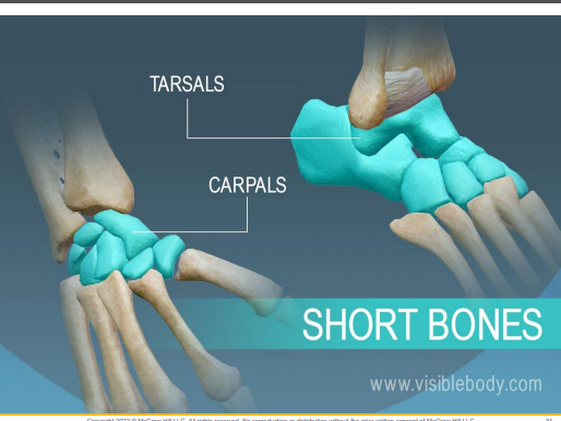
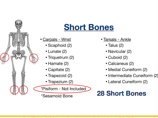
Short - wrist and ankle bones; are approximately as wide as they are long
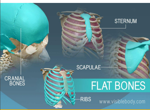
Flat - skull and sternum; has a relatively thin, flattened shape.
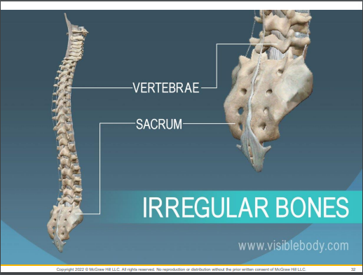
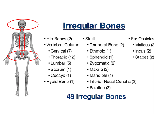
Irregular bones - vertebrae & facial bones; have shapes that do not fit readily into the other 3 categories.

Skeletal Terminology 1
Foramen - hole
Fossa - depression
Process - projection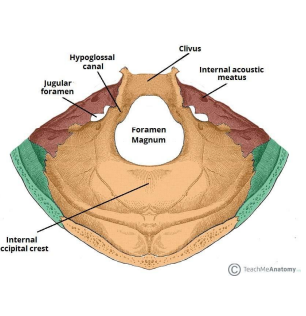
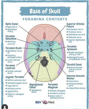
Condyle - smooth, rounded end
Meatus/canal - canal-like passageway
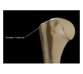
Tubercle/tuberosity - lump of bone
PROJECTIONS THAT ARE SITES OF MUSCLES AND LIGAMENT ATTACHMENT
Tuberosity = large rounded projection; may be roughened
Crest = Narrow ridge of bone; usually prominent
Trochanter - very large, blunt, irregularly shaped process (on femur)
Tubercle- small rounded projection/process
Epicondyle - raised area on/above a condyle
Spine - sharp, slender, often pointed projection
Process - Any bony prominence
PROJECTIONS THAT HELP FORM JOINTS
Head - bony expansion carried on a narrow neck
Facet - smooth, nearly flat articular surface
Condyle - Rounded articular projection
Ramus - Armlike bar of bone
DEPRESSIONS AND OPENINGS FOR PASSAGE OF BLOOD VESSELS AND NERVES
Groove - Furrow
Fissure - Round/oval opening through a bone
Notch - Indentation at the edge of a structure
OTHERS
Meatus - canal-like passage way
Sinus - bone cavity, filled with air and lined with mucous membrane
Fossa- shallow basinlike depression in a bone, often serving as an articular surface.
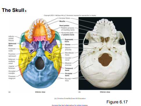
Axial skeleton - is composed of the skull, the vertebral column and the thoracic cage.
The skull has 22 bones
The bony structure of the face has 14 facial bones, and the braincase consists of 8 cranial bones. 13 of the facial bones are rather solidly connected to form the bulk of the face.
3 auditory ossicles in each middle ear (6 in total)
Cranial structures
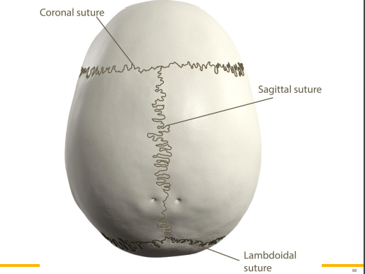
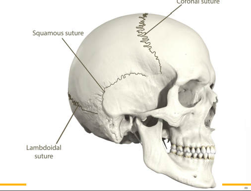
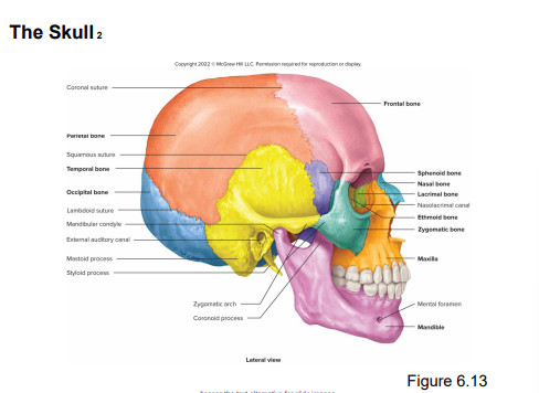
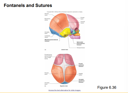
Sutures - immovable joints that connects the cranial bones.
4 principal sutures:
Coronal
Sagittal
Lamboid
Squamous
Cranial Bones
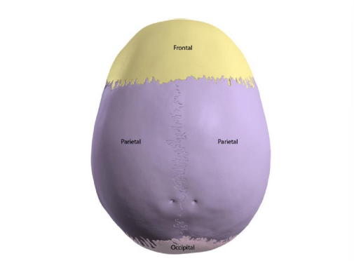
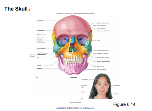
Frontal bone - anterior part of cranium, the ‘forehead’
Parietal bones - sides and roof of cranium.
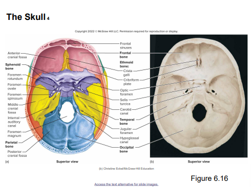
Occipital bones - posterior portion and floor of cranium.
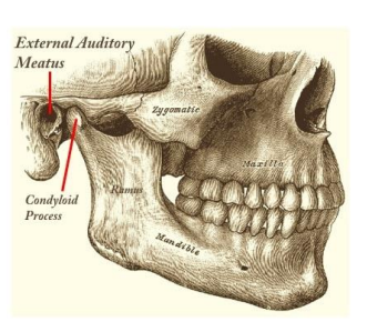
Temporal bones - inferior to parietal bones on each side of the cranium; Temporomandibular joint
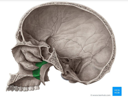
Sphenoid bone - forms part of the cranium floor, lateral posterior portions of eye orbits, lateral portions of cranium anterior to temporal bones; Sella turcica
Ethmoid bone - anterior portion of cranium, including medial surface of eye orbit and roof of nasal cavity; Nasal conchae
Facial Bones
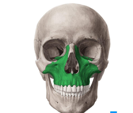
Maxillae - forms the upper jaw, anterior portion of hard palate, part of lateral walls of nasal cavity, floor of eye orbits; Maxillary sinus
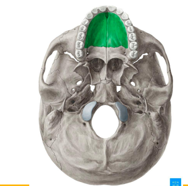
Palatine bones - forms posterior portion of hard palate, lateral wall of nasal cavity.
Facial bones
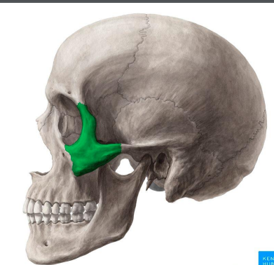
Zygomatic bones - cheek bones; also forms floor and lateral wall of each eye orbit
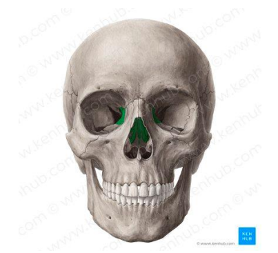
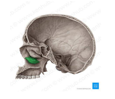
Lacrimal bones - medial surfaces of eye orbits
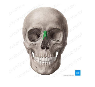
Nasal bones - forms bridge of nose
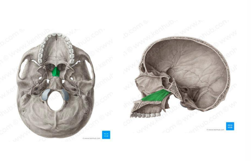
Vomer - in midline of nasal cavity; forms nasal septum with the ethmoid bone
Inferior nasal conchae - Attached to lateral walls of nasal cavity
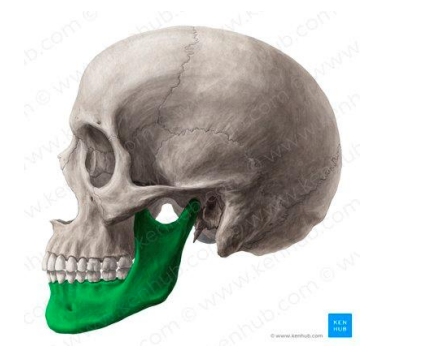
Mandible - lower jawbone; only movable skull bone
THE SKULL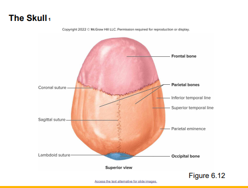
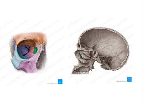
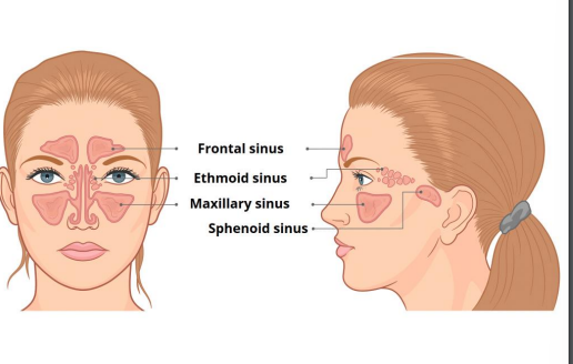
Paranasal Sinuses - several bones that are associated with the nasal cavity and has large cavities within them.
These are :
Rontal
Ethmoid
Sphenoid
Maxillary
Hyoid bone - an unpaired U-shaped bone that is not part of the skull and has no direct bony attachment to the skull or any other bones; it is the only bone in the body that does not articulate with another bone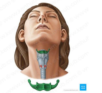

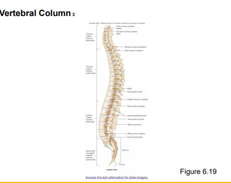
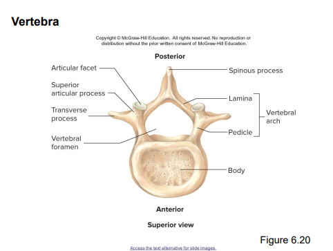
Vertebral Column - is the central axis of the skeleton, extending from the base of the skull to slightly past the end of the pelvis.
Consists o 26 individual bones, grouped into 5 regions
4 major curvatures :
Cervical region - curves anteriorly
Thoracic region - curves posteriorly
Lumbar region - curvers anteriorly
Sacral and coccygeal regions - together curve posteriorly
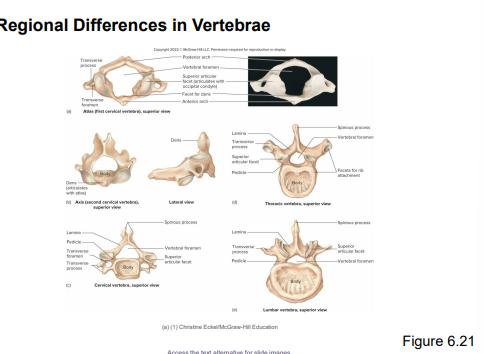
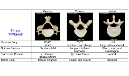
7 cervical vertebra
12 thoracic vertebra
5 lumbar vertebra
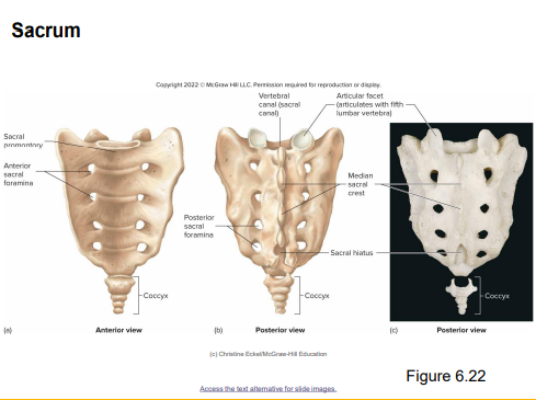
1 sacru
1 coccyx
Atlas
1st vertebra
Holds head
Axis
2nd vertebra
Rotates head
Functions of Vertebral Column
Supports body weight
Protects the spinal cord
Allows spinal nerves to exit the spinal cord
Provides a site for muscle attachment
Provides movement of

Thoracic cage 1- protects the vital organs; 12 pairs of ribs
Sternum : breastbone
True ribs : attach directly to sternum by cartilage
False ribs : attach indirectly to sternum by cartilage
Floating ribs : not attached to sternum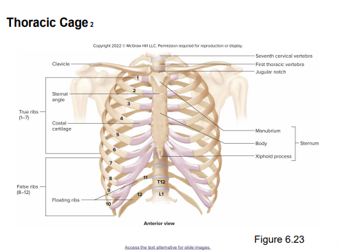
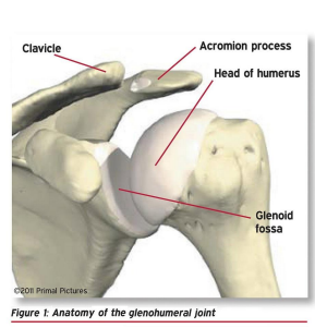
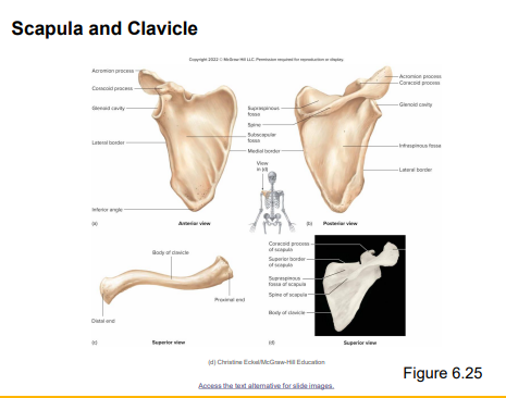
Pectoral Girdle and Upper Limb
Scapula - shoulder blade
Clavicle - collar bone
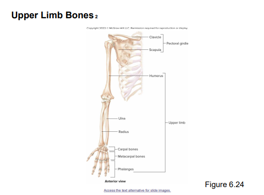 Humerus - upper portion of forelimb
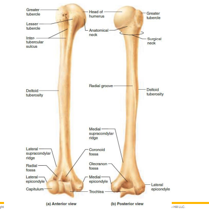
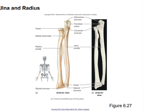
Ulna - forearm
Radius - forearm
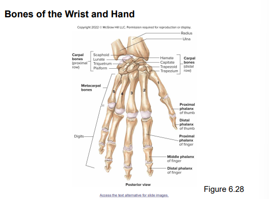
Carpals - wrist
Metacarpals - hands
Phalanges - fingers
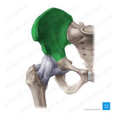
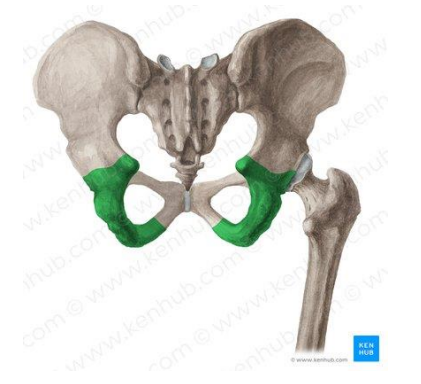
Pelvic Girdle - where lower limbs attach to the body
Pelvis - includes pelvic girdle and coccyx
Ischium - inferior and posterior region of hip bone
Ilium - most superior region of hip bone
Acetabulum - hip socket (joint) of hip bone
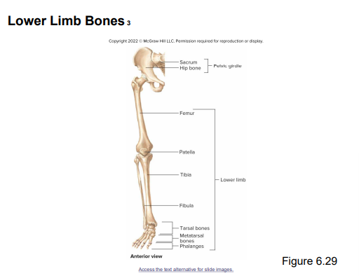
Lower Limb Bones:
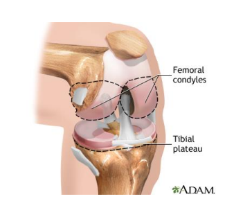
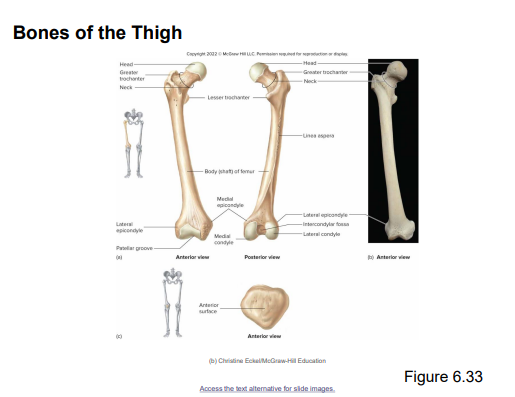
Femur - thigh
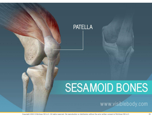
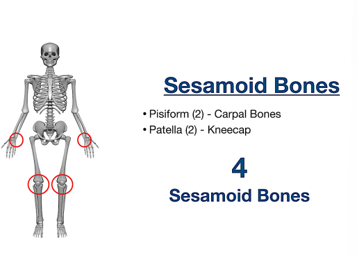
Patella - knee cap
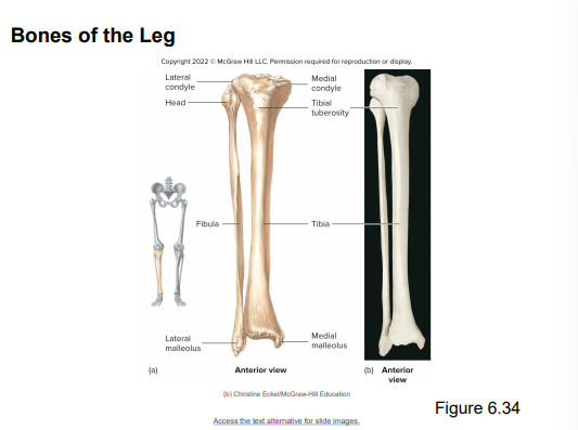
Tibia - Large bone of lower leg
Fibula - Smaller bone lower leg
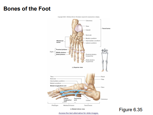
Tarsals - ankle
Metatarsals- foot
Phalanges - toes and fingers
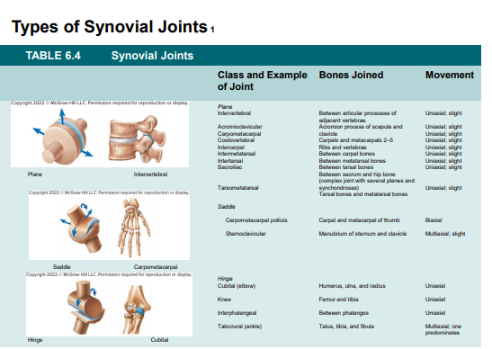
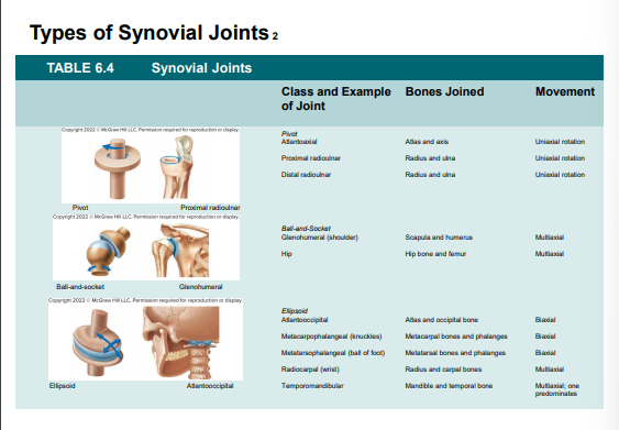
Articulations/ Joints
Are where 2 bones come together.
Classified as structurally fibrous, cartilaginous, or synovial according to the major connective tissue type that binds the bones together and whether a fluid-filled joint capsule is present.
Classified in functional categories according to their degree of motion as synarthroses, amphiarthrosis, diarthroses
Structural classification of joints:
Fibrous joint :
United by fibrous connective tissue; subclasses are sutures, syndesmosis & gomphoses
Cartilaginous :
United by means of cartilage; subclasses are synchondrosis & symphysis
Synovial
Joined by a fluid cavity; most joints of the appendicular skeleton
Surrounded by fluid filled fluid cavity; created by the joint capsule
The joint capsule consists of 2 layers:
An outer fibrous capsule
An inner synovial membrane
Functional Classification of Joints
Synarthosis:
Non-movable joint
Example - skull bone articulations
Amphiarthrosis
Slightly movable joint
Example - between vertebrae
Diarthrosis
Freely movable joint
Example - knee, elbow, and wrist articulations
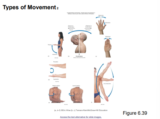
TYPES OF MOVEMENT:
Flexion - bending
Extension- straightening
Abduction- movement away from midline
Adduction - movement toward the midline
Pronation- rotation of the forearm with palms up
Supination - rotation of the forearm with palms up
Rotation- movement of a structure about the long axis