Intro to Anatomy
Anatomical features are often a representation of its properties; form follows function.
Study of bone structure & treatment of bone disorders = osteology.
4 components of the skeletal system;
1. Bone tissue
2. Appendicular skeleton
3. Axial skeleton
4. Joints
6 functions of skeletal system;
1. Support : structural framework, supports soft tissues, provides attachment points for tendons of skeletal muscles
2. Protection : internal organs form injury (like rib cage protects heart & lungs, cranial cavity protects brain)
3. Assistance in movement : most skeletal muscles attach to bones; contraction means they pull on bones to produce movement
4. Mineral homeostasis (storage & release) : tissue makes up 18% of weight of human body, storing several minerals {calcium – 99%, phosphorus} contributing to strength of bone, and bone can release minerals into blood to maintain critical mineral balances (homeostasis), distributing minerals to other parts of the body
5. Blood cell production : hemopoiesis (where connective tissue; red bone marrow, produces red & white blood cells & platelets), red bone marrow containing blood cells, adipocytes, fibroblasts & macrophages within network of reticular fibres. With age red ® yellow bone marrow
6. Triglyceride storage : yellow bone marrow (mainly adipose cells) storing triglycerides = potential energy reserve
Bone Tissue
Classified as a connective tissue
Categories;
1. Long bone; clavicle, humerus, radius, ulna, metacarpals, femur, tibia, fibula, metatarsals, phalanges {associated with movement}
2. Short bones; carpal & tarsal bones, sesamoid bones
3. Flat bones; skull, mandible, scapula, sternum, ribs
4. Irregular bones; vertebrae, sacrum, coccyx, pelvis, hyoid bone
Components;
Several tissues working together – bone (osseus) tissue = 80% compact/dense (cortical) + 20% spongy (trabecular)
1. Periosteum
2. Endosteum
3. Articular cartilage (associated with joints)
4. Blood vessels
5. Nervous tissue
6. Adipose tissue
Function
1. Supports soft tissue and provides attachment for skeletal muscles.
2. Protects internal organs.
3. Assists in movement, along with skeletal muscles.
4. Stores and releases minerals.
5. Contains red bone marrow, which produces blood cells.
6. Contains yellow bone marrow, which stores triglycerides (fats).
Composition & Mechanical Properties
1. Bone extracellular matrix; abundant, composed of 85-90% collagen type 1 fibres (scaffold for bone cells, contributing to mechanical properties i.e. tensile strength) integral for modelling and repair
2. Bone cellular matrix
Bone modelling & remodelling
Bone = complex & dynamic living tissue, continually remodelling; the building of new bone tissue and breaking down of old bone tissue
Bone tissue consists of both collagen fibres and mineralised salts, as well as roughly 15% water. Osteoclasts form a seal at the bones’ surface, and then carve out tunnels within old bone to dissolve bone proteins and minerals (resorption). Once the by-products of old bone are resorbed into blood capillaries, osteoblasts fill in the cavity with crystallized mineral salts (Ca Phosphate), which calcify within the collagen fibres initiated by osteoblasts, rebuilding the bone (deposition).
Remodelling = the building of bone tissue and breaking down of old bone tissue
- Bone resorption = removal of minerals & collagen fibres from by via osteoclasts
- Bone deposition = addition of minerals & collage fibres to bone via osteoblasts
This happens at different rates around the body, and can be affected by the levels of;
1. Minerals
2. Vitamins
3. Hormones
Demineralization; loss of calcium and other minerals from bone extracellular matrix, 30+ in females & 8%/10y, 60+ males & 3%/10y
Long bone anatomy
Features
1. Hollow shaft/diaphysis
2. Epiphyses; proximal (start) & distal (end) of bone
3. Metaphysis; between diaphysis and epiphyses containing an epiphyseal (growth) plate, or cartilage allowing bone to grow in length, later replaced by bone
4. Articular cartilage; covers epiphysis, reducing friction and shock of moving joints
5. Periosteum; connective tissue sheath that can increase thickness of bone, protecting & serving as attachment for ligaments, tendons and can nourish tissue
6. Medullary cavity; space within diaphysis containing fatty yellow bone marrow & blood vessels, reducing weight of bone
7. Endosteum; thin membrane lining medullary cavity & space inside bone, containing 1 layer of osteoprogenitor cells & some connective tissue
Diaphysis – collar of dense cortical bone around central medullary cavity
Metaphysis & epiphysis – composed of trabecular bone surrounded by thin shell of dense cortical bone
Exercise and Bone Tissue
1. Mechanical strain increases bone strength by increasing deposition of mineral salts and production of collagen fibres.
2. Removal of mechanical strain weakens bone through demineralization and collagen fibre reduction.
Appendicular skeleton
Primary function – movement, total of 126 bones [pectoral & pelvic girdle, upper & lower limb]
Skeletal mass & density (measured via DEXA scan; dual-energy x-ray absorptiometry)
Divisions of the Skeletal System
1. The axial skeleton consists of bones arranged along the longitudinal axis. The parts of the axial skeleton are the skull, auditory ossicles, hyoid bone, vertebral column, sternum, and ribs.
2. The appendicular skeleton consists of the bones of the girdles and the upper and lower limbs. The parts of the appendicular skeleton are the pectoral girdles, bones of the upper limbs, pelvic girdles, and bones of the lower limbs.
Homework Task:
Bone Landmarks
1. Proximal epiphysis
2. Diaphysis
3. Distal epiphysis
4. Spongy bone (red/yellow marrow)
5. Articular cartilage
6. Epiphyseal line
7. Periosteum
8. Compact bone
9. Medullary cavity
Cortical (hard/compact bone) & Trabecular (spongy/soft)
Cortical (compact/hard)
· Low porosity (5-10 %)
· Strong and dense (80% of the skeleton)
· Epiphysis (thin shell)
· Diaphysis (shaft, thicker)
Trabecular (cancellous/ spongy/soft)
· High porosity (30-90 %)
· Lighter and less dense than the cortical
· 20% of the skeleton
· Remaining space is filled with marrow and fat
Functions of Bone Cells
osteoBlasts Build bone, while osteoClasts Carve out bone
Osteoblasts: Make and deposit components of bone extracellular matrix, immature bone cells that secrete matrix compounds (osteogenesis)
Osteoclasts: Degrade and resorb bone for remodelling, secrete acids & protein-digesting enzymes, dissolve bone matrix & release stored minerals (osteolysis), derived from stem cells often found in endosteum lining marrow cavity
Osteocytes: “watcher cells” Sit in bone and monitor its current status, found between layers (lamellae) & maintain protein & mineral content of matrix, helping to repair if needed
Osteoprogenitor cells : Mesenchyme stem cells that divide to produce osteoblasts. Located in inner, cellular layer of periosteum and assist in fracture repair.
Function & Content of Bone Marrow
Red: produces blood cells (red, white, and platelets)
Yellow: contains adipose tissue which store triglycerides (fats) that can be used as energy for other tissues within the body
Stages of Endochondral Ossification (replacement of cartilage by bone)
1. Bone collar – cells within perichondrium change to osteoblasts, where a layer of bone forms around shaft which will then grow and compact
2. Cavitation – chondrocytes in centre of the cartilage will enlarge, form struts & calcify and then die leaving cartilage cavities
3. Invasion – periosteal bud brings blood vessels into the cartilage, and with it osteoblasts & osteoclasts, where spongy bone develops
4. Remodelling – remodelling and formation of medullary cavity/marrow, wherein bone replaces the cartilage & diaphysis elongates
5. Ossification – capillaries & osteoblasts enter epiphyses creating secondary ossification centres
6. Elongation – epiphyses fill will spongy bone with cartilage either end
· Ends of bone = articular cartilage
· Cartilage at metaphysis = epiphyseal cartilage
Bone adaptations through exercise;
When placed under strain, bone tissue becomes stronger through increased deposition of mineral salts and production of collagen fibres by osteoblasts.
· Wolff’s law holds that a bone grows or remodels in response to the forces which act upon it
· Changes in bone density in response to exercise
· Tension and compression forces must balance
Bone adaptations through ageing;
Loss of bone mass = demineralisation; loss of calcium (osteoporosis) and minerals from bone extracellular matrix.
Decreased rate of protein (collagen fibre) synthesis = brittleness, increased susceptibility to fracture.
Can lead to deformity, pain, loss of height and teeth.
LECTURE 2
Anatomical Terminology
Anatomical Position;
- Lying on your back = supine.
- Lying on your front = prone
- Front view = anterior
- Back view = posterior
Regional Terminology;
Positional Terms;
Directional Term | Definition |
Superior (cephalic/cranial) | Towards head/upper part of structure |
Inferior (caudal) | Away from head/lower part of structure |
Anterior (ventral) | Nearer to/at front of body |
Posterior (dorsal) | Nearer to/at back of body |
Medial | Nearer to midline (imaginary line dividing body externally to equal right & left sides) |
Lateral | Farther from midline |
Intermediate | Between 2 structures |
Ipsilateral | Same side as another structure within body |
Contralateral | Opposite side as another structure within body |
Proximal | Nearer attachment of limb to trunk/origination of structure |
Distal | Further from attachment of limb to trunk/origination of structure |
Superficial (external) | Towards/on surface of body |
Deep (internal) | Away from surface of body |
Thoracic Cavity:
- Ribs
- Chest muscles
- Sternum
- Thoracic portion of vertebral column
· Pericardial cavity; fluid space around heart
· Pleural cavity; fluid filled spaces around lungs
· Mediastinum; contains all thoracic organs except lungs (heart, oesophagus, trachea, thymus, & several large blood vessels that enter and exit the heart)
· Diaphragm; dome-shaped muscle separating thoracic cavity from abdominopelvic cavity
Planes (surface/slice) of the body;
1. Frontal/Coronal – cuts front/back, vertically
2. Transverse – cuts up/down, across
3. Midsagittal – cuts down midline
Why?
- Medical imagining (diagnosing
LECTURE 3
Anatomy of Pelvis & Hip Joint
Pelvis
2 hip bones unite anteriorly at the pubic symphysis and posteriorly at the sacrum to form the bony pelvis.
Pelvic Girdle =
- Right & left Hip bone
- Sacrum
- Pubic symphysis
- Obturator foramen
- Acetabulum
- Pelvic brim/inlet
· False pelvis (abdominal structures)
· True pelvis (bladder, prostrate)
- Sacroiliac joint
Main bone regions of pelvis =
1. Ilium – (superior) landmarks/muscles with ‘iliac’ in name
2. Ischium – (inferior, posterior) landmarks/muscles with ‘ischial’/’sciatic’ in name
3. Pubis – (anterior & inferior) landmarks/muscles with ‘pubic’ in name
Anterior and inferior part of the hip bone.
Movements of Pelvis =
1. Pelvis tilt (sagittal plane rotation)
2. Pelvis list (frontal plane rotation)
3. Pelvis rotation (transverse plane rotation)
 | |||
Femur (thigh bone) =
- Fovea capitis (connects head of femur to acetabulum)
- Greater trochanter (anterior to hollow side of hip)
- Neck
- Intertrochanteric line & crest
- Lesser trochanter (inferior, medial to Gt.)
- Gluteal tuberosity blends into linea aspera (tendon att. points)
- Anterior;
· Patella surface
· Lateral & medial epicondyle
· Lateral & medial condyle
· Adductor tubercle
The acetabulum is the socket for the head of the femur, where the three parts of the hip bone converge and ossify.
Ligaments (connective tissue, connect bone to bone & stabilise joints) of Hip =
- Iliofemoral (body’s strongest, prevents hyperextension of femur)
- Pubofemoral (prevents overabduction of femur, strengthens articular capsule)
- Ischiofemoral (slackens during adduction, tenses during abduction, strengthens articular capsule)
Hip Joint
Hip joint allows flexion, extension, abduction, adduction, lateral rotation medial rotation, and circumduction of the thigh
Components;
1. Articular capsule
2. Iliofemoral ligament
3. Pubofemoral ligament
4. Ischiofemoral ligament
5. Ligament of femur head
6. Acetabular labrum (cartilage rim of acetabulum, enhances ligament depth = reduces dislocation)
7. Transverse acetabular ligament
Movement (of thigh due to hip joint);
- Flexion
- Extension
- Abduction
- Adduction
- Lateral rotation
- Medial rotation
- Circumduction
Muscles of Hip Joint;
1. Psoas minor
2. Iliac crest
3. Iliacus
4. Psoas major
5. Pectineus
6. Adductor brevis
7. Adductor longus
8. Gracilis
9. Adductor magnus
Anterior – Hip Flexors | Posterior – Hip Extensors | Posterior-Lateral – External (lateral) Rotators | Medial/Internal Rotator Muscles | Abductor – Hip Muscles | Adductor – Hip Muscles |
Psoas major | Gluteus maximus | Gluteus maximus | Gluteus medius (anterior fibres) | Gluteus medius (all fibres) | Pectineus
|
Iliacus | Semitendinosus | Piriformis | Gluteus minimus (anterior fibres) | Gluteus minimus (all fibres) | Gracilis |
Rectus femoris | Semimembranosus | Obturator internus | Tensor fasciae latae | Tensor fasciae lata | Adductor longus |
Sartorius | Biceps femoris | Gemellus superior | Adductor longus |
| Adductor brevis |
| Adductor magnus | Gemellus inferior |
|
| Adductor magnus (anterior & posterior heads) |
|
| Quadratus femoris |
|
|
|
Movement | Description |
Gliding | Movement of relatively flat bone surfaces back-and-forth and side-to-side over one another; little change in angle between bones. |
Angular | Increase/decrease in angle between bones. |
Flexion | Decrease in angle between articulating bones, usually in sagittal plane or an anterior movement at a ball-and-socket joint. |
Lateral Flexion | Movement of trunk in frontal plane. |
Extension | Increase in angle between articulating bones, usually in sagittal plane or a posterior movement at a ball-and-socket joint. |
Abduction | Movement of bone away from midline, usually in coronal plane. |
Adduction | Movement of bone toward midline, usually in coronal plane. |
Circumduction | Flexion, abduction, extension, adduction, and rotation in succession (or in the opposite order); distal end of body part moves in circle. |
Rotation | Movement of bone around longitudinal axis; in limbs, may be medial (toward midline) or lateral (away from midline). |
Special | Occurs at specific joints. |
Elevation | Superior movement of body part. |
Depression | Inferior movement of body part. |
Protraction | Anterior movement of body part in transverse plane. |
Retraction | Posterior movement of body part in transverse plane. |
Inversion | Medial movement of sole. |
Eversion | Lateral movement of sole. |
Dorsiflexion | Bending foot in direction of dorsum (superior surface). |
Plantar flexion | Bending foot in direction of plantar surface (sole). |
Supination | Movement of forearm that turns palm anteriorly. |
Pronation | Movement of forearm that turns palm posteriorly. |
Opposition | Movement of thumb across palm to touch fingertips on same hand. |
Types, Functions & Properties of Muscle
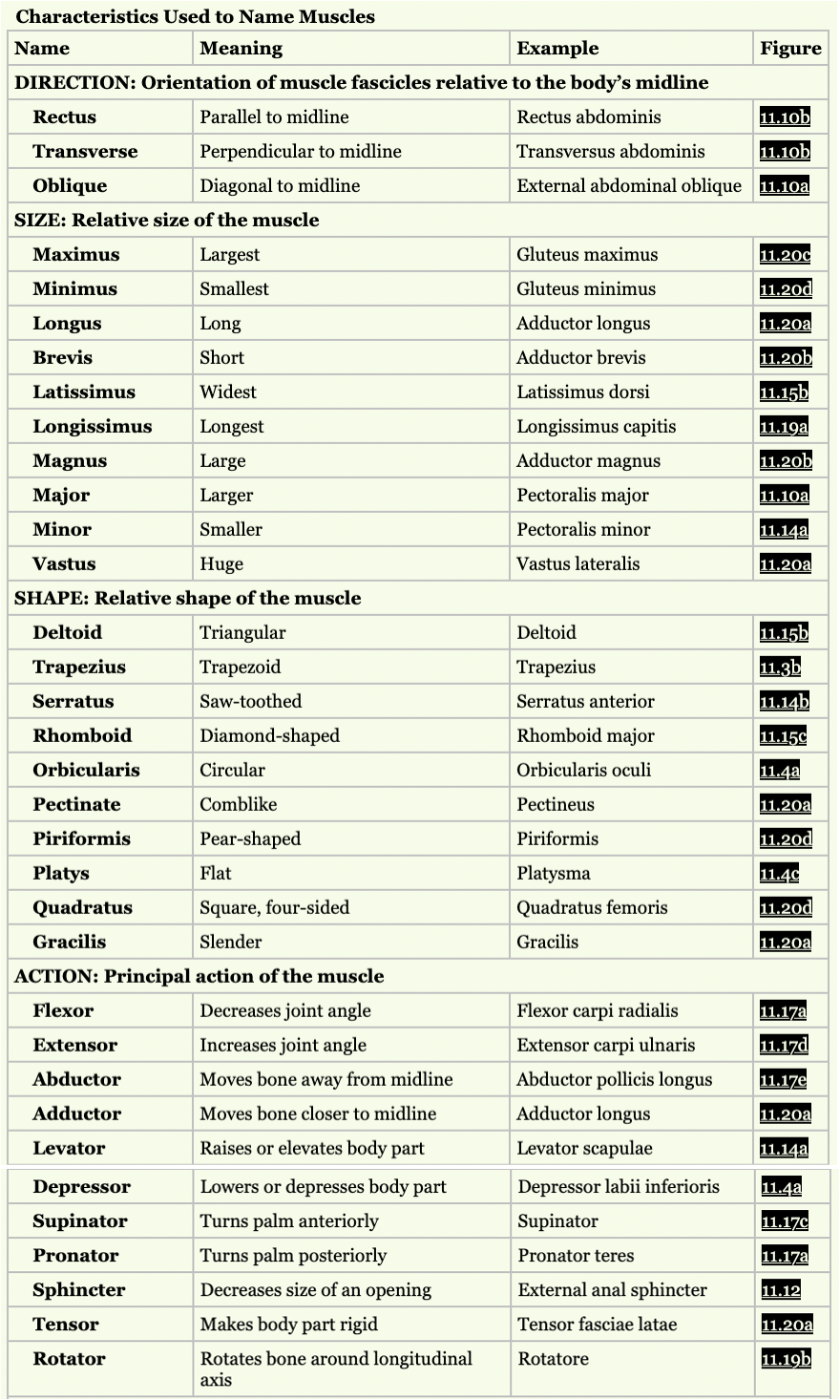
Muscles are built to ‘pull’ not push, producing force to bring their origin & insertion together, typically attaching to bone via tendons.
Types;
Smooth | Cardiac | Skeletal |
Not striated | Visible striation | Highly organised striation |
Single nuclei | Single nuclei | Multiple nuclei |
Autonomic control | Autonomic control | Voluntary control |
Doesn’t fatigue | Doesn’t fatigue | Function = generates movement of body parts |
Function = Internal organs | Function = allows heart to pump blood |
|
Functions;
- Movement of body parts
- Stabilisation of body
- Control organ volume (stomach, bladder)
- Move fluids (blood, lymphatic fluid)
- Thermoregulation (shivering)
- Energy storage (muscle glycogen)
Properties;
- Electrically excitable = responds to neutral input
- Contractility = generates tension
- Extensibility = lengthens without damage (limits)
- Elasticity = returns to original shape after stretch (in all directions)
Skeletal Muscle
- Typically produce movement by controlling rotation of joints
- Striated tissue
- Made up of muscle fascicles containing muscle fibres (myocytes), blood vessels & nerves, wrapped in epimysium (connective tissue layers)
Structure;
- Bone covered by periosteum
- Tendon
- Epimysium
- Muscle fascicle (fibres wrapped in perimysium)
· Endomysium
· Perimysium
- Muscle fibre
· Sarcoplasmic reticulum
· Sarcolemma
· Myofibril sarcoplasm
· Sarcomere
§ Z disc
§ Thick filament (myosin)
§ Thin filament (actin, tropomyosin, troponin)
· Nucleus
· T tubule
· Terminal cisterns
· Mitochondrion
Sliding Filament Mechanism
During muscle contractions, thin filaments move towards M line of each sarcomere. As these shorten, the muscle shortens too.
Sarcomere components =
1. Z discs; separates sarcomeres
2. A band; dark middle of sarcomere, includes thin filaments the length of sarcomere
3. I band; less dense, only thin filaments, Z disc passes through centre of each
4. H zone; thick centre of a band, only thick filaments
5. M line; H zone thick filament containing proteins
Types of muscle fibre/fascicle arrangement (all to longitudinal axis of muscle)
- Parallel
- Fusiform
- Circular/orbital
- Triangular
- Pennate (pennatus = ’winged’)
· Unipennate (one-directional)
· Bipennate (feather shaped/two-sided)
· Multipennate (bulging shape)