
DP2 Biology - topics: 6.1, D1 & D2
Why Digest?
make sure nutrients are small enough to fit in the cell
fuel for ATP production
Provide raw materials for building
Acquire nutrients like vitamins and minerals that the body needs
What process do we use?
Through Catabolism (Hydrolysis)
use H2O to breakdown polymers
the reverse of condensation rxn
cleave off one monomer at a time
H2O is split into H+ and OH–, then H+ & OH– attach to ends
Ex: (A---B (macromolecule) + H--O--H (water) ⇌ A--H+ + B--OH–) or ( H--O--H (water) ⇌ H+ + OH-)
requires enzymes
Why do we need Enzymes to digest?
Enzymes are needed to break down the larger, macromolecules into monomers. These smaller pieces can then be absorbed into the small intestine.
The digestive system is composed of circular and longitudinal muscles (smooth muscle made of short fibers). These muscles produce a moderate force along with short vigorous contractions to move food along the tube.
Mechanical vs Chemical digestion:
Mechanical: Mixing, via the muscles contracting
Chemical: Continued digestion from the stomach acid/enzymes and new secretions from the pancreas.
Mouth and Saliva Glands
Mechanical digestion using the teeth and tongue. Starting Chemical digestion using amylase from the saliva glands.
Esophagus
Moves food from mouth to stomach via peristalsis.
Peristalsis: muscles working together in a wave-like motion to move food from the mouth to the stomach
In addition to movement, peristaltic action aids in mechanical digestion.
Stomach
Structural description: J-shaped muscular organ located on the left side of the upper abdomen, as food reaches the end of the esophagus it enters the stomach through a muscular valve called the lower esophageal sphincter.
Job: Mechanical digestion by churning and mixing. Starts chemical digestion of proteins using
acid and enzymes.
Small Intestine
Structural description: Long (about 22 to 25 feet), highly convoluted, narrow, folded, or coiled tube extending from the stomach to the large intestine contained in the central and lower abdominal cavity.
Job: Mostly chemical digestion of lipids, carbohydrates, and proteins via enzymes secreted by accessory glands. The majority of nutrient absorption takes place here.
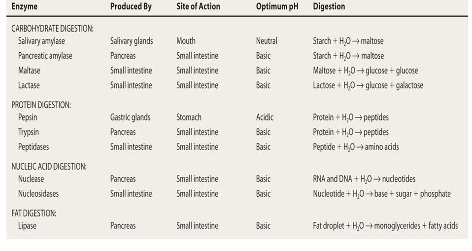
All chemical reactions experienced through the small intestine:
Nuclease: digests DNA & RNA into nucleotides
Maltase: digests maltose into 2 glucoses
Lactase: digests lactose into glucose & galactose
Sucrase: digests sucrose into glucose & fructose
Large Intestine
Structural description: The long, tube-like organ that is connected to the small intestine at one end and the anus at the other. Made of four parts: the cecum, colon, rectum, and anal canal.
Job: Water is re-absorbed, and some final digestion of carbohydrates. Feces are formed and stored here.
Pancreas
Structural description: made of two tissues - digestive tissue (exocrine) and hormonal tissue (endocrine)
Digestive tissue (exocrine): makes insulin which is critical for sugar regulation
Hormonal tissue (endocrine): makes 3 types of enzymes (lipase, amylase, and protease)
Job: Secretes lipase, amylase, and protease into the lumen of the small intestine for digestion. All the enzymes as secreted into the small intestine through the pancreatic duck.
Pancreatic amylase - breaks down starches
Lipase - breaks down triglycerides and phospholipids into glycerol and fatty acids
Protease (Pepsin) - protein and peptide digestion starts in stomach but continues in the small intestine.
Accessory Organ
Liver
Produces bile, which is a fat emulsifier to help break lipids down into small droplets. These are easier to absorb.
Accessory Organ
Gall Bladder
Job: For storage and regulation of bile.
releases bile into small intestine which coats fat, preventing small droplets form becoming large globules, increasing exposure to pancreatic lipase.
Accessory Organ
Essential idea: The structure of the small intestine allows it to move, digest, and absorb food.
The small intestined is filled with tiny folds called villi.
this us where the absorption of nutrients takes place.
the villi increase surface area which means more absorption.
Each villus (Villi - plural, Villus - singular) is covered in cells (Microvilli) that also have an increased surface area.
virtually all nutrients enter the body through the epithelial layer covering the villus. Nutrients like:
amino acids, sugars, vitamins and mineral ions.
the small bristles of the epithelial cells are the Microvilli.
most molecules will diffuse into the capillary network inside the villus, Aka enter the blood to be sent around the body.
Additional information about Capilaries: Capillaries are the smallest blood vessels in your vascular system. They are delicate blood vessels in charge of transporting transporting blood, nutrients and oxygen to cells in your organs and body systems.
Fats mobe into the lymphatic vessel (lacteal) which drains from the villus rapidly and enters the blood via thoracic duct. This is because they tend to remain larger molecules.
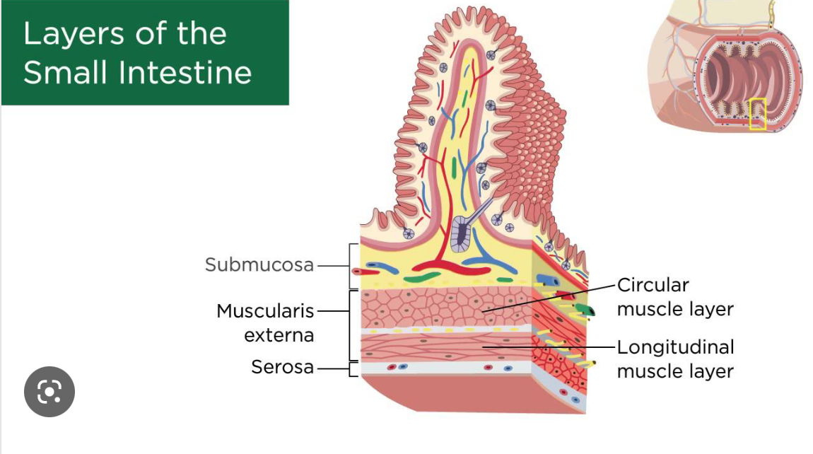
Names of layers in a close section of the small intestine: (You should be able to identify these layers in the test)
V = Villus, projections of mucosa
M = Mucosa, epithelial layer, responsible for nutrient absorption
SM = Sub-mucosa layer containing blood and lymph vessels (lacteal)
CM/LM = Circular Muscle/Longitudinal Muscle
S = Serosa, outer covering of intestine
Reminder from DP1!
Lipids (fats/oils) → Glycerol and Fatty acids
Polypeptides → Shorter peptides
Sugar (Starch) → Maltose and/or Glucose
Size, shape and charge are all factors in how molecules move.
Fats: To enter epithical cells, fatty acids and glycerol move across the membrane by diffusion. This is due to these monomers being small and nonpolar. Once in an epithelial cell, fatty acids and glycerol are recombined back into triglycerides and then have to leave the epithelial cell by exocytosis because they are packaged onto vesicle to be moved into the lacteal (or blood system)
Exocytosis
Nutrients (droplets of liquid) pass across the membrane by forming vesicles to bring inside. (active diffusion - energy is required)
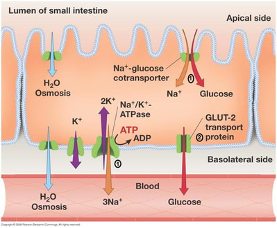
Glucose: it is polar therefore it is must cross the membrane through, facilitated diffusion (cotransport). However, to move through a protein into the epithelial cell it must move with Na+. In order for Na+ to diffuse into the epithelial cell and bring glucose with it, Na+ must be pumped out of the epithelial cell on the other end. This requires Active Transport because ions are being pumped against the gradient.
Simpler explanation:
A transport protein couples the active translocation of one molecule to the passive movement of another (co-transport)
Glucose and amino acids are co-transported across the epithelial membrane by the active translocation of sodium ions (Na+)
Diagram of Glucose diffusion:
Enzymes at work on Amylose:
Amylase breaks down starch into breaks down starches into Maltose (2 glucose) and maltotriose (3 glucose) because it can only break 1,4 bonds.
Amylopectin has both 1,4 & 1,6 bonds, so there is an additional group of enzymes needed called dextrinases
Cellular respiration requires sugars to be in the single monomer form (glucose)
Once absorbed where does it go? (you need to know this in the test)
Most nutrients are being used all the time…
Assimilation → The conversion of nutrients into fluid or solid parts of an organism
Assimilated into cellular structure (lipids, proteins, carbohydrates, nucleic acids, etc). AKA, makes us up!
Energy right now → Actively being used as energy (glucose shipped around the body for ATP production, mostly lipids and carbohydrates).
Storage → Shipped to storage locations (carbohydrates go to the liver, they are locked into inactive forms, Glycogen for the short-term). Fats get stored throughout the body, etc.
What is left over? (You need to know this for the test)
Although starch, glycogen, lipids and nucleic acids are digested, not all can be absorbed.
Cellulose (found in the cell walls of plants) is indigestive but we still need it.
We do not have the correct enzymes to break it down. But it helps to keep the system moving and clean as it scrapes along the intestines.
Where each digestion happens for each macromolecule
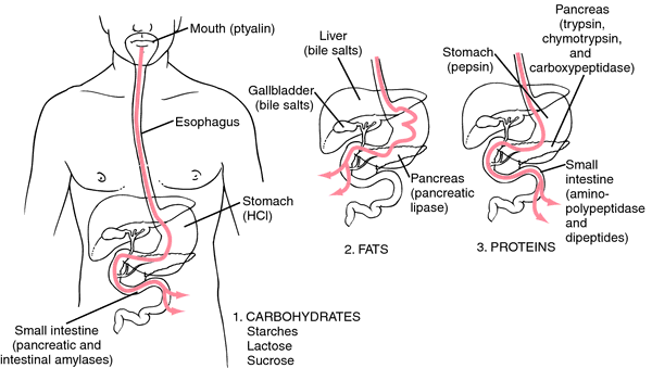
The most important digestive organs for…
…production of chemicals for carbohydrate digestion?
Mouth (salivary glands) and pancreas
…production of chemicals for protein digestion?
Stomach and pancreas
…production of chemicals for lipid digestion?
Pancreas and liver
…absorption?
Small (and large) intestine
http://www.old-ib.bioninja.com.au/standard-level/topic-6-human-health-and/61-digestion.html)
https://www.mrgscience.com/topic-61-digestion-and-absorption.html
Nutrients → chemical substance found in foods used bu the human body
There are six categories of nutrients → Carbohydrates, Lipids, Proteins, Vitamins, Minerals and Water.
Nutrients which our body can not make and thus can only be gaiend from ones diet, those types of nutrients are called essential.
Those include: vitamins, minerals, water, some amino acids, and some fatty acids
Non-essential nutrients can be made by the body or have a replacement nutrient which serves the same dietary purpose
Carbohydrates are not considered essential nutrients as human diets can obtain energy from other sources without ill effect
Vitamins | Minerals | |
|---|---|---|
Organic/Inorganic? | organic (carbon!) | inorganic |
What do they do? | help with grouth and development | maintain water balance, stong connective tissues/bones. blood clotting, carrying oxygen, etc. |
Examples: | A, B complex, C, D, E, K | Calcium, Magnesium, Iron, Zinc, Sodium, etc. |
What is malnutrition________?
→ Malnutrition can result from too few, too many or an imbalance of nutrients.
It can be caused by an improper dietary intake of nutrients – e.g. overnutrition (too much) or undernutrition (not enough)
It can be caused by the inadequate utilisation of nutrients by the body – e.g. due to illness or disease
Vitamin C: Some organism can make Vitamin C and some can’t, wich requires a dietary supply.
Role: Vitamin C is an antioxidant that helps protect your cells against the effects of free radicals — molecules produced when your body breaks down food or is exposed to tobacco smoke and radiation from the sun, X-rays or other sources. Free radicals might play a role in heart disease, cancer and other diseases.
What happens if Vitamin C is not acquired?
Vitamin C deficiency can lead to a disease called scurvy, which causes anemia, bleeding gums, bruising and poor wound healing.
Vitamin D:
What could happen form a Vitamin D and calcium deficiency?
Without enough vitamin D or calcium, your parathyroid glands compensate by producing too much of their hormone, a condition called hyperparathyroidism. That can lead to bone weakening (osteoporosis) and increased fracture risk
What are Omega-3 & Omega-6 fatty acids and what are their roles in the body?
They are Unsaturated fats, critical for brain and eye development.
10 out of 20 amino acids are essential. What if some of them are missing or in low quantities…How does it impact protein production?
Protein cannot be made if there is a shortage of one or more essential amino acids - this is known as protein deficiency malnutrition and can come from a low intake of protein in the diet or an imbalance of the types of protein consumed. PDM can cause a lack of blood plasma proteins which can result in tissue fluid retention.(Kwashiorkor syndrome)
Kwashiorkor Syndrome - nutritional disorder most often seen in regions experiencing famine. It is a form of malnutrition caused by a lack of protein in the diet. People suffering from kwashiorkor typically have an extremely emaciated appearance in all body parts except their ankles, feet, and belly, which swell with fluid.
The extreme lack of protein causes an osmotic imbalance in the gastrointestinal system causing swelling of the gut diagnosed as an edema or retention of water.
What if there is an excess of Phenylalanine?
Most of the time it’s not an issue as it is broken down into AA, then into smaller molecules. But..
Phenylketonuria (PKU) - a rare inherited disorder that causes the enzymes needed for breaking down Phenylalanine not to be produced. As a result, Phenylalanine builds up in the body
Treatment: managed diet through a strict low-protein diet.
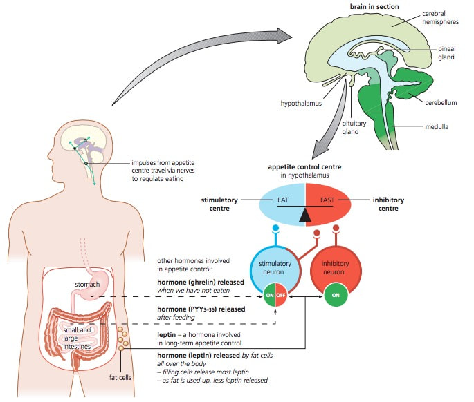
What controls appetite?
the hypothalamus. It also controls thirst and body temperature.
Hypothalamus detects an increase in hormones released from small intestine, adipose tissue, and insulin
How can both obesity and starvation lead to dire health consequences?
people with obesity are more likely to have high blood pressure and diabetes.
How does that happen?
diabetes -- the large intakes of sugar will lead to cells to start to ignore the excessive production of insulting as a result of the intake. This can be reversed through weight loss.
Starvation can lead to the breakdown of body tissue.
When there is restricted food intake the body will break down stored glycogen and muscle tissue to create energy.
How can anorexia nervosa lead to damaged heart tissue?
Anorexia nervosa is a complex condition that involves the extreme restriction of calories and a reduction in body mass.
As the body starts breaking down tissue for nutrients, it can cause deterioration in heart tissue too. In women, the menstrual cycle can be suspended.

https://ib.bioninja.com.au/options/option-d-human-physiology/d1-human-nutrition/
https://www.mrgscience.com/d1-human-nutrition-core.html
Nervous system:
When smelling food the nervous system will send a signal to your stomach to make gastric juice
if brain detects the presence of food in the stomach, it will trigger stomach cells to make gastrin (hormone)
Endocrine system:
Gastrin: hormone released from stomach to aid on making stomach acid.
Cholecystokinin (CCK) and secretin: stimulate the production of pancreatic guice and bile from liver.
What is an exocrine glad?
glands thatsecrete into a specific destination through a duct. (glands produce and secrete substances via a duct onto an epithelial surface)
Exocrine glads vs. endocrine
Endocrine glands secrete directly through ducts into your body’s surfaces while endocrine glands secrete their substances directly into the bloodstream.
Digestive fluids produced by exocrine glass:
Digestive fluid | Source | Composition |
|---|---|---|
Saliva | Salivary glands | water, electrolides, mucus, and salivary amylase |
Gastric Fluid | stomach (gastric glands in stomach wall) | water, mucus, pepsin, hydrochloric acid (HCL) |
Pancreatic Fluid | Pancreas (exocrine pumps -- the cells secrete them) | water, bicarbonate, enzymes: lipase, pancreatic amylase, proteases (trypsin & carboxypeptidase) |
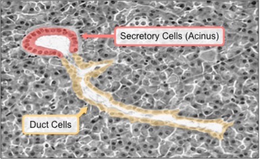
One acinus is the fundamental unit of a gland.
cluster of cells that form a hollow sphere
multiple acini (plural) are found in a gland
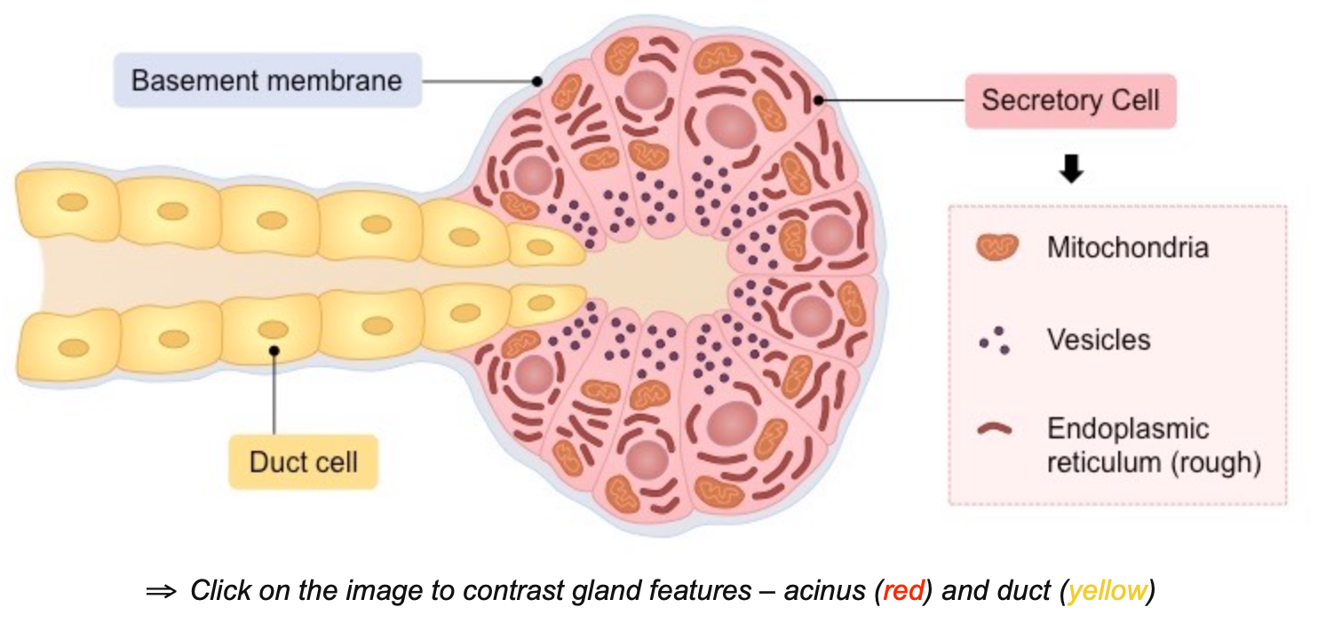
Mitochondria - support energy demands for protein production & other activities
Vesicles - filled with inactive enzymes
Rought ER - for enzyme synthesis
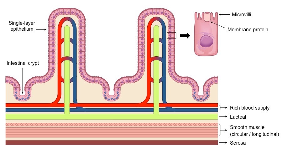
Intestinal Villy contain several structural features which facilitate the absorption of digestive products
Features of the intestinal villus:
__**M**__icrovilli -- riffing of epithelial layer to increase SA
__**R**__ich blood supply -- dense capillary network rapidly transports absorbed materials
__**S**__ingle layer epithelium -- minimizes diffusion distance between lumen & blood
__**L**__acteals -- absorve lipids from the intestine to the lympathic system
__**I**__ntestinal glands -- Exocrine pits release digestive juices
__**M**__embrane proteins -- Facilitates transport of digested materials into epithelial cells
__**P**__inocytotic vesicles -- may be present as well, since some nutrients are absorbed this way
Pinocytosis (‘cell-drinking’) is the non-specific uptake of fluids and dissolved solutes (a quick way to translocate in bulk)
These materials will be ingested via the breaking and reforming of the membrane and hence contained within a vesicle
Drawing of Villus:
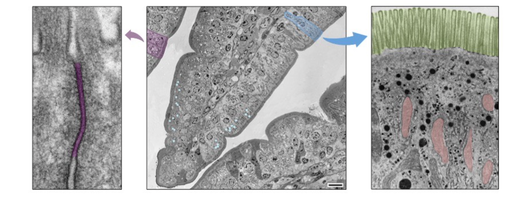
Tight Junctions -- purple
Pinocytotic vesicles -- light blue circles (second picture)
Mitochondria -- red
Microvilli -- green
Acid conditions of the stomach can be helpful in hydrolisys by:
disrupting the ECM holding cells together
denaturing proteins
killing most pathogens
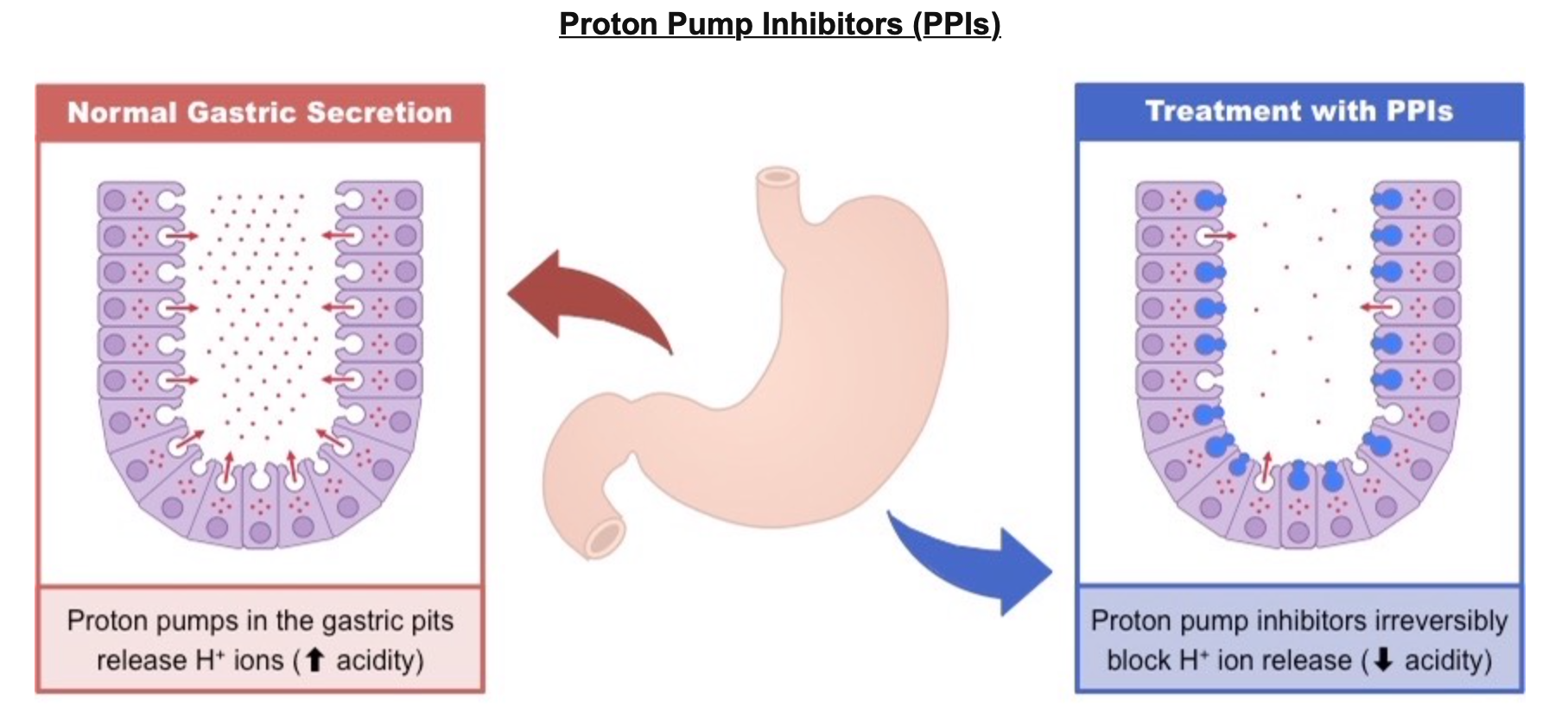
What are Proton Pump Inhibitors and how do they work?
PPI’s are medications that are used to treat acid refluch symptoms.
they work by reducing the amount of acid produced by glands within the stomach.
this is done through irreversibly binding to the proton pumps and prevent H+ ion secretion
This effectively raises the pH in the stomach to prevent gastric discomfort caused by high acidity (e.g. acid reflux)
Individuals taking PPIs may have increased susceptibility to gastric infections due to the reduction of acid secretion
Egestion → anything that can’t be absorbed is egested & leaves the body as fece.
Feces are made up of:
Bilirubin from broken down blood cells is present in fecal matter.
dietary fibre, such as cellulose and lignin
remains of intestinal epithelial cells, bile pigments and human flora (intestinal bacteria)
The human intestines function to complete the process of digestion and absorb digested products into the bloodstream
The small intestine absorbs usable food substances (i.e. nutrients – monosaccharides, amino acids, fatty acids, vitamins, etc.)
The large intestine absorbs water and dissolved minerals (i.e. ions) from the indigestible food residues
Why isn’t the molecular structure of fiber able to be digested by humans?
Humans lack the necessary enzymes to break down certain plant matter
plant materials like cellulose & lignin can’t be broken down and serve as ficer in our diets.
What is the role of dietary fiber in the process of egestion?
help fecal matter move along the large intestine to prevent constipation
also lowers the risk of colon and rectal cancer
promote the feeling of fullness to regulate food intake
slows absorption of sugars for blood sugar regulation
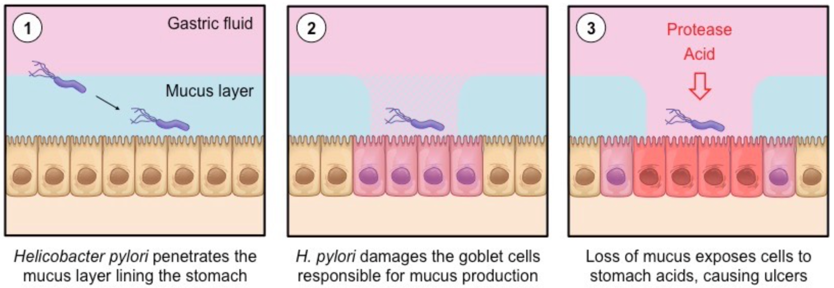
What are Ulcers?
Stomach ulcers are inflammed and damaged areas in the stomach wall, typically caused by exposure to gastric acids. They are often caused by a bacteria called Helicobacter pylori (H. pylori), a bacteria that can survive the acid conditions of the stomach by penetrating the mucus lining.
How does the virus cause the Ulcer? (Additional information)
H. pylori anchors to the epithelial lining of the stomach, underneath the mucus lining
An inflammatory immune response damages the epithelial cells of the stomach – including the mucus-secreting goblet cells
This results in the degradation of the protective mucus lining, exposing the stomach wall to gastric acids and causing ulcers
The prolonged presence of stomach ulcers may lead to the development of stomach cancer over many years (20 – 30 years)
H. pylori infections can be treated by antibiotics (previously, stomach ulcers were considered stress related and not treatable)
What is Cholera?
a bacterial pathogen that infects the intestines and causes acute diarrhoea and dehydration
Cholera can kill within hours unless treated with oral rehydration therapies.
How does choleara lead to dehydration?
V. cholerae releases a toxin that binds to ganglioside receptors on the surface of intestinal epithelium cells.
This toxin is internalised by endocytosis and triggers the production of cyclic AMP (a second messenger) within the cell
Cyclic AMP (cAMP) activates specific ion channels within the cell membrane, causing an efflux of ions from the cell
The build up of ions in the intestinal lumen draws water from cells and tissues via osmosis – causing acute diarrhoea
As water is being removed from body tissues, dehydration will result if left untreated
Condensed explanation: During the cholera, the virus release a powerfull toxing that attaches to the cell walls that line the small intestine and eventually enter the cells causing a suberting in the normall transport of chlotide ions and the water that flows these ions. This is caused by the virus stimulatin. a mass secretion of water containing the cholera bacteria, ions and bicacrbonates making the lymen hypertonic. This will lead to an been larger osmolar diffusion since the cells will not reabsorve the water secreted and thus causing diarrea and dehydration.
https://ib.bioninja.com.au/options/option-d-human-physiology/d2-digestion/exocrine-glands.html
Why Digest?
make sure nutrients are small enough to fit in the cell
fuel for ATP production
Provide raw materials for building
Acquire nutrients like vitamins and minerals that the body needs
What process do we use?
Through Catabolism (Hydrolysis)
use H2O to breakdown polymers
the reverse of condensation rxn
cleave off one monomer at a time
H2O is split into H+ and OH–, then H+ & OH– attach to ends
Ex: (A---B (macromolecule) + H--O--H (water) ⇌ A--H+ + B--OH–) or ( H--O--H (water) ⇌ H+ + OH-)
requires enzymes
Why do we need Enzymes to digest?
Enzymes are needed to break down the larger, macromolecules into monomers. These smaller pieces can then be absorbed into the small intestine.
The digestive system is composed of circular and longitudinal muscles (smooth muscle made of short fibers). These muscles produce a moderate force along with short vigorous contractions to move food along the tube.
Mechanical vs Chemical digestion:
Mechanical: Mixing, via the muscles contracting
Chemical: Continued digestion from the stomach acid/enzymes and new secretions from the pancreas.
Mouth and Saliva Glands
Mechanical digestion using the teeth and tongue. Starting Chemical digestion using amylase from the saliva glands.
Esophagus
Moves food from mouth to stomach via peristalsis.
Peristalsis: muscles working together in a wave-like motion to move food from the mouth to the stomach
In addition to movement, peristaltic action aids in mechanical digestion.
Stomach
Structural description: J-shaped muscular organ located on the left side of the upper abdomen, as food reaches the end of the esophagus it enters the stomach through a muscular valve called the lower esophageal sphincter.
Job: Mechanical digestion by churning and mixing. Starts chemical digestion of proteins using
acid and enzymes.
Small Intestine
Structural description: Long (about 22 to 25 feet), highly convoluted, narrow, folded, or coiled tube extending from the stomach to the large intestine contained in the central and lower abdominal cavity.
Job: Mostly chemical digestion of lipids, carbohydrates, and proteins via enzymes secreted by accessory glands. The majority of nutrient absorption takes place here.
All chemical reactions experienced through the small intestine:
Nuclease: digests DNA & RNA into nucleotides
Maltase: digests maltose into 2 glucoses
Lactase: digests lactose into glucose & galactose
Sucrase: digests sucrose into glucose & fructose
Large Intestine
Structural description: The long, tube-like organ that is connected to the small intestine at one end and the anus at the other. Made of four parts: the cecum, colon, rectum, and anal canal.
Job: Water is re-absorbed, and some final digestion of carbohydrates. Feces are formed and stored here.
Pancreas
Structural description: made of two tissues - digestive tissue (exocrine) and hormonal tissue (endocrine)
Digestive tissue (exocrine): makes insulin which is critical for sugar regulation
Hormonal tissue (endocrine): makes 3 types of enzymes (lipase, amylase, and protease)
Job: Secretes lipase, amylase, and protease into the lumen of the small intestine for digestion. All the enzymes as secreted into the small intestine through the pancreatic duck.
Pancreatic amylase - breaks down starches
Lipase - breaks down triglycerides and phospholipids into glycerol and fatty acids
Protease (Pepsin) - protein and peptide digestion starts in stomach but continues in the small intestine.
Accessory Organ
Liver
Produces bile, which is a fat emulsifier to help break lipids down into small droplets. These are easier to absorb.
Accessory Organ
Gall Bladder
Job: For storage and regulation of bile.
releases bile into small intestine which coats fat, preventing small droplets form becoming large globules, increasing exposure to pancreatic lipase.
Accessory Organ
Essential idea: The structure of the small intestine allows it to move, digest, and absorb food.
The small intestined is filled with tiny folds called villi.
this us where the absorption of nutrients takes place.
the villi increase surface area which means more absorption.
Each villus (Villi - plural, Villus - singular) is covered in cells (Microvilli) that also have an increased surface area.
virtually all nutrients enter the body through the epithelial layer covering the villus. Nutrients like:
amino acids, sugars, vitamins and mineral ions.
the small bristles of the epithelial cells are the Microvilli.
most molecules will diffuse into the capillary network inside the villus, Aka enter the blood to be sent around the body.
Additional information about Capilaries: Capillaries are the smallest blood vessels in your vascular system. They are delicate blood vessels in charge of transporting transporting blood, nutrients and oxygen to cells in your organs and body systems.
Fats mobe into the lymphatic vessel (lacteal) which drains from the villus rapidly and enters the blood via thoracic duct. This is because they tend to remain larger molecules.
Names of layers in a close section of the small intestine: (You should be able to identify these layers in the test)
V = Villus, projections of mucosa
M = Mucosa, epithelial layer, responsible for nutrient absorption
SM = Sub-mucosa layer containing blood and lymph vessels (lacteal)
CM/LM = Circular Muscle/Longitudinal Muscle
S = Serosa, outer covering of intestine
Reminder from DP1!
Lipids (fats/oils) → Glycerol and Fatty acids
Polypeptides → Shorter peptides
Sugar (Starch) → Maltose and/or Glucose
Size, shape and charge are all factors in how molecules move.
Fats: To enter epithical cells, fatty acids and glycerol move across the membrane by diffusion. This is due to these monomers being small and nonpolar. Once in an epithelial cell, fatty acids and glycerol are recombined back into triglycerides and then have to leave the epithelial cell by exocytosis because they are packaged onto vesicle to be moved into the lacteal (or blood system)
Exocytosis
Nutrients (droplets of liquid) pass across the membrane by forming vesicles to bring inside. (active diffusion - energy is required)
Glucose: it is polar therefore it is must cross the membrane through, facilitated diffusion (cotransport). However, to move through a protein into the epithelial cell it must move with Na+. In order for Na+ to diffuse into the epithelial cell and bring glucose with it, Na+ must be pumped out of the epithelial cell on the other end. This requires Active Transport because ions are being pumped against the gradient.
Simpler explanation:
A transport protein couples the active translocation of one molecule to the passive movement of another (co-transport)
Glucose and amino acids are co-transported across the epithelial membrane by the active translocation of sodium ions (Na+)
Diagram of Glucose diffusion:
Enzymes at work on Amylose:
Amylase breaks down starch into breaks down starches into Maltose (2 glucose) and maltotriose (3 glucose) because it can only break 1,4 bonds.
Amylopectin has both 1,4 & 1,6 bonds, so there is an additional group of enzymes needed called dextrinases
Cellular respiration requires sugars to be in the single monomer form (glucose)
Once absorbed where does it go? (you need to know this in the test)
Most nutrients are being used all the time…
Assimilation → The conversion of nutrients into fluid or solid parts of an organism
Assimilated into cellular structure (lipids, proteins, carbohydrates, nucleic acids, etc). AKA, makes us up!
Energy right now → Actively being used as energy (glucose shipped around the body for ATP production, mostly lipids and carbohydrates).
Storage → Shipped to storage locations (carbohydrates go to the liver, they are locked into inactive forms, Glycogen for the short-term). Fats get stored throughout the body, etc.
What is left over? (You need to know this for the test)
Although starch, glycogen, lipids and nucleic acids are digested, not all can be absorbed.
Cellulose (found in the cell walls of plants) is indigestive but we still need it.
We do not have the correct enzymes to break it down. But it helps to keep the system moving and clean as it scrapes along the intestines.
Where each digestion happens for each macromolecule
The most important digestive organs for…
…production of chemicals for carbohydrate digestion?
Mouth (salivary glands) and pancreas
…production of chemicals for protein digestion?
Stomach and pancreas
…production of chemicals for lipid digestion?
Pancreas and liver
…absorption?
Small (and large) intestine
http://www.old-ib.bioninja.com.au/standard-level/topic-6-human-health-and/61-digestion.html)
https://www.mrgscience.com/topic-61-digestion-and-absorption.html
Nutrients → chemical substance found in foods used bu the human body
There are six categories of nutrients → Carbohydrates, Lipids, Proteins, Vitamins, Minerals and Water.
Nutrients which our body can not make and thus can only be gaiend from ones diet, those types of nutrients are called essential.
Those include: vitamins, minerals, water, some amino acids, and some fatty acids
Non-essential nutrients can be made by the body or have a replacement nutrient which serves the same dietary purpose
Carbohydrates are not considered essential nutrients as human diets can obtain energy from other sources without ill effect
Vitamins | Minerals | |
|---|---|---|
Organic/Inorganic? | organic (carbon!) | inorganic |
What do they do? | help with grouth and development | maintain water balance, stong connective tissues/bones. blood clotting, carrying oxygen, etc. |
Examples: | A, B complex, C, D, E, K | Calcium, Magnesium, Iron, Zinc, Sodium, etc. |
What is malnutrition________?
→ Malnutrition can result from too few, too many or an imbalance of nutrients.
It can be caused by an improper dietary intake of nutrients – e.g. overnutrition (too much) or undernutrition (not enough)
It can be caused by the inadequate utilisation of nutrients by the body – e.g. due to illness or disease
Vitamin C: Some organism can make Vitamin C and some can’t, wich requires a dietary supply.
Role: Vitamin C is an antioxidant that helps protect your cells against the effects of free radicals — molecules produced when your body breaks down food or is exposed to tobacco smoke and radiation from the sun, X-rays or other sources. Free radicals might play a role in heart disease, cancer and other diseases.
What happens if Vitamin C is not acquired?
Vitamin C deficiency can lead to a disease called scurvy, which causes anemia, bleeding gums, bruising and poor wound healing.
Vitamin D:
What could happen form a Vitamin D and calcium deficiency?
Without enough vitamin D or calcium, your parathyroid glands compensate by producing too much of their hormone, a condition called hyperparathyroidism. That can lead to bone weakening (osteoporosis) and increased fracture risk
What are Omega-3 & Omega-6 fatty acids and what are their roles in the body?
They are Unsaturated fats, critical for brain and eye development.
10 out of 20 amino acids are essential. What if some of them are missing or in low quantities…How does it impact protein production?
Protein cannot be made if there is a shortage of one or more essential amino acids - this is known as protein deficiency malnutrition and can come from a low intake of protein in the diet or an imbalance of the types of protein consumed. PDM can cause a lack of blood plasma proteins which can result in tissue fluid retention.(Kwashiorkor syndrome)
Kwashiorkor Syndrome - nutritional disorder most often seen in regions experiencing famine. It is a form of malnutrition caused by a lack of protein in the diet. People suffering from kwashiorkor typically have an extremely emaciated appearance in all body parts except their ankles, feet, and belly, which swell with fluid.
The extreme lack of protein causes an osmotic imbalance in the gastrointestinal system causing swelling of the gut diagnosed as an edema or retention of water.
What if there is an excess of Phenylalanine?
Most of the time it’s not an issue as it is broken down into AA, then into smaller molecules. But..
Phenylketonuria (PKU) - a rare inherited disorder that causes the enzymes needed for breaking down Phenylalanine not to be produced. As a result, Phenylalanine builds up in the body
Treatment: managed diet through a strict low-protein diet.
What controls appetite?
the hypothalamus. It also controls thirst and body temperature.
Hypothalamus detects an increase in hormones released from small intestine, adipose tissue, and insulin
How can both obesity and starvation lead to dire health consequences?
people with obesity are more likely to have high blood pressure and diabetes.
How does that happen?
diabetes -- the large intakes of sugar will lead to cells to start to ignore the excessive production of insulting as a result of the intake. This can be reversed through weight loss.
Starvation can lead to the breakdown of body tissue.
When there is restricted food intake the body will break down stored glycogen and muscle tissue to create energy.
How can anorexia nervosa lead to damaged heart tissue?
Anorexia nervosa is a complex condition that involves the extreme restriction of calories and a reduction in body mass.
As the body starts breaking down tissue for nutrients, it can cause deterioration in heart tissue too. In women, the menstrual cycle can be suspended.
https://ib.bioninja.com.au/options/option-d-human-physiology/d1-human-nutrition/
https://www.mrgscience.com/d1-human-nutrition-core.html
Nervous system:
When smelling food the nervous system will send a signal to your stomach to make gastric juice
if brain detects the presence of food in the stomach, it will trigger stomach cells to make gastrin (hormone)
Endocrine system:
Gastrin: hormone released from stomach to aid on making stomach acid.
Cholecystokinin (CCK) and secretin: stimulate the production of pancreatic guice and bile from liver.
What is an exocrine glad?
glands thatsecrete into a specific destination through a duct. (glands produce and secrete substances via a duct onto an epithelial surface)
Exocrine glads vs. endocrine
Endocrine glands secrete directly through ducts into your body’s surfaces while endocrine glands secrete their substances directly into the bloodstream.
Digestive fluids produced by exocrine glass:
Digestive fluid | Source | Composition |
|---|---|---|
Saliva | Salivary glands | water, electrolides, mucus, and salivary amylase |
Gastric Fluid | stomach (gastric glands in stomach wall) | water, mucus, pepsin, hydrochloric acid (HCL) |
Pancreatic Fluid | Pancreas (exocrine pumps -- the cells secrete them) | water, bicarbonate, enzymes: lipase, pancreatic amylase, proteases (trypsin & carboxypeptidase) |
One acinus is the fundamental unit of a gland.
cluster of cells that form a hollow sphere
multiple acini (plural) are found in a gland
Mitochondria - support energy demands for protein production & other activities
Vesicles - filled with inactive enzymes
Rought ER - for enzyme synthesis
Intestinal Villy contain several structural features which facilitate the absorption of digestive products
Features of the intestinal villus:
__**M**__icrovilli -- riffing of epithelial layer to increase SA
__**R**__ich blood supply -- dense capillary network rapidly transports absorbed materials
__**S**__ingle layer epithelium -- minimizes diffusion distance between lumen & blood
__**L**__acteals -- absorve lipids from the intestine to the lympathic system
__**I**__ntestinal glands -- Exocrine pits release digestive juices
__**M**__embrane proteins -- Facilitates transport of digested materials into epithelial cells
__**P**__inocytotic vesicles -- may be present as well, since some nutrients are absorbed this way
Pinocytosis (‘cell-drinking’) is the non-specific uptake of fluids and dissolved solutes (a quick way to translocate in bulk)
These materials will be ingested via the breaking and reforming of the membrane and hence contained within a vesicle
Drawing of Villus:
Tight Junctions -- purple
Pinocytotic vesicles -- light blue circles (second picture)
Mitochondria -- red
Microvilli -- green
Acid conditions of the stomach can be helpful in hydrolisys by:
disrupting the ECM holding cells together
denaturing proteins
killing most pathogens
What are Proton Pump Inhibitors and how do they work?
PPI’s are medications that are used to treat acid refluch symptoms.
they work by reducing the amount of acid produced by glands within the stomach.
this is done through irreversibly binding to the proton pumps and prevent H+ ion secretion
This effectively raises the pH in the stomach to prevent gastric discomfort caused by high acidity (e.g. acid reflux)
Individuals taking PPIs may have increased susceptibility to gastric infections due to the reduction of acid secretion
Egestion → anything that can’t be absorbed is egested & leaves the body as fece.
Feces are made up of:
Bilirubin from broken down blood cells is present in fecal matter.
dietary fibre, such as cellulose and lignin
remains of intestinal epithelial cells, bile pigments and human flora (intestinal bacteria)
The human intestines function to complete the process of digestion and absorb digested products into the bloodstream
The small intestine absorbs usable food substances (i.e. nutrients – monosaccharides, amino acids, fatty acids, vitamins, etc.)
The large intestine absorbs water and dissolved minerals (i.e. ions) from the indigestible food residues
Why isn’t the molecular structure of fiber able to be digested by humans?
Humans lack the necessary enzymes to break down certain plant matter
plant materials like cellulose & lignin can’t be broken down and serve as ficer in our diets.
What is the role of dietary fiber in the process of egestion?
help fecal matter move along the large intestine to prevent constipation
also lowers the risk of colon and rectal cancer
promote the feeling of fullness to regulate food intake
slows absorption of sugars for blood sugar regulation
What are Ulcers?
Stomach ulcers are inflammed and damaged areas in the stomach wall, typically caused by exposure to gastric acids. They are often caused by a bacteria called Helicobacter pylori (H. pylori), a bacteria that can survive the acid conditions of the stomach by penetrating the mucus lining.
How does the virus cause the Ulcer? (Additional information)
H. pylori anchors to the epithelial lining of the stomach, underneath the mucus lining
An inflammatory immune response damages the epithelial cells of the stomach – including the mucus-secreting goblet cells
This results in the degradation of the protective mucus lining, exposing the stomach wall to gastric acids and causing ulcers
The prolonged presence of stomach ulcers may lead to the development of stomach cancer over many years (20 – 30 years)
H. pylori infections can be treated by antibiotics (previously, stomach ulcers were considered stress related and not treatable)
What is Cholera?
a bacterial pathogen that infects the intestines and causes acute diarrhoea and dehydration
Cholera can kill within hours unless treated with oral rehydration therapies.
How does choleara lead to dehydration?
V. cholerae releases a toxin that binds to ganglioside receptors on the surface of intestinal epithelium cells.
This toxin is internalised by endocytosis and triggers the production of cyclic AMP (a second messenger) within the cell
Cyclic AMP (cAMP) activates specific ion channels within the cell membrane, causing an efflux of ions from the cell
The build up of ions in the intestinal lumen draws water from cells and tissues via osmosis – causing acute diarrhoea
As water is being removed from body tissues, dehydration will result if left untreated
Condensed explanation: During the cholera, the virus release a powerfull toxing that attaches to the cell walls that line the small intestine and eventually enter the cells causing a suberting in the normall transport of chlotide ions and the water that flows these ions. This is caused by the virus stimulatin. a mass secretion of water containing the cholera bacteria, ions and bicacrbonates making the lymen hypertonic. This will lead to an been larger osmolar diffusion since the cells will not reabsorve the water secreted and thus causing diarrea and dehydration.
https://ib.bioninja.com.au/options/option-d-human-physiology/d2-digestion/exocrine-glands.html

