Vision: From Eye to Brain - Detailed Notes
Vision: From Eye to Brain
Introduction
The lecture covers the journey of vision from the eye to the brain, detailing the functions of various components and potential disorders.
Blindsight Explained
G.Y., a patient with damage to the occipital cortex, was clinically blind.
Despite claiming to see nothing, G.Y. could accurately point to a light source 99% of the time when asked to guess.
This phenomenon is known as blindsight, where individuals can react to visual stimuli without conscious awareness.
Anatomy of the Eye and Retina
Key structures include the cornea, sclera, pupil, iris, lens, ciliary muscle, choroid, and retina.
The retina contains photoreceptors (rods and cones), horizontal cells, bipolar cells, amacrine cells, and ganglion cells.
Photoreceptors: Rods and Cones
Rods:
Contain rhodopsin, a photopigment extremely sensitive to light.
Function well in dim light but not in bright light.
Do not distinguish color.
Located in the peripheral retina.
Cones:
Contain iodopsin, which requires bright light to function.
Work well in daylight but not in dim light.
Three types of iodopsins respond to different wavelengths (red, blue, green).
Located in the central retina (fovea).
Lateral Processing Cells
Amacrine cells: Contact bipolar and ganglion cells.
Horizontal cells: Contact photoreceptors and bipolar cells.
Neural Signals
All cell types except ganglion cells generate graded potentials.
Ganglion cells fire action potentials.
Receptive Fields
Receptive field: The area of the visual world that a receptor can "see."
Cones:
Small receptive fields.
Fewer cones per ganglion cell.
In the fovea, each cone has its own ganglion cell.
High visual acuity (ability to see details).
Rods:
Large receptive fields.
Many rods share a ganglion cell.
Enhances light sensitivity but reduces acuity.
Phototransduction
Rhodopsin Activation:
Rhodopsin consists of opsin and retinal.
When light strikes rhodopsin, retinal is activated.
Transduction Process:
In the dark, Na^+ channels are open, allowing sodium ions to enter the cell.
Light activates rhodopsin, which activates transducin (G-protein).
Transducin activates phosphodiesterase, which hydrolyzes cGMP.
Reduced cGMP closes Na^+ channels, causing hyperpolarization and turning rods off.
Genetic Vision Disorders
Leber’s Congenital Optic Degeneration:
Caused by a defective RPE65 gene, leading to photoreceptor degeneration.
Gene therapy can be used as a treatment.
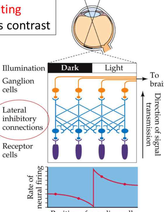
Mach Bands and Lateral Inhibition
Mach Bands: Optical illusion where changes in light intensity are exaggerated due to lateral inhibition.
Lateral Inhibition:
Inhibiting one's neighbors produces contrast.
Enhances the perception of edges and boundaries.
Receptor cells and ganglion cells are involved.
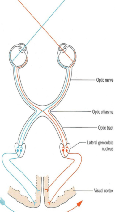
Visual Pathways
The visual pathway consists of:
Retina
Optic chiasm
Lateral geniculate nucleus (LGN) in the thalamus
Visual cortex (occipital lobe)
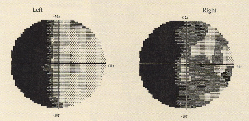
Hemianopia
Hemianopia: Loss of vision in half of the visual field.
A case study discussed a 29-year-old man with worsening vision in his left visual field due to a temporal lobe tumor.
Resection of the tumor, followed by chemotherapy and radiation, initially showed a good response, but the patient developed left homonymous hemianopia.
Receptive Fields in the Retina: On-Center and Off-Center Cells
On-Center/Off-Surround Cells (Type 1):
Excited by light in the center of their receptive field and inhibited by light in the surround.
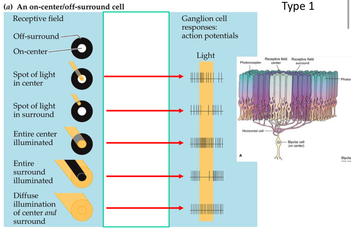
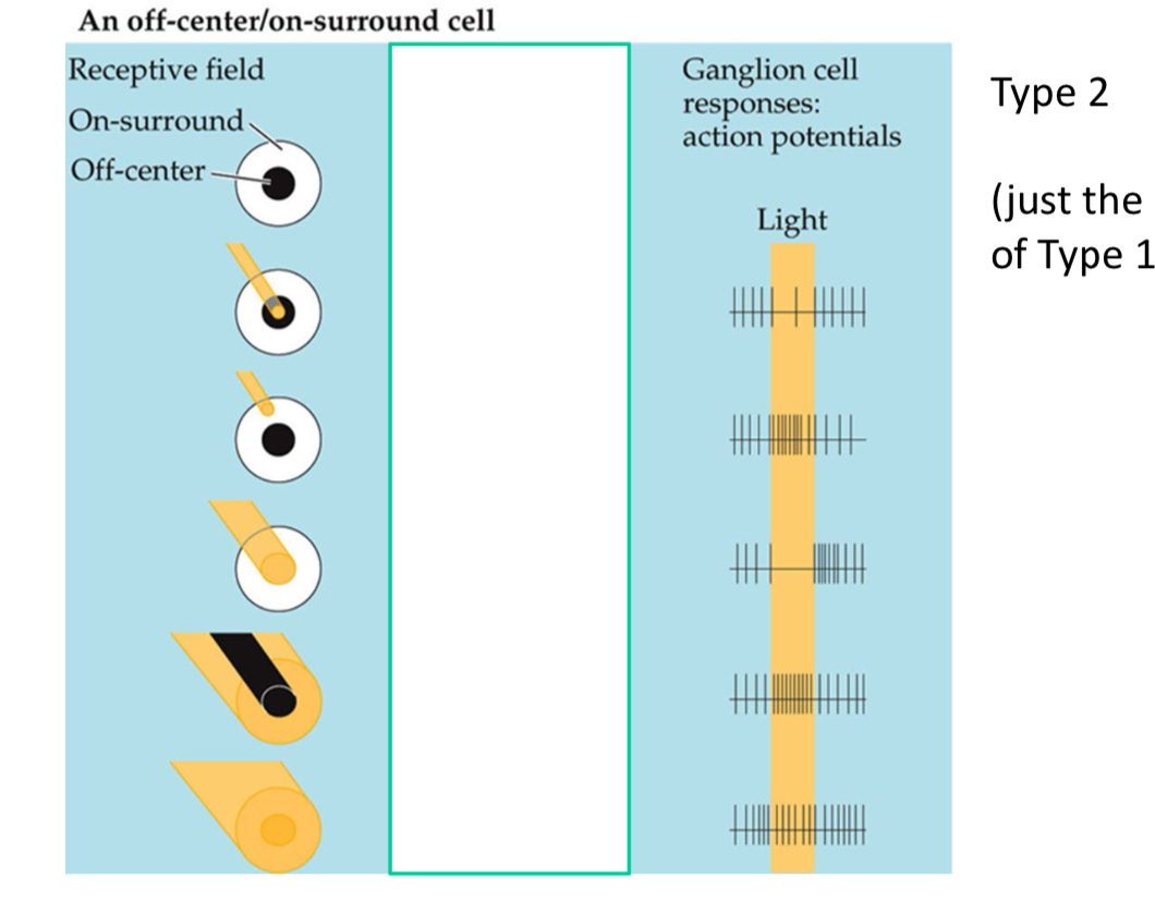
Off-Center/On-Surround Cells (Type 2):
Inhibited by light in the center and excited by light in the surround (reverse of Type 1).
Lateral Geniculate Nucleus (LGN)
The LGN in the thalamus has three cell types:
Parvocellular: Small cells, small receptive fields.
Magnocellular: Large cells, large receptive fields.
Koniocellular: Very small cells, located between the main layers.
Primary Visual Cortex (V1)
Most visual information first arrives at the primary visual cortex (V1) in the occipital lobe.
Brain maps of visual space are mostly devoted to the fovea.
Cortical Cells
Simple Cortical Cells: Respond to an edge or bar of a particular width, orientation, and location.
Complex Cortical Cells: Respond to a bar of a particular width and orientation but can be anywhere in the visual field.
Visual Cortex Processing
V1 is needed to form all visual images.
V2, V4, and the inferior temporal lobe perceive complex forms.
V5 is specialized for motion perception.
V1 Function
V1 breaks down the visual image into components: color, shape, and location, processing these inputs simultaneously.
V2 Function
V2 'fills in the gaps,' extrapolating and predicting from what is actually seen.
V4 Function
V4 cells respond to concentric and radial stimuli.
V4 is involved in color perception.
Motion Detection
Motion blindness (akinetopsia) occurs when the brain cannot process motion.
The brain determines if an object is moving before identifying it.
The retinal periphery (rods) is sensitive to motion and projects to V5.
Inferotemporal Cortex (IT)
Cells in the inferotemporal cortex respond to complex forms, including forms learned through recognition.
Receptive fields in IT cortex develop through experience and learning.
IT cortex is the final stage of visual processing.
Visual Processing Hierarchy
The visual processing hierarchy moves from simple edges and borders (V1) to complex forms (IT).
Three Channels from Retina to Higher Visual Cortex
M Channel (Magnocellular Pathway):
Orientation selective, directional sensitive for movement.
No color sensitivity.
Analysis of object motion.
P-IB Channel (Parvocellular Interblob Pathway):
High orientation sensitivity, no color sensitivity.
Small receptive fields.
Analysis of object shape.
Blob Channel (Parvocellular Blob and Koniocellular Pathway):
No orientation sensitivity, color sensitivity.
Analysis of object color.
Cortical Processing Disorders
Synesthesia: A condition where stimulation of one sense triggers experiences in another sense.
Example: A person might see colors when hearing sounds.
Alice in Wonderland Syndrome:
Characterized by micropsia (objects appear smaller than they are) and macropsia (objects appear larger than they are).
Occurs in migraine, EBV infection, and is common in children.
Integrative Agnosia:
Inability to attend to more than a small area of the visual field despite normal visual acuity.
Patients can see individual objects but cannot perceive a visual scene as a whole.
Palinopsia:
Patients see afterimages, both as a reduced amount of time required to form an afterimage and an increased duration of the afterimage.
Visual Agnosia:
Sensation intact and speech normal, but patients are unable to name objects, although they know how to use them.
Example: Patient GS.
Stroop Test
A test demonstrating interference in the reaction time of a task; for example, naming the color of a word when it differs from the word's meaning.
Blindsight Revisited
Patients with damage to the primary visual cortex can tell where an object is, although they claim they cannot see it.
In patient G.Y., blindsight is thought to be due to minor pathways into the extrastriate cortex that bypass V1.