Integumentary System
Intro to Skin
Made up of various tissues
Considered an organ
Functions of the Skin
Barrier against bacteria
Prevents excessive loss or absorption of water
Contains pigments that protect against ultraviolet rays (melanin)
Relays sensations from surroundings to the central nervous system
Helps maintain a constant body temperature
Storage place for water, fats, glucose
Production of vitamin D3
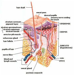
Composition
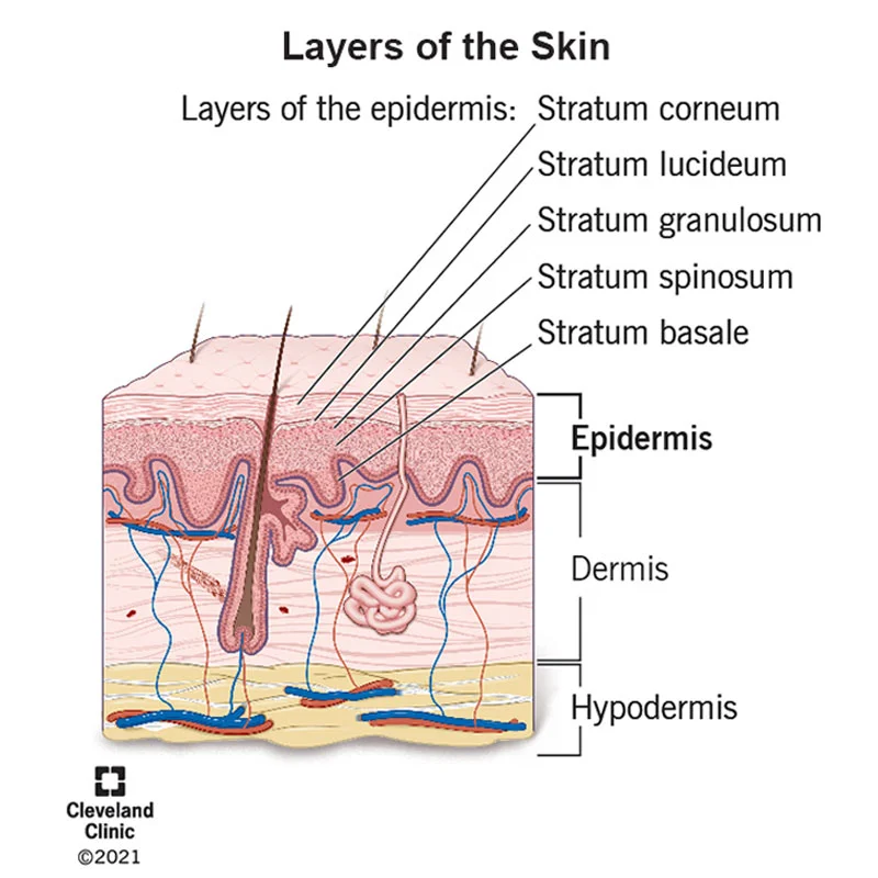
3 Major layers
Epidermis - outer epithelial layers
Dermis - middle connected tissue layers
Subcutaneous (hypodermis) - deeper adipose region, not considered true skin
Epidermal Layers
Stratum Corneum - Outermost Layers
Keratinized Layer - means the layer is hard and serves as protection
Stratum Lucidum – only on the palms and the soles of the feet.
(Skin is thickest here. Friction occurs most here)
Stratum Ganulosum
Stratum Spinosum
Stratum Germinativum ( or Basal, Basale)
Cells divide here and push their way to the top of the epidermis over 2 to 3 weeks
Specialized Epidermal Cells
Keratinocytes – Cells which produce keratin, a protein that makes both skin and hair elastic
Melanocytes – Located only in the Basal Layer
Produces Melanin, a protein that gives colour to the skin, hair, and eyes
Langerhans Cells - Help other cells of the immune system recognize invading pathogens
Easily damaged by UV light
Merkel Cells - Located in the Basal Layer
Detect Touch
Melanin
Based on the evolutionary advantage of having more absorbing melanin to prevent cell damage
Amount of melanin present makes for differences in skin color
Dark skin has more active melanocytes
Suntan – comes from the increased activity of melanocytes
A protective reaction to too much exposure to UV radiation
WEAR YOUR SUNSCREEN!
Albinos – organisms without melanin
White skin, hair
Red eyes due to blood passing through vessels of the retina
Dermis
Lies directly under epidermis
Connective tissue
Two Layers of the Dermis:
Papillary Layer – Lies directly under epidermis
Bumps along the top edge called Dermal Papillae
Fingerprints
Reticular Layer – deepest part of “true skin”
Elastic, connective tissue
Forms structure and framework for skin
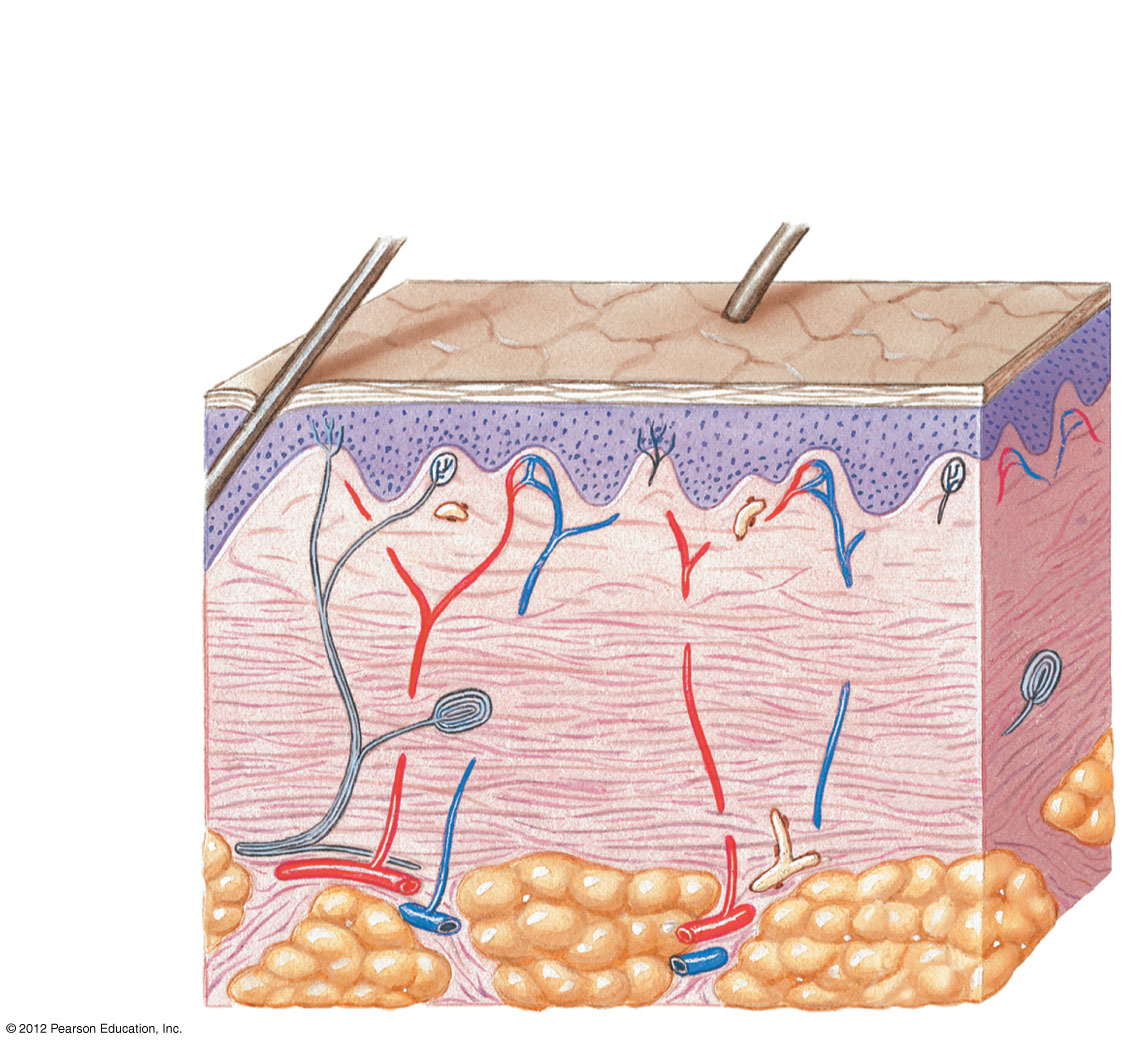
Characteristics of the Dermis
Well-supplied with blood vessels, lymph vessels, nerves, and accessory structures
High number of capillaries
When these capillaries dilate, blood rushes to the area and the skin reddens – blushing
Blood vessels come out of the heart and get smaller until they begin to return to the heart
Arteries (Largest) → Aterioles → Capilliares (Smallest; where exchanges happens) → Venules → Veins
Subcutaneous Region
Area below dermis
Also called Hypodermis
Adipose (fat) tissue stored her
Contains superficial fascia
Superficial Fascia is a connective tissue covering over the fat and connects the skin to the underlying bone or deep fascia
Adipose provides warmth and cushioning for underlying structure
Ringworm
Fungus – Dermatophyte (Disease is called Dermatophytosis)
Same fungus “family” as athlete’s foot: Tinea pedis
Can appear anywhere on the body
Red, bumpy, itchy ring
Very contagious
Treatments (Tx):
Antifungal creams – OTC
Antifungal medications from doctor
Doctor diagnoses using Wood’s light
Special UV light and if it is ringworm, the ring will glow
Usually clears up with treatment in one or two weeks
Sweating
Sweating - releases heat
Heat causes vessels in dermis to dilate
Blood rushes to skin surface, where heat can be released
Water (in the form of sweat from sweat glands) is brought to the surface of skin and evaporates
Takes heat with it
Skin Day 2
Skin Cancer
Cancer Vocab
Malignant = cancerous
Invades other tissues and damage is widespread
Benign = non-cancerous
Cells stay put and don’t invade other tissues
Oma = tumor
Carcin = Cancerous
Melan = Melanin
General Info
Skin cancer incidence increasing
Ozone layer thinning, more direct UV rays
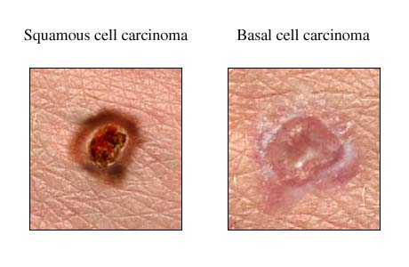
Cell Type
Most common type of skin cancer:
Basal cell carcinoma (Deepest Layer of Epidermis)
Most often found on face, head, neck, and hands
Mohs surgery to remove
Most deadly type:
Malignant Melanoma (affects melanocytes)
Treatment with surgery - wide margins
Severity of cancer depends on division
Rated as 1 – 5, with 1 being early stage and 5 being severe
Rank of Skin
Fair Skin
Blue or green eyes
Blistering sunburn before the age of 12
High level of sun exposure
Genetics
Freckles from the sun?
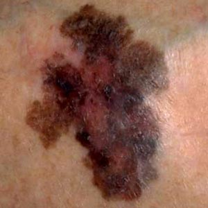
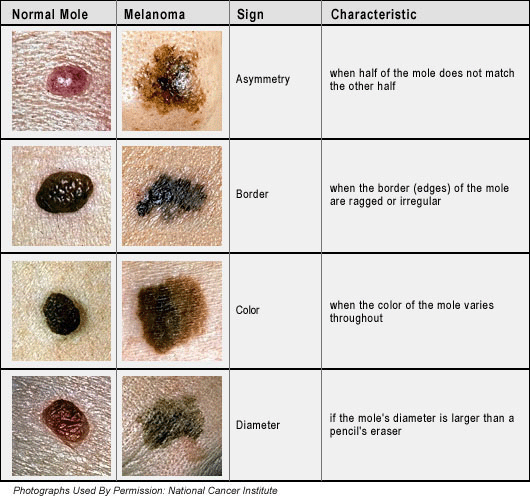
Moles
Possibly indicates malignant melanoma
Look for the following:
A – asymmetric shape
B – Border that is irregular or diffuse
C – Color that is pearly or multicoloured
D – Diameter larger than 5 mm (pencil eraser)
E – Elevation above the skin
Prevention
Early detection
Watch your moles
Avoid exposure to excessive amounts of sun, especially between the hours of 10:00 and 2:00
Wear sun protective clothing
Wear sunscreen
Australian Campaign:
Slip – on a shirt
Slap – on a hat
Slop – on sunscreen
DON’T USE TANNING BEDS
75% increase in skin cancer incidence
If in a high risk category, see your dermatologist
Parts of Hair
Shaft – visible part above skin surface
Root – part below skin surface
Hair bulb – expanded portion at base of root
Follicle – membranous enclosure
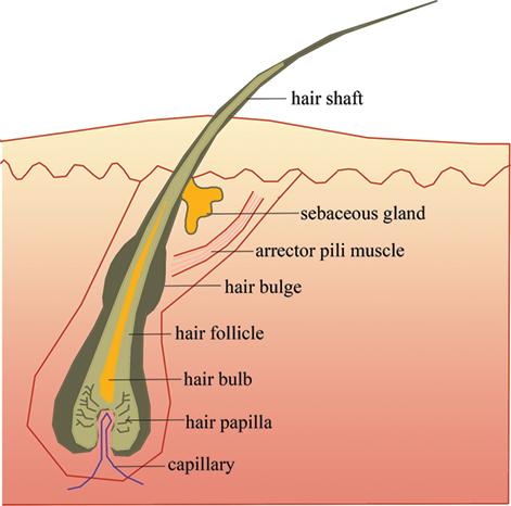
Arrector Pili Muscle – attached to each follicle and contracts as a result of cold or fright – goose bumps
Nerve receptor sac – wrapped around hair bulb
Sebaceous gland – located in dermis. Open to hair follicle and secretes substance for natural oil of hair
Hair Facts
Hair covers entire body except palms of hands and soles of feet
Vellus – light colored hair covering most of skin (peach fuzz)
Lanugo – hair covering a developing baby in utero
Visible hair is dead – can be cut, styled without pain
During pregnancy, hair grows thick and strong. Tends to fall out after labor and delivery due to the trauma to the body
Hair Life Cycle
Anagen – growth phase
Telogen – rest period following growth
During the rest period, hair becomes weak and may fall out (especially during brushing, etc.)
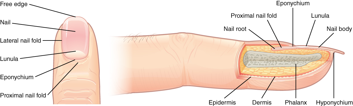
Nails
Nail plate composed of Keratin
No color due to no melanin
Pink due to capillaries below
Nail Parts
Nail Bed – groove in which nail plate lies
Nail Matrix – supporting structure under the plate
Nail Root – responsible for nail growth
Lunula – half moon area at base of nail
Eponchyeum – cuticle
Proximal Nail fold – fold of skin at base of nail
Nail Facts
Grow about 1/8 inch per month
3 or 4 weeks to grow from root to visible point
3 to 5 months to reproduce entirely
Nail plate is dead tissue which is why you can cut it without pain
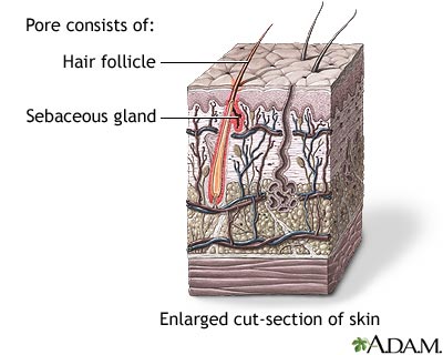
Sebaceous Glands
Oil glands attached to hair follicles
Produces sebum
Sebum is acidic & prevents skin infections
Sebum holds in moisture
Source of most acne
Present on all parts of body except palms of the hands and soles of the feet
Sweat Glands
Coiled, tubular glands within the dermis
Eccrine –
High number over entire body
Especially numerous on palms, soles of feet, and forehead
Passageway for sweat
Sweat composed of water and some salts
Sweat a response to emotional stress &/or thermal stress
Palms and soles respond more to emotional stress
Apocrine
Attached to hair follicles
Found mostly in axillary & anogenital region
Secrete sweat that contains fatty substances and proteins
Secretions may look milky in color
If bacteria feed on the secretions, foul odor occurs (body odor)
Generally start secretions at puberty due to influence of sex hormones
Lactation/milk/glands are apocrine
Ceruminous Glands – in the outer ear & secrete cerumen or ear wax
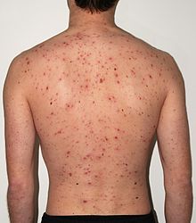
Varicella Zoster/Herpes Zoster
Virus
In children, causes Chicken Pox (Varicella Zoster)
If immune system compromised, causes shingles (Herpes Zoster)
Must have been exposed to Chicken Pox first to get shingles
Chicken Pox
Small, itchy bumps
Rare to have a recurrence
Immune System suppresses the virus, but it lies hidden in your body
Was very common until vaccinations began in 1995
The virus often stays hidden until after age 50
Lies dormant in cranial and spinal nerves
Something triggers the virus to become active again
Age
Exposure to excessive sunlight
Lowered immune system (including stress!)
Shingles
Red, itchy, runny, painful bumps
Usually on the torso
Very painful, as the virus is attacking the nerve root
Rash will clear within 4 to 6 weeks
Pain may continue for up to 6 months
May or may not have recurrences
Anything that suppresses the immune system after initial viral exposure may cause a breakout of shingles…