Localized Brain Damage
Overview:
Neuroscientists’ “Systems” Perspective: neural circuits in terms of function
Neuroscientists’ systems’ perspective contrasts neurologist’s systems’ perspective — the brain as an organ.
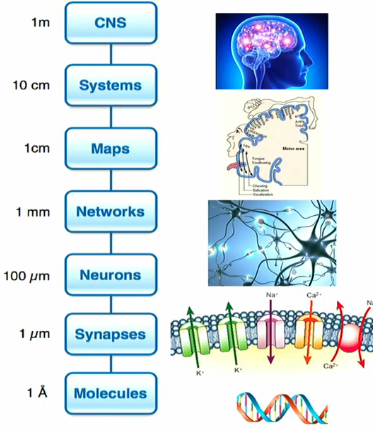 Looking at the nervous system from different scales of analysis.
Systems Neuroscience is the study of neural circuits in terms of their functions. This implies the involvement of many neurons and often includes consideration of multiple brain regions; e.g., neural processing in the visual cortex, motor coordination in the cortex and cerebellum, decision-making in the parietal and frontal cortex, and memory consolidation mediated by the hippocampus.
Due to emphasis on specialized function of large chunks of the brain, the systems’ level is healful for understanding the effects of localized brain damage. For various different disorders, different levels of analysis (different spatial scale) will be most useful as a leverage point depending on what' level of depth is necessary.
The leverage point for localized brain damage is the Systems neuroscience.
Other cases where the better leverage point is individual neurons (e.g., epilepsy)
Or on a molecular scale when talking about genetic causes of disorders.
Neuropsychology covers the relationship of brain damage and cognitive function.
Neuropsychological Study of Cognition ***
Localization of function in the visual system
Working with different regions, and seeing what happens with localized damage in those various brain regions: what is the cause of this localized damage? TBI, cerebrovascular (stroke), cancer (brain tumor).
Inouye observed wounded individuals as a result of war (injury: bullet that caused localized damage in the brain).
In this study he observed:
no two parties are ever identical in presentation of impairment.
The most common outcome of visual cortex damage is some variety of visual impairment.
The portion of visual space affected is usually contralateral from the damage.
If the right cortex is damaged, the left half of our visual field is impaired.
The specific nature of impairment depends on the part of visual cortex that is damaged.
Agnosia is an inability to process sensory information, not resulting from an elementary sensory defect (e.g., loss of an eye).
Brain damage in the primary visual cortex (V1)
The V1 receives the largest projection of visual input from the periphery of any area of cortex (via LGN in the thalamus). It is the dominant source of input (direct and indirect) for most of the other visual areas in the cortex. What happens when there is damage to V1?
V1 lesions result in the loss of conscious visual percepts in the corresponding region of visual space.
loss of conscious visual perceptions is only as bad as how much visual space is not being consciously perceived — the brain can usually fill in the void. This is a scotoma.
Brain damage to the MT
MT neurons are strongly sensitive to visual motion in a preferred location of visual space and of a preferred direction. Artificial stimulation of MT neurons in nonhuman animal models alters behavioral reports about the direction of visual motion.
if you stimulate neurons that prefer a rightward direction then animals will more likely report things as moving rightward even if its not. Essentially causing illusion of motion.
MT lesions in nonhuman animal models results in impairments in visual motion processing.
Perform poorly when asked to report the direction of motion.
MT lesions in humans can result in selective loss of motion perception with otherwise preserved vision — motion blindness. Motion perception is more than just recognition that something has been displaced in place.
imagine looking at life in screenshots over routine time intervals.
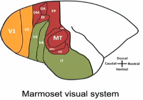
Organization of the visual system — at the level of the cortex, there are multiple distinct regions devoted to visual processing, including V1, V2, V3, V4, and MT (medial temporal). This suggests individual visual regions may have functional specialization.
These maps are distinct, each region (V1, V2, V3, V4, and MT) has a full map of visual space.
There are neurons in each of these regions that are selective for different parts of visual space.
The neurons modulate their responses based on the different features of the visual environment.
V1 neurons are selective (sensitive) to the orientation (angle) of a visual edge (bar)
MT neurons are sensitive to the direction of visual motion in a particular area of the visual space.
Blindsight — people with loss of conscious vision perform at higher than chance levels when guessing the direction of an object that is moving in front of them in their blind spot. This most frequently occurs with lesions on a single side of the visual cortex. These results suggest that other pathways bypass V1.
Specialized visual processing of faces — humans are able to detect differences in upright faces with a high degree of sensitivity.
Selective impairment for visual processing of faces: faces of different individuals are highly similar and therefore challenging to recognize.
In the human brain, the fusiform face area (FFA) shows strong activity in response to facial stimuli. Damage at and around the FFA can result in selective impairment in facial recognition — prosopagnosia. Being able to identify someone is a higher level order of functioning, so there are still ways a person with prosopagnosia is able to identify others using other course characteristics (e.g., voice)
Impaired spatial awareness with parietal damage (Hemispatial neglect)
Hemispatial neglect is characterized by a deficit in awareness (consciousness) and attention (focus) for one side of the visual space. It is most commonly caused by damage in the parietal cortex; more often on the right (left-side neglect).
Neglect is a complex cognitive impairment. Individuals can improve over time (e.g., strategic adjustment
This type of impairment implies that different brain regions make distinct contributions to cognitive processing. The parietal cortex is important for cognition related to spatial awareness. Keep in mind other brain regions are likely important for this function and the parietal cortex is important for other functions as well.
Impaired executive function with frontal damage.
Phineas Gage — suffered major frontal lobe damage as a rod went completely through his head but he survived; however, he changed. Before the incident, he was considered hard-working, responsible, intelligent, and held in high regard. After the incident, he was less responses, more obstinate, more impulsive. He wasn’t able to keep his job.
This pattern is fairly consistent with other patients with frontal damage (MT). Also common for slow, progressive degeneration frontal cortex or TBI to the frontal cortex.
TBI could be external trauma or stroke
This suggested that the frontal cortex played a critical role in executive function.
Localization function in the domain of memory.
The effects of localized brain damage extend into the cognitive domain — impairment of memory due to hippocampal damage. The hippocampus is a specialized structure in the cerebrum. It connects to the cortex but is a distinct structure. There is one on each side of the brain.
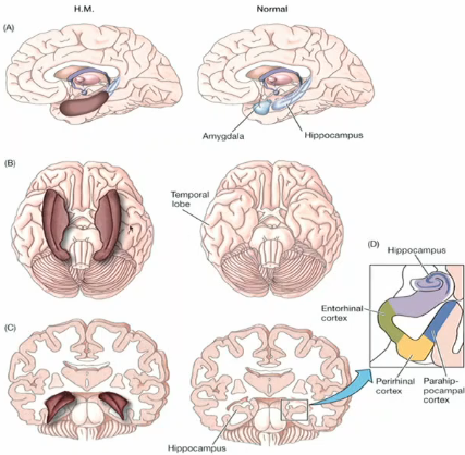
Example: HM (Henry Molaison) had brain surgery to treat epilepsy. They removed 2/3rds of both hippocampi + surrounding brain structure. What he removed from his surgery resulted in more debilitating symptoms than what was caused by his epilepsy. He had severe and specific memory impairments.
He was studied for +50 years until death in 2008. His non-memory cognitive faculties were preserved but suffered from severe anterograde amnesia — (inability to make new long-term memories). He had retrograde memory (memories prior to surgery) and short-term memory (sec to mins).
His anterograde memory deficit was specific for declarative memories (you are conscious about and can easily explain) while having preserved the ability to form new procedural memories (not easily explained, but can show you).
This suggests that the hippocampus is not necessary for having long-term memories, but is essential for forming new long-term memories → memory consolidation. This also suggests that memory is composed of multiple systems.
Specific language impairments with localized brain damage.
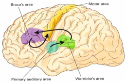 Language is hearing things, deciphering what they mean, and then producing new words to communicate back. There are area of the brain where localized cortical damage can result in specific impairments to language capacities (aphasia). Aphasia can apply to all types of aspects about speech: comprehension, production, etc. In contrast to hemispatial neglect, the left hemisphere of the cerebral cortex
In most people, the left hemisphere of the cerebral cortex appears to be more important for language capacities. This can be determined through a test — injecting a compound through the carotid artery that temporarily reduces activity on one side of the brain and seeing whether language is impaired or not depending on what side of the brain is affected.
There are individual differences. In most people (typical of right-handed people), the left hemisphere of the cerebral cortex appears to be more important for language capacities. This is opposite from the side where damage is most likely to cause demispatial neglect.
Left-handed people exhibit more distributed activity along the cerebral cortex when engaging in language capacities.
However, it can be distributed or even right hemisphere specific for right-handed individuals.
When we talk about spatial neglect and parietal cortex it is commonly a result of damage to the right parietal cortex.
Notice this is opposite from the side where damage is most likely to cause hemispatial neglect
Wernicke’s area (green area) is a part of the temporal lobe of the cortex that receives input from the primary auditory cortex.
primary auditory cortex → temporal lobe → Wernicke’s area
Broca’s Area (purple)— located in the frontal lobe. This area has neurons and axons that are sent to the motor area; the majority of axons remain in Broca’s area, but few go from Broca’s area to the primary motor cortex. Ir receives prominent axonal projections from Wernicke’s area.
Auditory processing: primary auditory cortex, Wernicke’s Area, Broca’s Area, motor cortex, and so on.
What happens if there is damage to each of these areas?
Example: Wernicke’s Aphasia — difficulties in language comprehension. From the distance, you would not be able to tell this person is speaking nonsense. You would likely think they were holding a normal conversations until you start to listen to the contents.
This suggests that Wernicke’s area may have a specialized role in processing language in part via input from the primary auditory cortex. Auditory signals are received and sent to Wernicke’s area where they are processed and comprehended. It likely not the only area of the brain involved in language processing, but it plays a crucial role.
Example: Broca’s Aphasia — The participant suffered from a stroke which resulted in permanent damage to Broca’s area. People with this condition have difficulties with language production but they can fully understand the conversations around them.
This suggests that Broca’s area may have a specialized role in producing language that is communicated to the musculature via the primary motor cortex.
Language is a complex interactive undertaking with comprehension and production working in tandem — you listen, you talk. There are a variety of aphasia that are related to damage to connections between regions (Wernicke’s area, Broca’s area, primary auditory area, motor area). The deficit caused by damage does not mean that region is only associated with that area and function, that region of the brain can be involved in other processes. Damage to connections between Wernicke’s and Broca’s area can also result in language deficits, possibly due to the need for complex coordination between comprehension and production.
Learning Objectives
Specialization in cortical visual processing
Laterality of cortex
Blindsight
Motion blindness
Prosopagnosia
Hemispatial neglect
Effects of (pre)frontal damage.
Impaired memory with hippocampal damage
Broca’s and Wernicke’s aphasia
Improvement over time (plasticity) and its limitation