knowt logo

CELL BIO UNIT 2 NOTES

Chapter 14

  • Oxidative phosphorylation - ADP + Pi ->ATP THIS IS INNER MEMBRANE

  • Electron transport equation- NADH + ½ O2+H+->NAD+ +H2O NET OF INNER TRANSPORT

  • Biological oxidation is important so humans don’t combust spon

-famously and the energy in the bonds are in small dosages

  • O2 is important for biological oxidation so there is a place for

Energy to go

  • Citric acid cycle> NADH dehydrogenase>Quinone>cytocromeC(REDUCED)>cytocrome C oxidase(OXIDIZE)
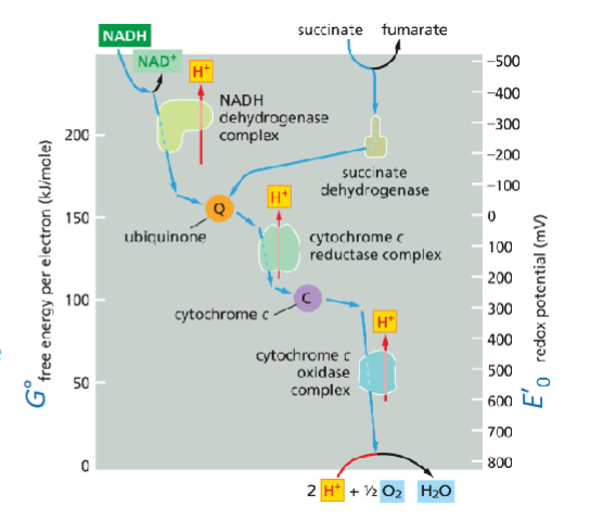
MEMORIZE THIS

Chapter 14BCONT

  • First is reduced and then oxidized

  • redox(mV) to free energy(kJ/mole)

  • E’delta=mV, G0=kJ/mole

  • The more positive redox potential the stronger acceptor more NEGATIVE BETTER DONOR

  • n=#ofelectrons

  • CAVEAT redox potential can be influenced by redox conc3entration

  • Heme group attached to c cytocrome

  • Copper is critical to complex 4

  • Iron sulfur cluster

  • Q(noOH), QH(1OH), QH2(2OH)

  • None, semi, all

  • MATRIX ARM -electron transport

  • MEMBRANE ARM-proton pumping

  • Proton highways are good and 40X fast but its non-selective , we dont know if its unidirectional

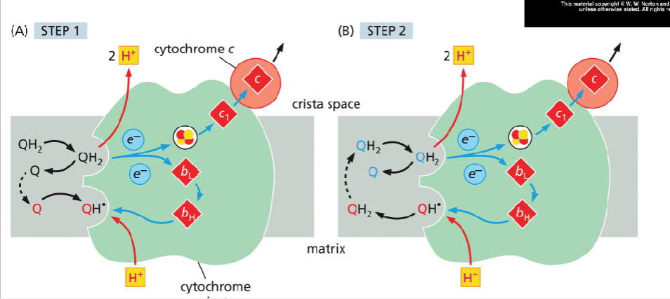
  • When 2 H+ joins Q it becomes QH2

  • Through cytocrome reductase QH2>iron sulfur cluster>cytocrome b>hemebh>heme bL>cytocrome c1>cytocrome c

  • none>semi then semi>all

  • Cytocrome C passes through cu and then heme

  • iron/sulfur>su>heme

  • Cytocreme C can indicate cell death

  • FAD from succinate dehydrogenase donates to Q

CH 14C

  • 1 glucose > 2 pyruvate+2NADH(1.5ATP)+2ATP GLYCOLYSIS

  • 2pyruvate>2acetyl coA+2NADH(2.5 ATP per) PYRUVATE DEHYDROGENASE

  • 2acetyl CoA>6NADH+2FADH2+2GTP CAC

  • 2pyruvate > 8NADH+2FADH2 (1.5 ATP)+2GTP NET MITOCONDRIA

  • 1 palmitoyl CoA-> 8Acetyl CoA +7NADH +7 FADH2 FATTY ACID OXIDATION

  • 8 Acetyl CoA.>24 NADH + 8 FADH2 +8 GTP CAC

  • 1 palmitoyl CoA >31 NADH +15FADH2 +8 GTP NET RESULT MITOCONDRIA

  • NADH has 2 values because LOCATION CHANGES
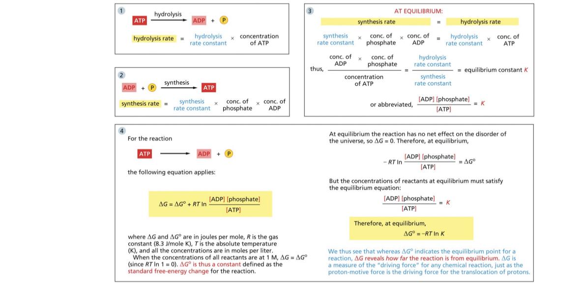
  • ATP hydrolysis is -30.5kJ/mole

  • -54kJ is Gdelta or 1ATP

  • Nadh to oxygen is spontaneous

  • 14 protons in total

  • Only 61.4% of energy reaches the next stage

  • - delta G is coupled with positive and for example NADH oxidation and creation of the proton gradient are energetically coupled

  • Diffusion of the proton gradient-and generation of atp synthase+

  • Long arm is stator in ATP synthase

  • There is a rotor stalk

  • As the protons move they loose their negative and is pushed into the matrix

  • The stator holds F1 stable so the F1 can catalyzize atp synthesis

  • Each bump in the images is created by hairpin protien (F0)

  • 13-15 hairpins but can be as low as 8-9 or 21-23(F0)

  • You need 1 proton for every hairpin

  • The difference in charge drives the rotor around

  • There are dimers that stabilize the atp syntheses

  • There is a sink for atp synthase

  • 8000 revolutions per minute,3 atp per turn

  • ADP/ATP cotransporter

  • The inter membrane space will be the more positive side

  • ATP being negative drives directionality

  • Bacteria use proton to pump nutrients into the cell and ATP

  • In anerobic atp pumps and Na but no gradient is established

CHAPTER 14 D

  • Stroma = matrix

  • Thylakoids= cristae

  • ATP in plants is produced in the stroma and consumed
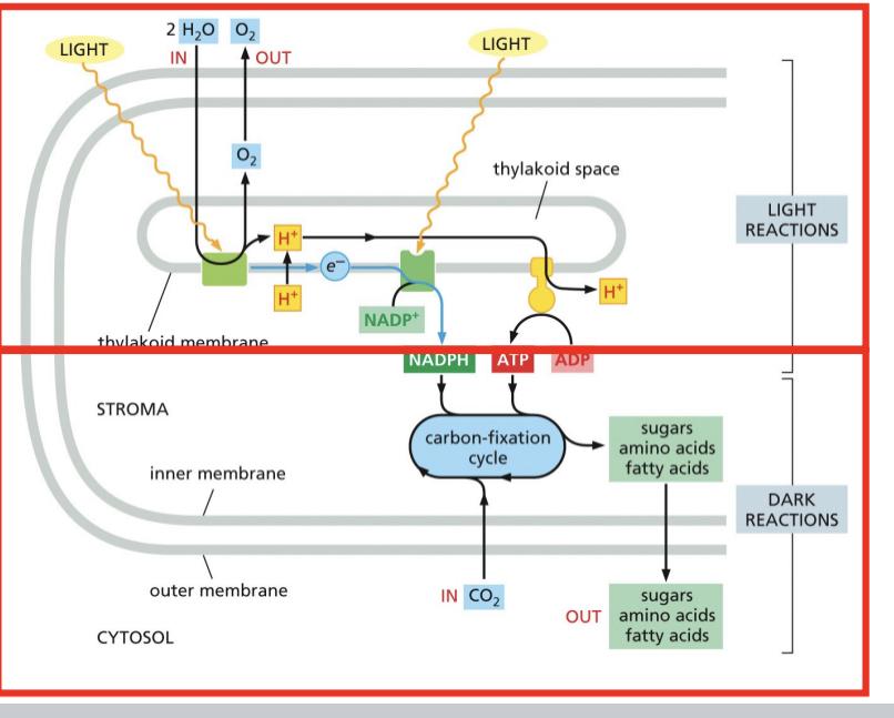
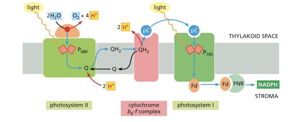
  • The high level electrons get excited and water replaces those electrons

  • Dark reactions are dependent on the products of light
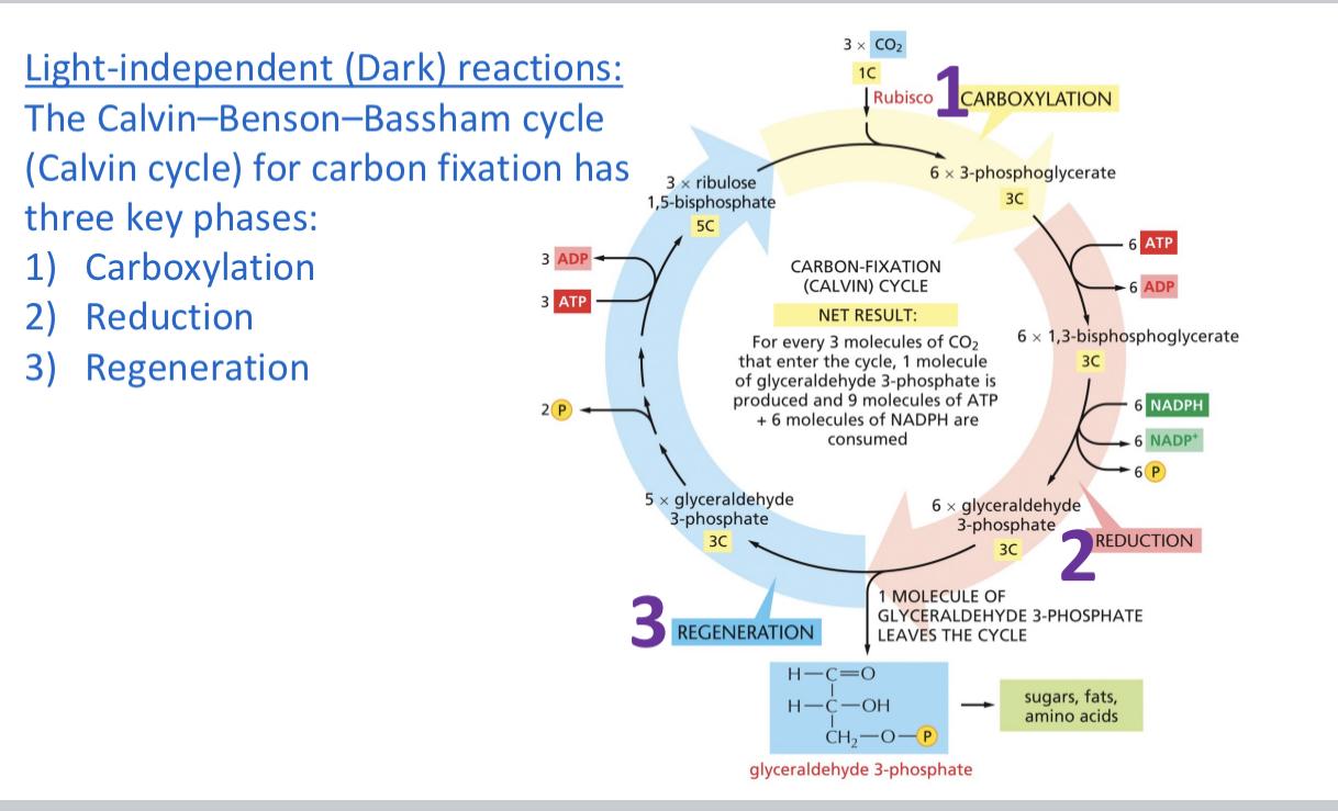
  • Carboxylation, take the product 5C +1C=6C/2=3C molecules

  • Rubisco fixes the carbon

  • Reduction is gain of electrons and remodeling of bonds 6 3 Carbon molecules NADPH is in the bond energy , 5 continue because we keep one to break down

  • REGENERATION 5 * 3C +ATP from light

  • Carboxylation is CO2+ribulose 1,biphosphate>w/rubisco intermediate +H2O2 molecules of 3-phosphoglycerate
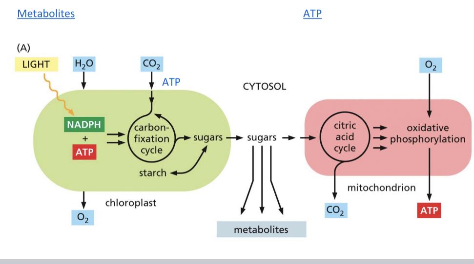
  • Review the photo system video

  • 100 per second

  • Mn cluster splits the water for electrons to be excited into the zscheme

  • Photo system 2 only has one pathway that’s used rapid
  • The alpha rotor has 12 subunits=12 - charged residues

  • Every time a proton enters the hairpin is moved one over

  • How many protons are needed to turn =how many subunits there are

  • Every 12 protons 3 ATP

  • Ancient origin ATP synthesis (used originally for protons not ATP) first then ETC and then they’d are combined

  • Started from ancestral fermenting minimal atp and H2S synthesis’s

  • Purple non sulfur bacteria used something like cytochrome c but it wasn’t

CHAPTER 16A

  • Freeway system made out of ants

  • Cytoskeleton maintains org,cell structure , cell shape changes,cell movement, and intracellular trafficking of vesicles/organelles, cell division

  • Cytoskeleton react in response to a lot of signals

  • Repolarize-rebuild cell skeleton

  • Actin and microtubules are dynamic to refunctionung

  • All filaments need 1000 monomeric subunits and though they are structurally simple prokaryotes have ace steal versions of these filaments

  • Actin fibers can be like a fishing line, very versatile

  • Microtubules have + or - ends

  • All of these filaments can interact to make and maintain a mature cell cytoskeleton

  • Intermediate filaments are made of a-helical region its coiled together until dimer becomes a Teramer and it is no longer parallel

  • This is a really strong 32 protein chains and it is a very strong

  • Mesenchymal cells muscle and keratin

  • Nuclear laminate give tremendous structural support unless it’s phosphorylated and it falls apart

  • Epidermal IF keratin filaments connect through desmosomes or hemidesmosomes

  • EBS rapid blistering due to tetramers not being able to form into protofilaments

  • Axons IFs-

  • Actin filaments, most abundant proteins in animals, all cells need it, 7nm , used as a reference control in everything and thin mesh work

  • Repeat overly short lived , creates striated muscles

  • Dynamic burst of polymerazation makes the cell move really quickly

  • G-Actin -globular actin is a mono subunit

  • It is a spherical contact had atp Imbedded inside the gactin

  • Factin- there is a + and - end

  • Suitable pool of charged g-actin ATP

  • Add more readily to the + end

  • You cannot depolymerize from the middle

  • Polymerization happens in one configuration

  • Origin of actin actin and myosin bonds and

  • Myosin creates a barb and the plus end is more barbed

  • The process of polymerizing 1.lag phase2.growth phase3.equilibrium phase lots of gactin to factin

  • Lag phase - nucleation it’s rryl long

  • The growth phase occusd as a monomer add to the exposed ends of the growing filaments,causing filament elongation

  • If a gactin needs to be added then it needs to be removed, balanced

  • We run out of free subunits and it hits an equilibrium

  • Disassembly is more favorable at the - end

  • Once they are in the polymer there is no difference in the - and + surface

  • The plus end is more stable because they ATP binds initially (glue) that does not happen to the negative end

  • If depolymerazes because of a time differential

  • Critical concentration is higher for the ATP bound + side then the ADP bound - side

  • Hydrolysis is not nessecarg

  • Treadmilling when the actin filaments results when assembly at the plus end is concomitant with depolymerization at the minus end

CHAPTER 16 B

  • Arp2 and 3 very actin related

  • Arp3/2 bind to NPF and clicks both into a active state so it can bind to a uncleared actin filament (ON MINUS END)

  • ARPS kind of glue onto the edge (70° angle) these make branched formations

  • ADP it’s pushed into the NPF and the actins dissasociatw.

  • Profilin is opposed by thyomisin

  • Forms clamp the actin and myosin to the membrane (associated with the plus end)

  • Uniform Z disks lead to striating

  • Tropomodilin and CapZ prevent polymerization and depolarizing

  • We can use coffilin to break off actin filament ends(induces severe twisting)

  • Creates new ends to deassamble more rapidly

  • Filament is gel forming and brain relies on it

  • I. Fibrin it’s exclusively structural

  • Periventrical heterotrophia-seisures

  • Myosin a ton binding protien 

  • Listeria bacteria-pathogenic food poisoning coops actin array and pull the subunits to breaking 

  • You can generate force by the actin by densly pushing things to the front

  • Focal adhesions via integrins to move the leading edge

  • All of the cell Mobility is myosin dependent 

  • Cell moves like this

  • Coffilin will twist the actin filaments to have Arp fill in to make it very large 

  • Amobeid cell membrane is facilitated by external sensing and feedback, they hunt for pathogens and commit eating so it can use the actins from them 

  • Ratchet is used to describe the actin filaments moving forward and not back 

  • Profilin recruits actins

  • Capping protien binds to plus end to stop it Arp is the branch

  • These factor give polarity and allow the cell to move 

  • A lot of feedback crll regulation

  • Myosin is an allosteric binding protien that depends on polarity and atp Adele activity, + end directed, actin dependent

  • Myosin and actin work together to perform cellular functions: vesicle translocation,cytoplasmic streaming,cell locomotion, and muscle contraction

  • Allosteric-protien with a binding site so the structure can conform to it

  • Atp and ADP stay in their pocket tnad it effect the relative shape

  • Myosin is highly concerned

  • 2myosinnprotiens will be interwoven

  • Myosin 1-the bilayer is attaching to the cortical actin to hydrolyze atp

  • Myosin 5- the carbon binding protien da drag and grab vesicles by “walking” and it has a 35nm  distance(draw this )

  • Myosin 2- has 2 heavy subunits with a coil coil terminus

  • The neck regions diversify the force

  • These are regulated , from the center there is a bare zone 

  • 50-60 myosin heads 

    CHAPTER 19

  • Cadenerin based protiens respond to adherin junctions

  • How one cell is connected to another

  • Cells anchor to other junctions based off cadaherins

  • High calcium is needed for cadaherims to link and then anchor to the cytoskeleton

  • Under tension protiens are extended to reduce stress on the molecule

  • The actin support cell connections, important and weak, useless without adherin junction

  • Adherin junction can generate force and bend and pull tubes out

  • Desmosomes cell cell anchoring junctions they are embedded within the plasma membrane with

  • Integrins is like cadaherins but it binds to the extracellular material

  • Tight Junction- elaborate array of protien, function is to seperate spaces that are created by a layer of cells, separates interstitial and blood stream(Tupperware lid) creates a seal

  • Series of protiens stitched together (clodins and aculin ) with lateral attachments across

  • Channel forming junctions or gap junctions- connects cytosol , chemical synapses, only 1000 daltons can pass small ions and molecules can pass

  • Gap junctions that are small stack together

  • The ECM is diverse and complicated mixture, its secreted and it can be structural or space filling,

  • Makes you durable and promotes compression resistance

  • Fibrous protiens provide tensile resistance

  • Hyluranan forms the backbone for bigger complex of things like heparin that ca. dock on the hyluran complex

  • Collagen- an example of a fibrous protien that can provided tensile strength via GAG and it’s usually made of a chain of amino acid, fhe partner chains are woven together

  • Hydroxylation gives collagen distinctive folding patterns.

  • Ex bone defects,dwarfism, nasal laminate will fail in the kidenehs , and all sorts of problems that come from a lack of calllahdn

  • Collagen is dependent on vitamin C as a cofactor

  • Elastin- forms cross links and are coiled up to allow streachimess and relaxation and prevents the other end without tearing ping or ripping

  • Finromectim and other multi adhesive protiens all of domains can be switched out via alternitive splicing

  • Fibromectins bind integrins

  • Arg protiens allow the integrity protiens allow the cell being knit into the and then show it to. E

  • Basal lamina is exclusive to the epithelial cells

  • The key assembly factor that is growing into the cell did 3 rd protien call dystroglucan,indigenous,,perfect

  • Integrity bind the cell to the extracellular ,atria

  • Connected to cell with intermembrane filaments, and anchoring junctions

  • Inactive integrity- makes internal and external signals on the alpha and beta site inactive anchoring

  • Lateral cross linking protiens mediate binding

LC

CELL BIO UNIT 2 NOTES

Chapter 14

  • Oxidative phosphorylation - ADP + Pi ->ATP THIS IS INNER MEMBRANE

  • Electron transport equation- NADH + ½ O2+H+->NAD+ +H2O NET OF INNER TRANSPORT

  • Biological oxidation is important so humans don’t combust spon

-famously and the energy in the bonds are in small dosages

  • O2 is important for biological oxidation so there is a place for

Energy to go

  • Citric acid cycle> NADH dehydrogenase>Quinone>cytocromeC(REDUCED)>cytocrome C oxidase(OXIDIZE)

MEMORIZE THIS

Chapter 14BCONT

  • First is reduced and then oxidized

  • redox(mV) to free energy(kJ/mole)

  • E’delta=mV, G0=kJ/mole

  • The more positive redox potential the stronger acceptor more NEGATIVE BETTER DONOR

  • n=#ofelectrons

  • CAVEAT redox potential can be influenced by redox conc3entration

  • Heme group attached to c cytocrome

  • Copper is critical to complex 4

  • Iron sulfur cluster

  • Q(noOH), QH(1OH), QH2(2OH)

  • None, semi, all

  • MATRIX ARM -electron transport

  • MEMBRANE ARM-proton pumping

  • Proton highways are good and 40X fast but its non-selective , we dont know if its unidirectional

  • When 2 H+ joins Q it becomes QH2

  • Through cytocrome reductase QH2>iron sulfur cluster>cytocrome b>hemebh>heme bL>cytocrome c1>cytocrome c

  • none>semi then semi>all

  • Cytocrome C passes through cu and then heme

  • iron/sulfur>su>heme

  • Cytocreme C can indicate cell death

  • FAD from succinate dehydrogenase donates to Q

CH 14C

  • 1 glucose > 2 pyruvate+2NADH(1.5ATP)+2ATP GLYCOLYSIS

  • 2pyruvate>2acetyl coA+2NADH(2.5 ATP per) PYRUVATE DEHYDROGENASE

  • 2acetyl CoA>6NADH+2FADH2+2GTP CAC

  • 2pyruvate > 8NADH+2FADH2 (1.5 ATP)+2GTP NET MITOCONDRIA

  • 1 palmitoyl CoA-> 8Acetyl CoA +7NADH +7 FADH2 FATTY ACID OXIDATION

  • 8 Acetyl CoA.>24 NADH + 8 FADH2 +8 GTP CAC

  • 1 palmitoyl CoA >31 NADH +15FADH2 +8 GTP NET RESULT MITOCONDRIA

  • NADH has 2 values because LOCATION CHANGES

  • ATP hydrolysis is -30.5kJ/mole

  • -54kJ is Gdelta or 1ATP

  • Nadh to oxygen is spontaneous

  • 14 protons in total

  • Only 61.4% of energy reaches the next stage

  • - delta G is coupled with positive and for example NADH oxidation and creation of the proton gradient are energetically coupled

  • Diffusion of the proton gradient-and generation of atp synthase+

  • Long arm is stator in ATP synthase

  • There is a rotor stalk

  • As the protons move they loose their negative and is pushed into the matrix

  • The stator holds F1 stable so the F1 can catalyzize atp synthesis

  • Each bump in the images is created by hairpin protien (F0)

  • 13-15 hairpins but can be as low as 8-9 or 21-23(F0)

  • You need 1 proton for every hairpin

  • The difference in charge drives the rotor around

  • There are dimers that stabilize the atp syntheses

  • There is a sink for atp synthase

  • 8000 revolutions per minute,3 atp per turn

  • ADP/ATP cotransporter

  • The inter membrane space will be the more positive side

  • ATP being negative drives directionality

  • Bacteria use proton to pump nutrients into the cell and ATP

  • In anerobic atp pumps and Na but no gradient is established

CHAPTER 14 D

  • Stroma = matrix

  • Thylakoids= cristae

  • ATP in plants is produced in the stroma and consumed

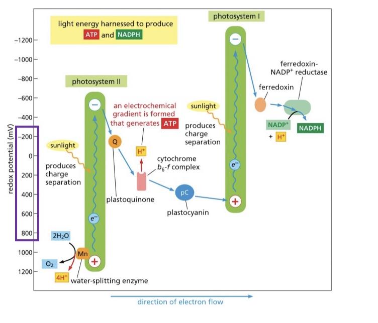
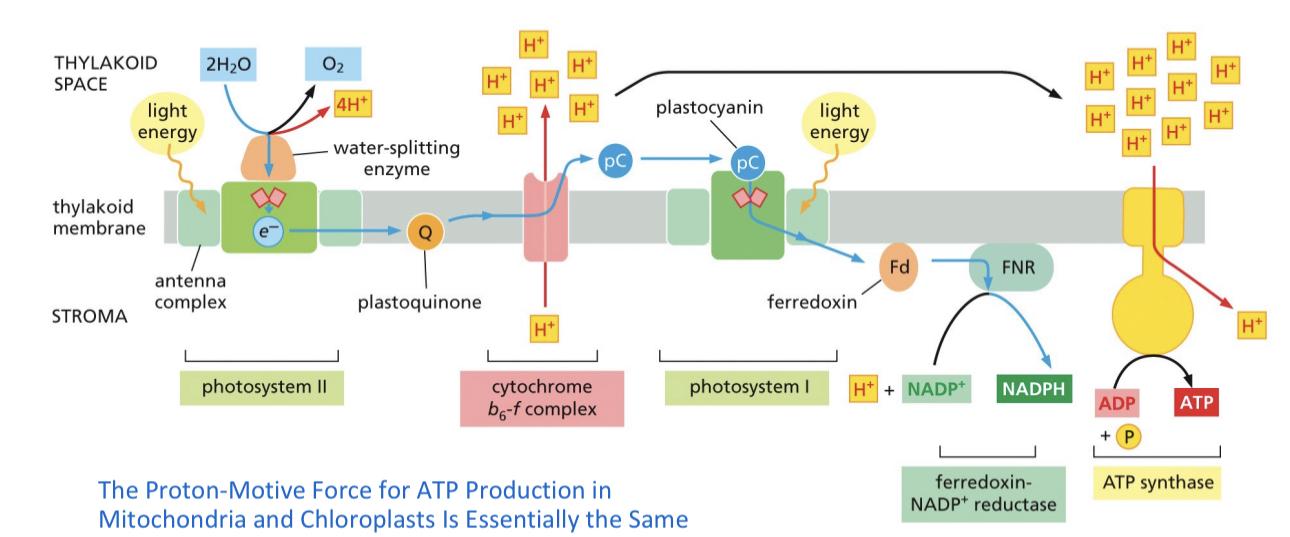
  • The high level electrons get excited and water replaces those electrons

  • Dark reactions are dependent on the products of light

  • Carboxylation, take the product 5C +1C=6C/2=3C molecules

  • Rubisco fixes the carbon

  • Reduction is gain of electrons and remodeling of bonds 6 3 Carbon molecules NADPH is in the bond energy , 5 continue because we keep one to break down

  • REGENERATION 5 * 3C +ATP from light

  • Carboxylation is CO2+ribulose 1,biphosphate>w/rubisco intermediate +H2O2 molecules of 3-phosphoglycerate

  • Review the photo system video

  • 100 per second

  • Mn cluster splits the water for electrons to be excited into the zscheme

  • Photo system 2 only has one pathway that’s used rapid
  • The alpha rotor has 12 subunits=12 - charged residues

  • Every time a proton enters the hairpin is moved one over

  • How many protons are needed to turn =how many subunits there are

  • Every 12 protons 3 ATP

  • Ancient origin ATP synthesis (used originally for protons not ATP) first then ETC and then they’d are combined

  • Started from ancestral fermenting minimal atp and H2S synthesis’s

  • Purple non sulfur bacteria used something like cytochrome c but it wasn’t

CHAPTER 16A

  • Freeway system made out of ants

  • Cytoskeleton maintains org,cell structure , cell shape changes,cell movement, and intracellular trafficking of vesicles/organelles, cell division

  • Cytoskeleton react in response to a lot of signals

  • Repolarize-rebuild cell skeleton

  • Actin and microtubules are dynamic to refunctionung

  • All filaments need 1000 monomeric subunits and though they are structurally simple prokaryotes have ace steal versions of these filaments
  • Actin fibers can be like a fishing line, very versatile

  • Microtubules have + or - ends

  • All of these filaments can interact to make and maintain a mature cell cytoskeleton

  • Intermediate filaments are made of a-helical region its coiled together until dimer becomes a Teramer and it is no longer parallel

  • This is a really strong 32 protein chains and it is a very strong

  • Mesenchymal cells muscle and keratin

  • Nuclear laminate give tremendous structural support unless it’s phosphorylated and it falls apart

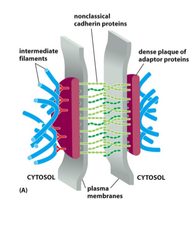
  • Epidermal IF keratin filaments connect through desmosomes or hemidesmosomes

  • EBS rapid blistering due to tetramers not being able to form into protofilaments

  • Axons IFs-

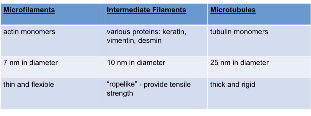
  • Actin filaments, most abundant proteins in animals, all cells need it, 7nm , used as a reference control in everything and thin mesh work

  • Repeat overly short lived , creates striated muscles

  • Dynamic burst of polymerazation makes the cell move really quickly

  • G-Actin -globular actin is a mono subunit

  • It is a spherical contact had atp Imbedded inside the gactin

  • Factin- there is a + and - end

  • Suitable pool of charged g-actin ATP

  • Add more readily to the + end

  • You cannot depolymerize from the middle

  • Polymerization happens in one configuration

  • Origin of actin actin and myosin bonds and

  • Myosin creates a barb and the plus end is more barbed

  • The process of polymerizing 1.lag phase2.growth phase3.equilibrium phase lots of gactin to factin

  • Lag phase - nucleation it’s rryl long

  • The growth phase occusd as a monomer add to the exposed ends of the growing filaments,causing filament elongation

  • If a gactin needs to be added then it needs to be removed, balanced

  • We run out of free subunits and it hits an equilibrium

  • Disassembly is more favorable at the - end

  • Once they are in the polymer there is no difference in the - and + surface

  • The plus end is more stable because they ATP binds initially (glue) that does not happen to the negative end

  • If depolymerazes because of a time differential

  • Critical concentration is higher for the ATP bound + side then the ADP bound - side

  • Hydrolysis is not nessecarg

  • Treadmilling when the actin filaments results when assembly at the plus end is concomitant with depolymerization at the minus end

CHAPTER 16 B
  • Arp2 and 3 very actin related

  • Arp3/2 bind to NPF and clicks both into a active state so it can bind to a uncleared actin filament (ON MINUS END)

  • ARPS kind of glue onto the edge (70° angle) these make branched formations

  • ADP it’s pushed into the NPF and the actins dissasociatw.

  • Profilin is opposed by thyomisin

  • Forms clamp the actin and myosin to the membrane (associated with the plus end)

  • Uniform Z disks lead to striating

  • Tropomodilin and CapZ prevent polymerization and depolarizing

  • We can use coffilin to break off actin filament ends(induces severe twisting)

  • Creates new ends to deassamble more rapidly
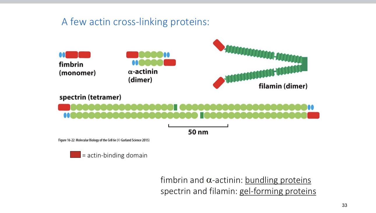
  • Filament is gel forming and brain relies on it

  • I. Fibrin it’s exclusively structural

  • Periventrical heterotrophia-seisures

  • Myosin a ton binding protien 

  • Listeria bacteria-pathogenic food poisoning coops actin array and pull the subunits to breaking 

  • You can generate force by the actin by densly pushing things to the front

  • Focal adhesions via integrins to move the leading edge

  • All of the cell Mobility is myosin dependent 

  • Cell moves like this

  • Coffilin will twist the actin filaments to have Arp fill in to make it very large 

  • Amobeid cell membrane is facilitated by external sensing and feedback, they hunt for pathogens and commit eating so it can use the actins from them 

  • Ratchet is used to describe the actin filaments moving forward and not back 

  • Profilin recruits actins

  • Capping protien binds to plus end to stop it Arp is the branch

  • These factor give polarity and allow the cell to move 

  • A lot of feedback crll regulation

  • Myosin is an allosteric binding protien that depends on polarity and atp Adele activity, + end directed, actin dependent

  • Myosin and actin work together to perform cellular functions: vesicle translocation,cytoplasmic streaming,cell locomotion, and muscle contraction

  • Allosteric-protien with a binding site so the structure can conform to it

  • Atp and ADP stay in their pocket tnad it effect the relative shape

  • Myosin is highly concerned

  • 2myosinnprotiens will be interwoven

  • Myosin 1-the bilayer is attaching to the cortical actin to hydrolyze atp

  • Myosin 5- the carbon binding protien da drag and grab vesicles by “walking” and it has a 35nm  distance(draw this )

  • Myosin 2- has 2 heavy subunits with a coil coil terminus

  • The neck regions diversify the force

  • These are regulated , from the center there is a bare zone 

  • 50-60 myosin heads 

    CHAPTER 19

  • Cadenerin based protiens respond to adherin junctions

  • How one cell is connected to another

  • Cells anchor to other junctions based off cadaherins

  • High calcium is needed for cadaherims to link and then anchor to the cytoskeleton

  • Under tension protiens are extended to reduce stress on the molecule

  • The actin support cell connections, important and weak, useless without adherin junction

  • Adherin junction can generate force and bend and pull tubes out

  • Desmosomes cell cell anchoring junctions they are embedded within the plasma membrane with
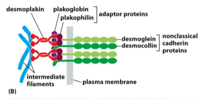
  • Integrins is like cadaherins but it binds to the extracellular material

  • Tight Junction- elaborate array of protien, function is to seperate spaces that are created by a layer of cells, separates interstitial and blood stream(Tupperware lid) creates a seal

  • Series of protiens stitched together (clodins and aculin ) with lateral attachments across

  • Channel forming junctions or gap junctions- connects cytosol , chemical synapses, only 1000 daltons can pass small ions and molecules can pass

  • Gap junctions that are small stack together

  • The ECM is diverse and complicated mixture, its secreted and it can be structural or space filling,
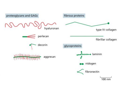
  • Makes you durable and promotes compression resistance

  • Fibrous protiens provide tensile resistance
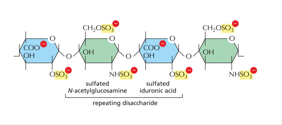
  • Hyluranan forms the backbone for bigger complex of things like heparin that ca. dock on the hyluran complex

  • Collagen- an example of a fibrous protien that can provided tensile strength via GAG and it’s usually made of a chain of amino acid, fhe partner chains are woven together

  • Hydroxylation gives collagen distinctive folding patterns.

  • Ex bone defects,dwarfism, nasal laminate will fail in the kidenehs , and all sorts of problems that come from a lack of calllahdn

  • Collagen is dependent on vitamin C as a cofactor

  • Elastin- forms cross links and are coiled up to allow streachimess and relaxation and prevents the other end without tearing ping or ripping

  • Finromectim and other multi adhesive protiens all of domains can be switched out via alternitive splicing

  • Fibromectins bind integrins

  • Arg protiens allow the integrity protiens allow the cell being knit into the and then show it to. E

  • Basal lamina is exclusive to the epithelial cells

  • The key assembly factor that is growing into the cell did 3 rd protien call dystroglucan,indigenous,,perfect

  • Integrity bind the cell to the extracellular ,atria

  • Connected to cell with intermembrane filaments, and anchoring junctions

  • Inactive integrity- makes internal and external signals on the alpha and beta site inactive anchoring

  • Lateral cross linking protiens mediate binding

robot