Eukaryotic cells
Larger and more complex than prokaryotes
Can be unicellular or part of multicellular organisms
Have linear DNA molecules packaged as chromosomes enclosed in nucleus
Chromosomes number, ploidy (number of copies of the genome), and C-value (amount of DNA in cell) widely variable.
Have membrane bound organelles
Unicellular eukaryotes
Some of the most complex eukaryotic cells
Need to be able to perform all functions in harsh environments
Some exist as unicellular organisms when food supplies available, but aggregate and specialise to form primitive multicellular organisms when food is scarce

Structure of the cell membrane
Phospholipids are a key component of cell membranes
They are amphipathic in nature, polar head, hydrophobic tail
They form closed bilayers headgroups facing out, hydrophobic tails in the middle.
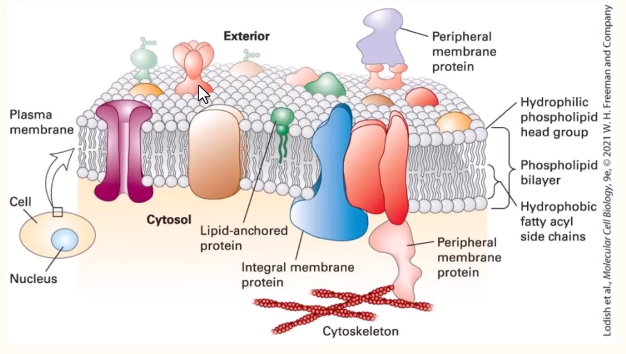
They are complex with different components embedded into the plasma membrane ie proteins.
The cell membrane is asymmetric as some are only displayed on one face of the membrane. The lipids and proteins can be shown in one face or both.
Function of plasma membrane
Regulation of transport - nutrients into cell, waste out of cell
Maintains balance of chemical conditions in the cell such as pH- homeostasis
Provides a site for chemical reactions not likely to occur in an aqueous environment
Detects signals in the extracellular environment
Interacts with other cells or the extracellular matrix - in multicellular organisms
Organelles
Nucleus
A membrane-bound structure usually visible by light microscopy, contains the cell’s genetic material
Electron microscopy reveals two membranes each about 5-7 nm thick spaced by 11-40nm
Inner membrane is in contact with the nuclear contents
Outer membrane appear to be continuous with endoplasmic reticulum
Contents are in contact with cytoplasm via nuclear pores which pass theough both membranes
Large dense region- Nucleolus - visible within nucleus by electron microscopy
Rich in protein and RNA- involved in synthesis of ribosomal RNA (rRNA) and ribosomes
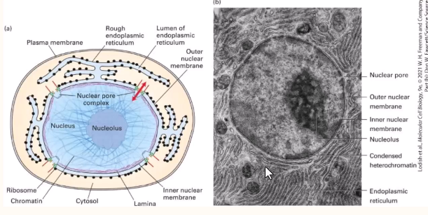
The nucleus separates DNA from cytosol
Transcription from translation separated in space.
Key features- outer membrane, inner membrane, nuclear pores, nucleolus
Endoplasmic reticulum
Extensive membrane structure forming interconnected sacs and tubules
Responsible for most lipid synthesis
Most membrane protein synthesis
Ca2+ ion storage
Detoxification
Rough ER
Ribosomes attached to surface
Plays a role in synthesising membrane-bound and secreted proteins
Extensive in cells synthesising these proteins
Smooth ER
Carries no ribosomes
Plays a role in producing lipids eg membrane lipids and steroid hormones
Ribosomes
Complex multi-subunit structures comprised of toughly 50% proteins and 50% ribosomal RNA (rRNA)
rRNA are key to the structure and function of ribosomes
Involved in the synthesis of proteins
Eukaryotic ribosomes consist of 40S and 60S subunits- assembled give 80S
Often associated with endoplasmic reticulum or as cytoplasmic polyribosomes
Golgi complex/ apparatus
Stack of flattened membranous sacs which vary in number
Sacs form from parts of rough ER which breaks off and fuse
Inner cis face is close to the nucleus
membrane at nuclear face 5-7nm thick like nuclear membrane
At outer trans face it is 7-9nm thick like plasma membrane ensures that vesicles budding off outer face can fuse with plasma membrane
packages lysosomal proteins and proteins to be secreted from the cell
composed of 3 regions cis (entry), medial, trans (exit)
each region contains different sets of modifying enzymes
Mitochondria
Typically 0.5-10 µm (size of typical bacterium), but can vary in shape from cylindrical to almost spherical
Contains DNA and ribosomes which are smaller than normal eukaryotic type
Can direct production of some of own proteins
Self-replicating; reproduces via binary fission
Multiple mitochondria per cell 1 to1000
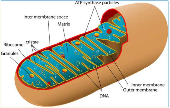
Aqueous environment
Site of ATP production via aerobic metabolism
Plays important role in apoptosis- programmed cell death
structures
Outer membrane
Intermembrane space
Inner membrane
Matrix
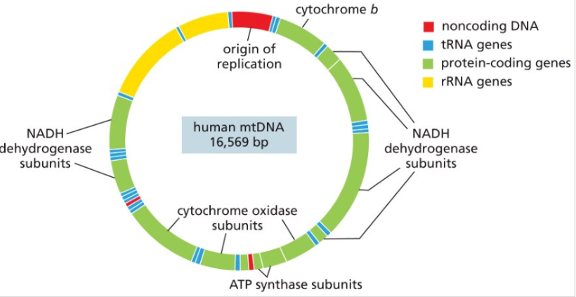
Mitochondrial DNA
Contains multiple mtDNA molecules
Genes in mtDNA exhibit cytoplasmic inheritance and encode rRNA and tRNA and some mitochondrial proteins
The size and coding capacity of mtDNA varies considerably in different organisms
The products of mitochondrial genes are not exported
Mutations in mtDNA cause several genetic diseases in humans ie Leigh syndrome, Leber Hereditary Optic Neuropathy
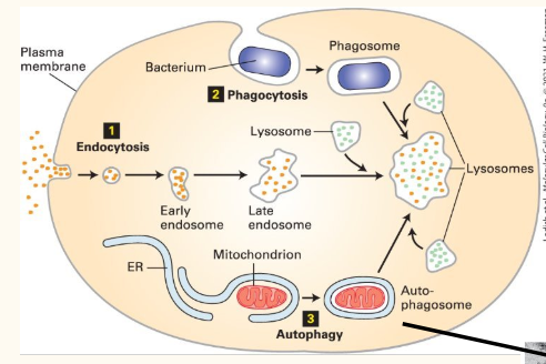
Lysosomes
Appear as electron-dense bodies up to 400nm by EM
Single membrane bound organelles containing hydrolytic enzymes
Degrade materials taken up by endocytosis and cell debris and organelles
Degrade damaged newly synthesised proteins
Interior pH is low about 3.5-5.0
Enzymes eg acid hydrolases, they contain could cause serious cell damage but cannot function at neutral cytoplasmic pH
If a lysosomal enzyme is missing due to genetic defect, results in accumulation of material: lysosomal storage disease- usual fatal in adolescence
Not present in plant cells- vacuole plays some roles of lysosomes in plants
Peroxisomes
Single membrane-bound organelles
Can be diverse in size and enzyme composition
Contains catalase and urate oxidase
Breaks down very long chains of fatty acids via beta oxidation spiral
Oxidation of toxins (alcohol)
Cytosol
The portion of the cell enclosed by the plasma membrane but not part of any organelle.
Not static, the contents of the cell are continuously moving
Key features include;
Cytoskeleton
Polyribosomes
Metabolic enzymes
Cytoskeleton
Lattice like array of filaments and fine tubules
Involved in cell movement, cell division, maintenance of cell shape, Intracellular trafficking or organelles, coordinated movement of tissues
3 major components: microfilaments, microtubules and intermediate filaments
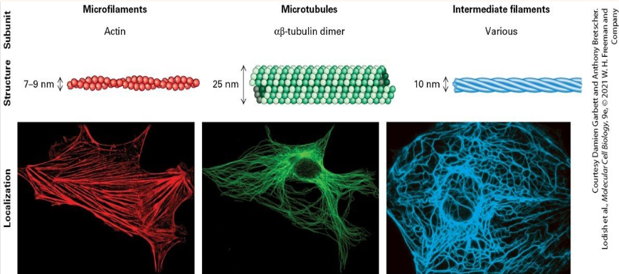
Microfilaments
F-actin filaments are double helices of polymerised G-actin subunits
Fibers expand and contract by further polymerisation and depolymerisation
ATP dependent
Interact with other filaments and motors to create movement: contraction can cause shape change
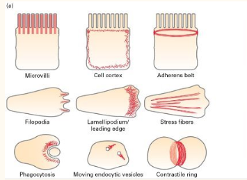
Actin + myosin
Actin microfilaments work with myosin in muscle fibres
Myosin filaments walk along the tethered actin hence pulling the filaments towards the centre to cause muscle contraction
Microtubules
Tubes of tubulin: grow by polymerisation from specific organising centres
Can form trackways in cells along which motor proteins (kinesins) drag vesicles, organelles etc
Fundamental role in portioning the chromatids in cell division
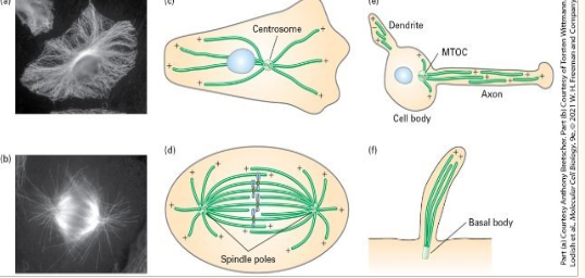
Motor protein complexes carry cellular components along the microtubule tracks
Different motors for different directions of movement
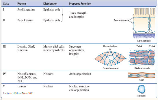
Intermediate filaments
Structures about 10nm in diameter
Many different types that differ in composition and function
May have role in maintaining cell shape, tissue integrity
Chloroplasts- plant cells only
Double membrane structure found in photosynthetic plant cells
Up to 10µm by 2µm- larger than mitochondria
Contain their own DNA molecules
Stacked inner thylakoid membranes contain green pigment chlorophyll and other pigments that absorb light and generate NADPH and ATP during photosynthesis- used to produce organic molecules.
Vacuoles- plant cell
Occupy up to 80% of the plant cell
Stores water, ions and nutrients and degrade macromolecules
Inflow of water by osmosis causes vacuole expansion and maintenance of turgor pressure
Expansion of vacuoles involved in cell elongation
Plasmodesmata- plant cells
Plant cells are surrounded by a rigid cell walls designed to withstand turgor pressure
Comprised mainly of cellulose cross-linked by hemicellulose, pectin and lignin-insoluble
Plasmodesmata directly connect the cytosol of adjacent cells in higher plants
Specialised peroxisomes
One type is found in the leaves where it involved in photorespiration- utilises oxygen and generated carbon dioxide.
Glyoxysomes are found in germinating seeds- carry out glyoxylate cycle to convert fatty acids into sugars