AP Bio Unit 4 Review Notes
Today's Plan
Cell Communication
Cell Cycle
Practice Questions
Q&A
Note: Questions can be posted in the chat, but there may be a 30-second to 1-minute lag. Participants are encouraged to help each other by answering questions in the chat.
Cell Communication
Forms a significant portion of Unit 4.
Cell communication is a broad topic. The exam will present unfamiliar scenarios, requiring application of known principles.
Steps of Cell Communication (Signal Transduction Pathway)
Reception
A signaling molecule (ligand) binds to a receptor.
The receptor is a protein, and binding causes a conformational shape change.
Examples of Receptors
G Protein-Coupled Receptor (GPCR)
Works with a G protein.
Activated by binding GTP (Guanosine-5'-triphosphate).
Initiates a downstream pathway.
Ligand-Gated Ion Channels
Receptor normally closes the ion channel.
Ligand binding causes a conformational change, opening the channel for ion flow.
Tyrosine Receptor Kinase (TKRs)
Two receptors form a dimer.
Kinases add phosphate groups to tyrosine amino acids.
Activates multiple transduction pathways.
Ligands
Steroid Hormones
Nonpolar (based on Unit 1 knowledge).
Receptor is located inside the cell (intracellular receptor).
Examples: testosterone, estrogen.
Enter the cell via simple diffusion.
Protein Hormones
Receptor must be on the cell membrane (extracellular receptors).
Proteins can't pass through the membrane via simple diffusion.
Released via exocytosis: transport vesicle moves to the plasma membrane and releases contents.
Example: insulin.
Insulin binds to a receptor on the membrane, signaling body cells to take up glucose.
Transduction
The intermediate stage between reception and response; cascade of events relaying the signal.
The signaling molecule itself does not move through the transaction, the message does.
Involves relay molecules that amplify and modify the signal.
Two Main Ways of Transduction
Phosphorylation Cascade
Protein kinases phosphorylate relay proteins.
Adding PO_4 to proteins to move the message through.
Secondary Messengers
Small molecules in cytosol that move to continue the cascade.
Examples:
Calcium (Ca^{2+})
Stored in the smooth ER (sarcoplasmic reticulum in muscle cells).
In muscle contraction, an electrical signal opens an ion-gated channel on the smooth ER membrane.
Calcium is released and binds to troponin and tropomyosin, allowing actin and myosin to interact/ muscle contraction.
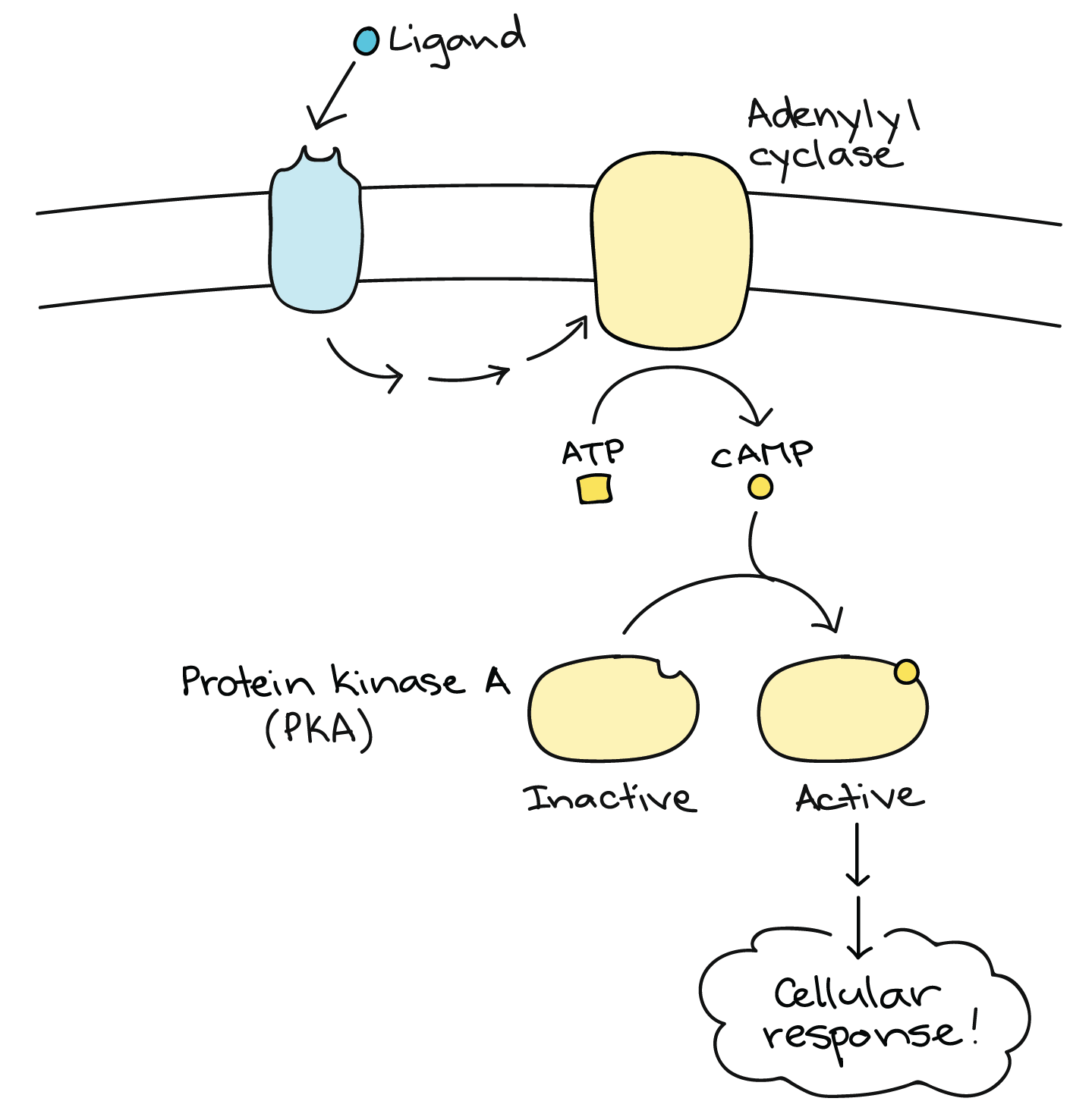
Cyclic AMP (cAMP)
Cyclic adenosine monophosphate.
Adenylyl cyclase converts ATP to cAMP.
{ATP \rightarrow cAMP}
cAMP activates protein kinase, leading to a phosphorylation cascade.
Response
The final outcome of the signal.
Potential cellular responses:
Cell growth
Secretion of molecules
Modification of gene expression (turning genes on or off)
Alteration of cell function or phenotype
Apoptosis (programmed cell death)
Receptor as Enzyme?
Some receptors are enzymes (e.g., tyrosine receptor kinase).
Others are simply proteins that bind to other molecules (e.g., G protein-coupled receptors).
Difference between cyclic AMP and ATP: ATP has three phosphates, while cyclic AMP has one phosphate and a cyclic structure.
Cellular Communication Examples
2013 Exam
The diagram was on the 2013 exam, and they asked you to tell us what the three steps were and what their functions were:
Reception: a protein hormone binds to the receptor on the plasma membrane.
Transduction: the binding causes a conformational shape change that causes a cascade that will transmit the signal from one relay molecule to the next.
Response: the protein will interact with DNA to turn on or off a gene.
2021 Exam
Scientists added an inhibitor for pMEC to cells, and the exam asks to determine what change would occur in the relative ratio of ERK to PERK compared to cells without the inhibitor if pMEC is to speed the reaction between ERK to PERK.
The ratio of ERK to PERC will increase because pMEC is unable to allow for the phosphorylation of ERK to PERC, which lead to a buildup of ERK because PERC is not being made.
2018 Exam
Inactive caspase one is cleaved into active caspase one which then will cleave the gastroderman, which allows it to go into the membrane. The active caspase also activates the interleukin.
If step three is inhibited, then the formation of pores would not form because the gastroderman cannot be converted. And there will be no difference in the release of interleukin because step three has no effect on step two.
Different Types of Signaling
Paracrine Signaling:
Signaling to nearby cells.
Synaptic Signaling:
Specific signaling between a neuron and another cell (e.g., muscle, another neuron, endocrine gland).
Occurs at the synapse, the space between the axon terminal of the neuron and the postsynaptic cell.
Considered a type of paracrine signaling specific to neurons.
Endocrine Signaling:
Hormones are secreted into the bloodstream and reach receptors in far away cells
Juxtaposition Signaling:
Involves direct communication between adjacent cells through gap junctions, plasmodesmata, allowing for rapid exchange of ions and small molecules.
Cell Cycle
Mitosis and the life cycle of a cell.
Steps:
Interphase
G1 phase
S phase
G2 phase
Mitotic Phase
Mitosis
Cytokinesis
Interphase
Consists of G1, S, and G2 phases.
Cell grows throughout all phases.
G1 Phase (Gap 1)
Duplication of organelles.
Synthesis of proteins needed for the new cell.
Signal transduction and communication take place.
RNA and building blocks are synthesized.
S Phase
Replication of DNA.
Replication of centrosomes (organizing centers for genetic material).
G2 Phase
Cell continues to grow.
Synthesis of proteins and RNA.
Continued organelle production.
Reorganization of cellular components.
G0 Phase
A non-dividing state where the cell exits the cell cycle and "chills".
Examples: neurons, muscle cells, liver cells (can exit G0 based on environmental stimuli).
Mitosis
Phases
Prophase
Metaphase
Anaphase
Telophase
Cytokinesis follows mitosis.
Prophase
Cell prepares to divide.
Chromatids condense into sister chromatids, attached by cohesion molecules at the centromeres.
Nuclear envelope disintegrates.
Centrosomes organize microtubules.
Metaphase
Sister chromatids align on the metaphase plate (middle of the cell).
Microtubules attach to the sister chromatids.
Alignment is maintained by a "tug of war" of forces to ensure proper alignment before separation.
Anaphase
Sister chromatids are pulled apart to opposite sides of the cell.
The cohesin molecule is broken down by the separase enzyme.
Each chromatid is attached to a microtubule, pulling it toward the pole.
Telophase
Two new nuclei form.
The nuclear envelope reforms around each set of chromosomes.
Chromatids (now chromosomes) decondense.
Cytokinesis
The cytoplasmic division.
In animal cells, a cleavage furrow forms due to an actin ring contracting.
In plant cells, vesicles release and build a cell plate, which forms the cell wall.
Regulation of the Cell Cycle
Checkpoints
Ensure everything proceeds correctly.
G1 Checkpoint
Determines whether the cell should divide.
Checks for:
Growth factors (paracrine signals binding to receptors).
Sufficient resources.
DNA damage.
If the cell doesn't pass, it enters G0 (nondividing state).
Cyclin activates, leading to a phosphorylation cascade, and then the s phase will begin the DNA replication
G2 Checkpoint
Occurs at the end of interphase.
Checks for:
Complete and accurate DNA replication.
If problems are detected, the cycle halts, and DNA is repaired.
p53 is involved in stopping the cycle for DNA repair.
M Checkpoint
Occurs during metaphase.
Ensures all sister chromatids are attached to microtubules.
Cohesin holds chromatids together; separase breaks them apart.
Oncogenes and Tumor Suppressor Genes
Oncogenes:
Normal genes involved in cell division.
When mutated, they can become hyperactive.
Example: RAS protein (when hyperactive, it constantly signals for division even without growth factors).
Tumor Suppressor Genes:
Halt the cell cycle.
Example: p53 (if mutated, cells with DNA errors will continue to divide unchecked, leading to cancer).
CDK (Cyclin Dependent Kinase):
It's a kinase dependent on cyclin.
Practice Questions
Multiple Choice
Insulin (protein hormone) binds to receptors on liver cells, stimulating a phosphorylation cascade that causes glucose transporters to move to the plasma membrane. What is the role of insulin in this liver cell?
Answer: Ligand (signaling molecule that binds to a receptor).
Which scenario demonstrates negative feedback in the endocrine system?
Answer: After a meal, elevated blood glucose levels cause the pancreas to release insulin, which converts excess glucose to glycogen in the liver, reducing blood glucose levels.
Long FRQ (2022 #1)
A pathway that shows a extracellular ligand binding to a g protein coupled receptor, which will active the g protein. Once the g protein is active it will activate the adenylyl cyclase, which will convert ATP to cyclic AMP which will activate protein kinase.
Part A
Describe one characteristic of a membrane that requires a channel to be present for chloride ions to pass across the membrane.
Plasma membrane is nonpolar due to the hydrophobic fatty acids in the interior.
Explain why the movement of chloride ions out of the cell leads to water loss.
The outside of the cell becomes hypertonic, so water rushes out to make it equal.
Part B
Identify the independent variable in the experiment.
The presence or absence of GTP and Cholera toxin.
Identify a negative control.
Sample 1 (no GTP or cholera).
Justify why sample three has been to include one of the control treatments in this experiment.
We are comparing with a sample that has the cholera toxin and the GTP.
Part C
Based on the data, describe the effect of the cholera toxin on the synthesis of cyclic AMP.
Cholera has no effect on cyclic AMP production in the absence of GTP.
Cholera toxin will increase cyclic AMP with GTP presence.
Calculate the percent change in the rate acetic AMP production due to the presence of the cholera toxin in sample four compared to sample two.
Formula: ((Final - Initial) / Initial )* 100
((27-10)/10)*100 = 170%