Week 4 notes
Brain Development Overview
The development of the human brain is a complex process that occurs in several stages, involving various structures and biological mechanisms.
Brain Weight and Age
Brain Weight: The weight of the human brain increases significantly from birth to adulthood, reaching about 1400 grams.
Development Timeline: Brain weight is represented against age, showing a steady increase beginning from around 25 weeks gestational age until adulthood, indicating the critical phase of brain growth.
Major Parts of Human Brain
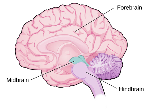
The human brain consists of three primary parts:
Forebrain: This is the largest part of the brain and includes the two cerebral hemispheres.
Cortex: The outer layer of the cerebral hemispheres, highly folded (wrinkled), which increases surface area.
Gyri and Sulci: A ridge on the surface is called a gyrus, while a groove is termed a sulcus.
Midbrain: A smaller part located below the forebrain primarily involved in processing sensory information.
Hindbrain: It includes structures that regulate functions such as breathing and heart rate.
Stages of Central Nervous System (CNS) Development
Brain development occurs through six distinct stages:
Neurogenesis: Formation of neurons from precursor cells.
Migration: Neurons move to their destined positions in the brain.
Differentiation: Cells mature and specialize into various types of neurons and glial cells.
Synaptogenesis: Formation of synapses between neurons.
Neuronal Cell Death: Removal of excess neurons.
Synaptic Refinement: Fine-tuning the connections between neurons.
CNS Development Process
The CNS begins as a flat plate composed of three layers: ectoderm, mesoderm, and endoderm, which eventually rolls into a neural tube.
The ectoderm develops into the neural structures while mesoderm and endoderm will form other body systems.
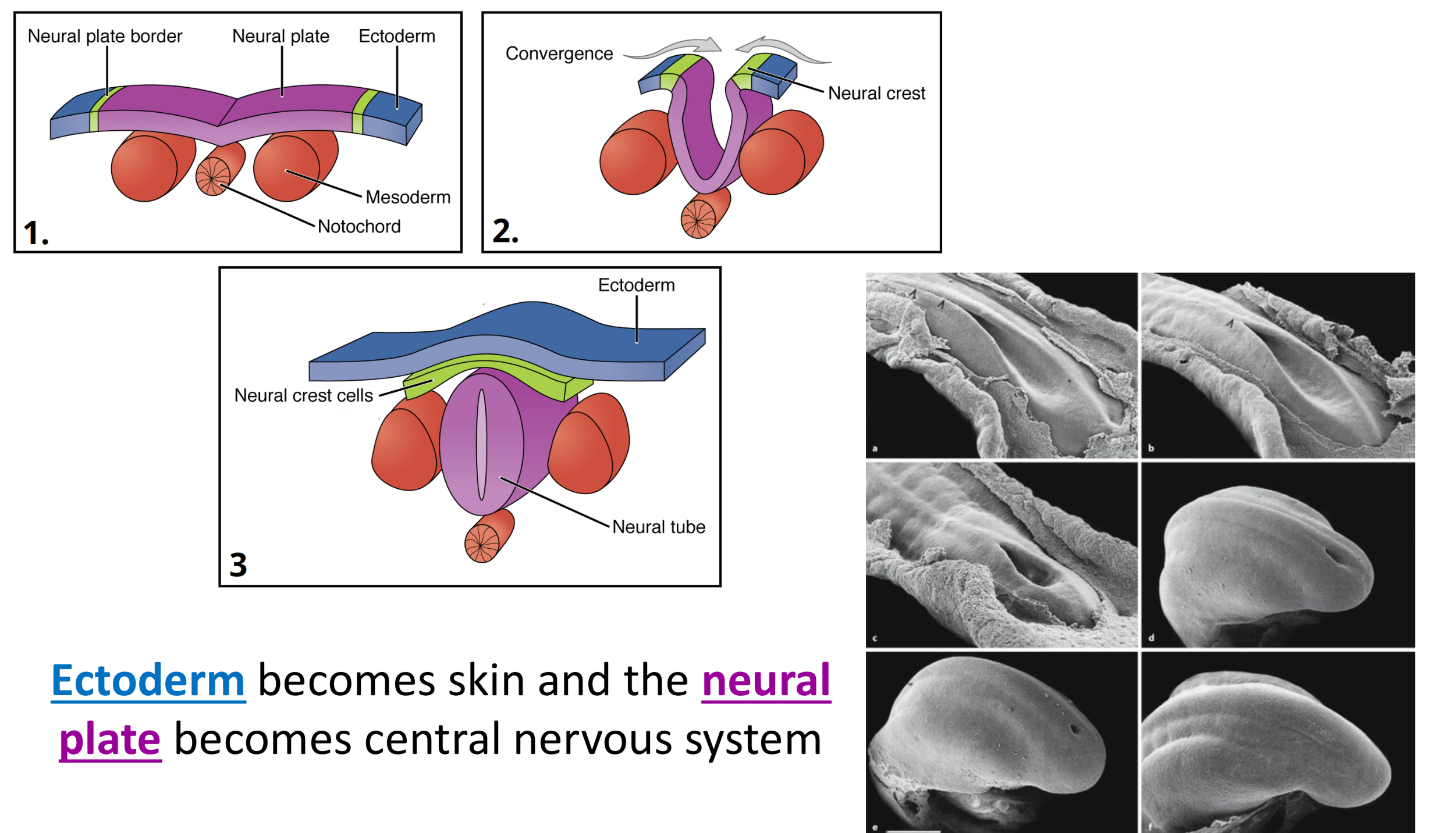
Neurulation in Zebrafish
Zebrafish serve as a model organism to study neurulation, the process by which the neural tube forms during embryonic development.
Clinical Case Study: Tuberous Sclerosis
A case of a junior high school student with seizures was evaluated, highlighting conditions like Tuberous Sclerosis, associated with abnormal brain development. Too many glial cells cause Sclerosis.
Neurogenesis Details
Neurogenesis involves the mitotic division of precursor (stem) cells. In this step, neurons are formed with limited time in the ventricular zone (inner layer of neural tube.) Failure in mitosis can lead to disorders such as microcephaly. In microcephaly, the brain is so small because there are not enough neurons.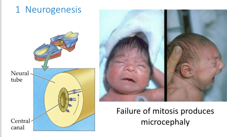
Neurons and glial cells form next to the central canal (ventricle).
Migration
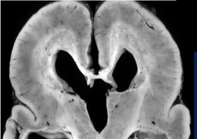
During migration, cells in the neural tube (soon to be the brain), move out of ventricular zone to the surface. Disruptions during the migration stage of neuron development can result in structural brain malformations, e.g., pachygyria.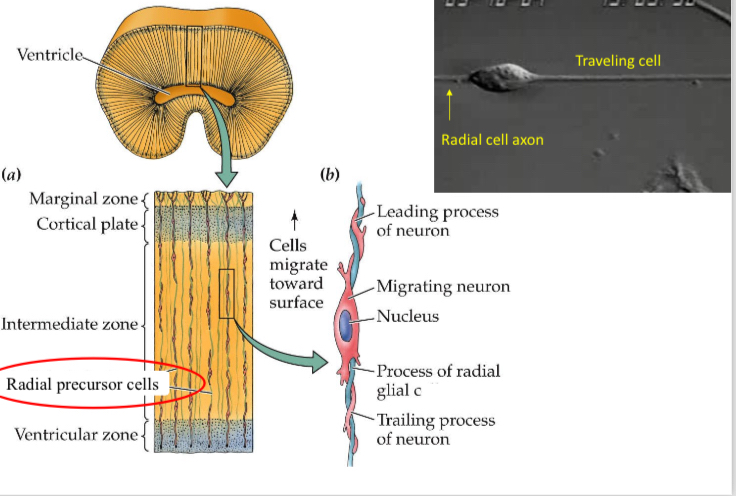
AXONS ARE GUIDED BY CHEMO ATTRACTANTS (attract certain growth cones), and CHEMOREPELLANTS (repel growth cones) released by target cells.
Growth cones: Sensory motile organelles at tip of growing axons and dendrites.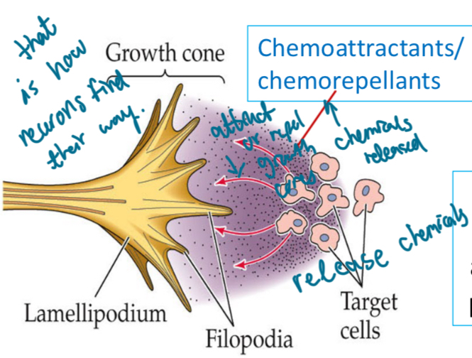
Differentiation Mechanisms
During differentiation, notochird releases protein, Sonic hedgehog, that guide precursor cells to develop into specific types of motoneurons in the SPINAL CORD. The rest become sensory neurons. 
Synaptogenesis
Synapses form rapidly during early life, with a high number of synapses created during fetal development, infancy, and childhood followed by refinement in adolescence and adulthood.
For 2 weeks, you make lots of synapses, then you stop because you don’t need them.
Neuronal Cell Death and Apoptosis
Neurons undergo a natural cell death process called apoptosis, influenced by various factors, including cell signaling pathways.
Caspases cut up proteins and DNA (kills neurons). Calcium enters the cell, mitochondria releases Diablo. Diablo attaches itself to IAP’s (Inhibitors of Aptosis) which inhibit caspases. So now the cell is killing all neurons(suicide).
Diablo doesn’t do anything directly, it instigates cell suicide.
Survival of the fittest: Cells compete for cell food NERVE GROWTH FACTOR (NGF) and BRAIN DERIVED Neurotrophic FACTOR (BDNF) and synaptic connections.
NGF and BDNF are produced by targets and eaten by neurons. These neurons that eat them survive and the rest die.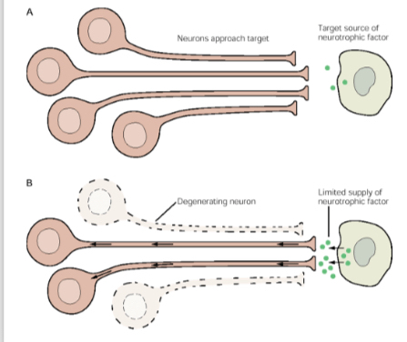
Disorders like Fragile X syndrome are linked to improper neuronal pruning (eliminating extra synapses not needed) and development. There is a genetic mutation on the X chromosome and it is linked to a specific gene (FMR1 gene). Resulting in a lack of FMRP protein. They have too many synapses (larger brain.)
Synaptic Refinement
This involves the elimination of excessive synapses, ensuring that the most effective connections are maintained in the brain. (Brain will change itself bit by bit)
Amblyopia: One eye does not work properly. The body begins ignoring that eye as there is little to no activity from it. Can eventually lead to blindness.
Sensitive Periods in Development
Certain experiences, such as early visual exposure, can lead to significant and lasting effects in brain organization and function.
Autism Spectrum Disorders
Clinical Features: Autism is characterized by difficulties in communication, social interaction, and repetitive behaviors.
Genetic Factors: Abnormalities on specific chromosomes and environmental influences play significant roles in the development of autism.
Neurodevelopmental Aspects: There are observed structural differences in the brains of autistic individuals, indicating atypical neural connectivity.
Syvan syndrome? Autistic people who have very good to excellent functioning?
In autism, the brain has to many connections.
Vaccination Myths
Despite ongoing discussions, extensive research shows no causal link between vaccinations and autism. Misunderstandings and influences from public figures contribute to the persistence of these myths.
Rett Syndrome
Rett syndrome is a neurodevelopmental disorder characterized by normal early development followed by a regression of skills, predominantly impacting girls.