Anatomy of Diencephalon
DIENCEPHALON
Overview
The diencephalon is a critical region located at the central core of the forebrain, completely surrounded by the cerebral hemispheres except at its ventral surface, where it connects to the midbrain.
It contains a cavity known as the 3rd ventricle, which plays an essential role in the circulation of cerebrospinal fluid (CSF).
Components of Diencephalon
Thalamus
The thalamus is the largest component of the diencephalon, generally resembling a pair of egg-shaped masses of grey matter embedded deep within the white matter of the cerebral hemisphere.
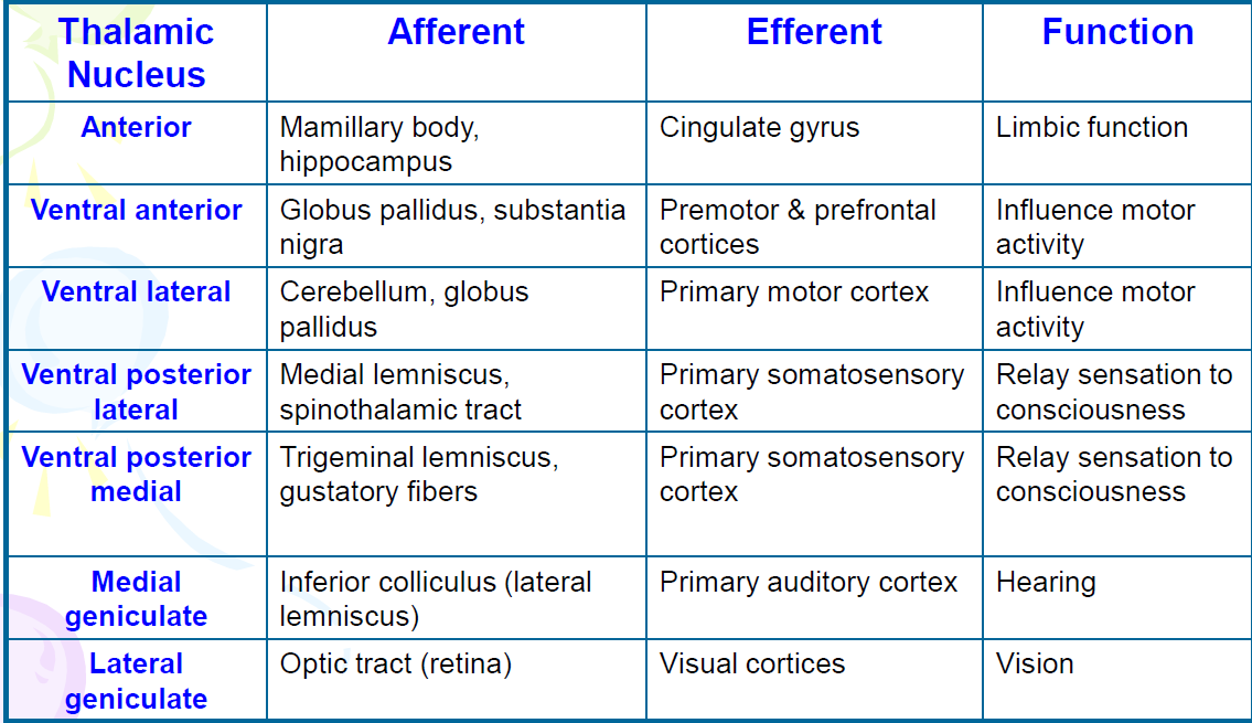
It is subdivided into numerous nuclei, each responsible for different afferent/efferent connections, facilitating various sensory and motor functions.
Hypothalamus
The hypothalamus occupies the side walls and the floor of the third ventricle and is vital for maintaining homeostasis by integrating autonomic, endocrine, and limbic functions.
It directly connects to the pituitary gland via the pituitary stalk and regulates multiple physiological processes.
Epithalamus
Positioned dorsal to the thalamus, the epithalamus includes structures such as the pineal gland, habenula, and stria medullaris thalami.
The pineal gland secretes melatonin, which is crucial in regulating sleep-wake cycles and circadian rhythms.
Subthalamus
Located ventral to the thalamus, the subthalamus functions as an integral part of the basal ganglia.
It consists of structures such as the subthalamic nucleus and zona incerta, contributing to motor control and coordination.
THALAMUS
Structure
The thalamus is characterized by two thalami separated by the 3rd ventricle, connected through the interthalamic adhesion.
It lies adjacent to the midbrain, contributing significantly to sensory processing.
Functions
Sensory Relay Station
Serves as a relay for all sensory inputs (excluding smell) to the cerebral cortex, effectively processing and transmitting sensory information.
Motor Relay Station
Relays motor information from the cerebellum and basal ganglia to the cerebral cortex, assisting in voluntary motor control.
Cognitive Function
Involvement in arousal and memory processing through the intralaminar thalamic nuclei, linking sensory perception and attention.
Relations
Anterior End: Faces the interventricular foramen
Posterior End: Faces the subarachnoid space
Medial Surface: Forms the wall of the 3rd ventricle
Dorsal Surface: Forms the floor of the central part of the lateral ventricle
Lateral Surface: Surrounded by the external medullary lamina
Inferior Surface: Lies atop the hypothalamus medially and the subthalamus laterally.
Medullary Lamina of Thalamus
Internal Medullary Lamina (ILM)
Y-shaped band of myelinated fibers within thalamus
Divide thalamus into 3 compartments
Contain nerve fiber connecting different nuclei
External Medullary Lamina (EML)
White matter band that covers most of thalamus lateral surface.
Lies deep to internal capsule.
Contain nerve fibers leaving/entering thalamus to/from internal capsule.
THALAMIC NUCLEI
Anatomical Classification:
Anterior Group: Contains the anterior nucleus
Medial Group: Includes the medial dorsal nucleus
Lateral Group: Further subdivided into dorsal and ventral tiers
Ventral Tier: Comprises Ventral Anterior (VA), Ventral Lateral (VL), and Ventral Posterior (VP) (further divided into VPM and VPL), which play roles in sensory and motor functions.
Dorsal Tier: Includes Lateral Dorsal (LD), Lateral Posterior (LP), and Pulvinar (P), associated with visual and somatosensory processing.
Functional Organization of Thalamic Nuclei:
Specific Relay Nuclei: Project to the respective areas of the cerebral cortex, including sensory, motor, and
limbic relay nuclei.
Association Nuclei: Exhibit widespread connections with cortical association areas, facilitating complex cognitive functions.
Non-Specific Nuclei: Provide diffuse projections to the cerebral cortex, playing a role in arousal and attention.
HYPOTHALAMUS
Location and Structure
Although weighing only around 4g, the hypothalamus is crucial for homeostasis and survival, positioned beneath the thalamus, forming the floor and part of the lateral wall of the 3rd ventricle.
It integrates various physiological processes crucial for body regulation.
Functions
Controls various vital physiological processes:
Control Center for the Autonomic Nervous System: Regulates involuntary bodily functions such as heart rate and digestion.
Control Center for the Endocrine System: Influences hormone secretion through its connection with the pituitary gland.
Regulates Body Temperature: Responds to changes in internal temperature to maintain homeostasis.
Regulates Food and Water Intake: Involves mechanisms for hunger and satiety.
Regulates Biological Rhythms: Influences circadian rhythms and sleep cycles.
Emotional Responses: Integrates emotions with physical responses.
Sexual Arousal: Plays a role in reproductive behaviors and reproductive hormone release.
Divisions
Divided into two primary zones via the column of fornix:
Medial Zone: Contains well-defined nuclei, such as the ventromedial nucleus that regulates hunger.
Lateral Zone: Features poorly organized nuclei, including the lateral hypothalamic nucleus, which is instrumental in hunger signaling.
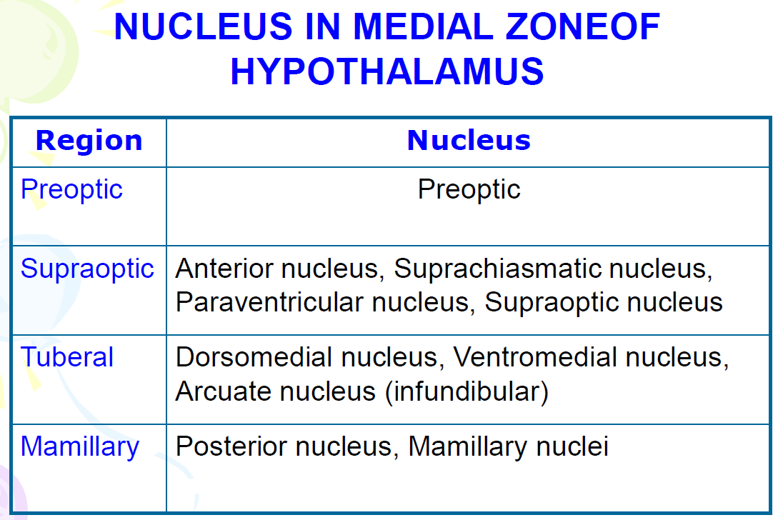
Preoptic Region
Most rostral part of hypothalamus
Contain medial & lateral preoptic nucleus
Medial preoptic nucleus: secrete gonadotrophin releasing hormone.
Supraoptic (Suprachiasmatic) Region
Located above optic chiasm.
Nucleus: supraoptic , suprachiasmatic, anterior, paraventricular
Tuberal Region
Extends from infundibulum to mamillary body
Nucleus: ventromedial (center for satiety), dorsomedial, arcuate.
Mamillary Region
Mamillary nucleus
Located at mamillary bodies
Mammillothalamic tract arise here
Posterior nucleus
Located above mamillary body
Main source of descending hypothalamic fibers to brain stem.
Lateral Zone of Hypothalamus
Lies lateral to fornix & mamillothalamic tract.
Lateral hypothalamic nucleus: feeding center
Contains large diffuse population of neurons called lateral hypothalamic nucleus and smaller condensation of neurons in its ventral portions called lateral tuberal & tuberomamillary nuclei
Lateral hypothalamic center: feeding center
EPITHALAMUS
Structure
Located dorsomedial to the thalamus and inferior to the splenium of the corpus callosum, the epithalamus is composed of several key structures:
Pineal Gland: A pine-cone structured responsible for synthesizing and releasing melatonin, which is vital in circadian rhythms.
Habenula: A triangular structure with medial and lateral nuclei that coordinates responses to stress and reward.
Stria Medullaris Thalami: A bundle of nerve fibers that connect the habenula to other areas within the forebrain.
SUBTHALAMUS
Location and Function
Situated inferior to the thalamus and between the thalamus and midbrain tegmentum, the subthalamus is functionally part of the basal ganglia and plays a role in movement regulation.
Components
Subthalamic Nucleus: Lens-shaped mass of gray matter, located dorsolateral to substantia nigra
Zona Incerta: Is actually an extension of brain stem reticular formation in subthalamus
Fields of Forel: Refers to nerve bundles containing pallidal & cerebellar efferents to thalamus
Summary
The diencephalon plays an instrumental role in the human brain, comprising critical structures such as the thalamus, hypothalamus, epithalamus, and subthalamus. Each component contributes to sensory and motor processing, endocrine regulation, and limbic function, thereby ensuring homeostasis and essential survival mechanisms, making it vital for maintaining the body's equilibrium and response to psychological and physiological stresses.