Topic 3: Classifications
VISUALIZING CELLS: STAINING TECHNIQUES
stains make it easier to observe cellular details under a light microscope
tains typically consist of salts where one of the ions acts as a chromophore (chromophore carries color)
Acidic dyes carry a negative charge and bind to positively charged cellular components
ex. congor red
Basic dyes have a positive charge and bind to negatively charged structures
crystal violet
some dyes do not bond to cellular components but instead work based on solubility principles.
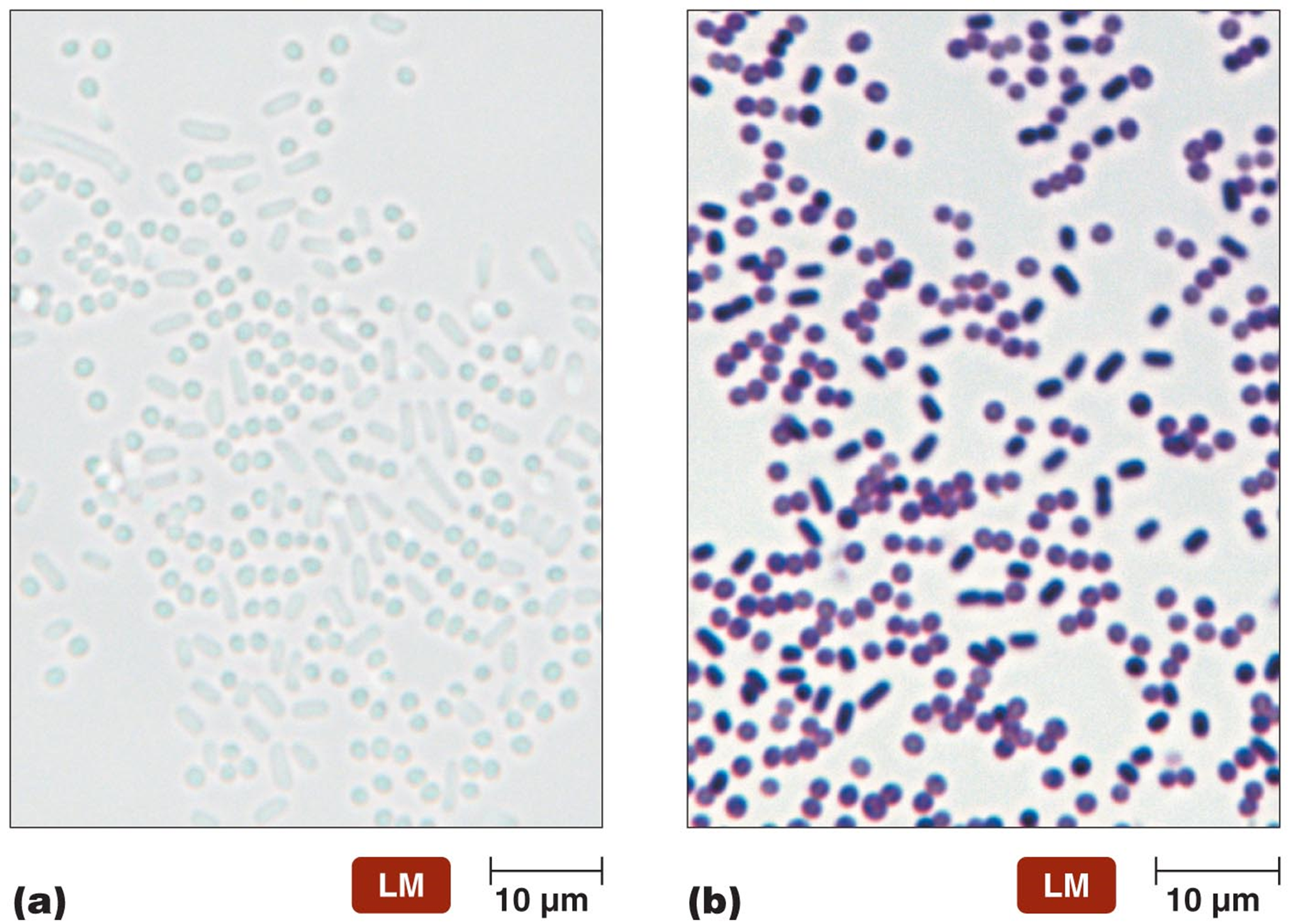
SIMPLE STAIN - No difference between G+ and G-
involves single basic dye to show size and shape of cells
ex. crystal violet, safranin, methylene blue
procedure: thin smear prepared on a slide, cells fixed onto cell by either heat or chemicals, soak smear with dye for 30 sec - 1 min
DIFFERENTIAL STAINS
uses more than 1 dye to differentiate cells based on characteristics
cell types: G+/G-
structures: flagella
chamicals - glycocalyx
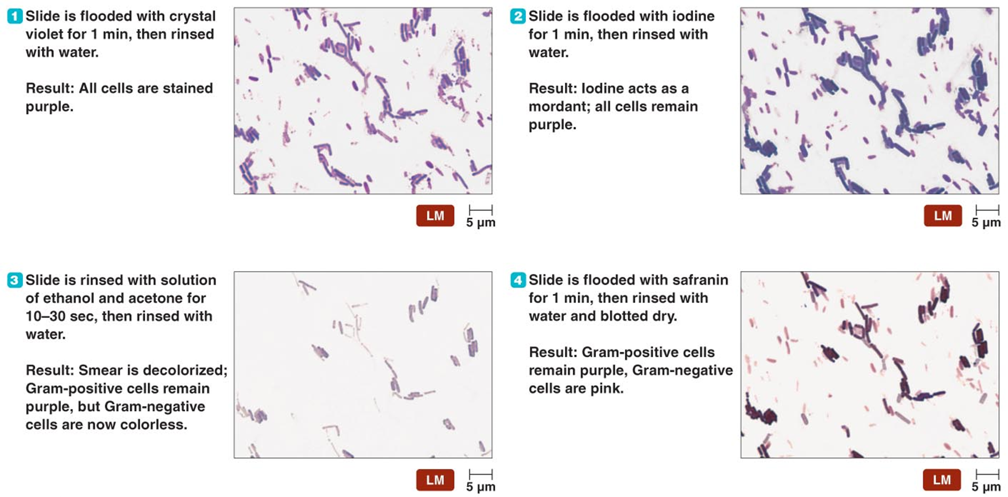
GRAM STAIN - differentiates between G+ and G-
procedure ****
Flood the slide with the primary stain, let sit, then rinse with water - CV penetrates PDG in G+ cells
Flood the slide with iodine. let sit, then rinse with water - iodine acts as a mordant → strengthens connection between CV and G+ PDG
Rinse the smear with a mixture of ethanol & acetone for 15-20 sec (if more than everything will be erased- decolorizing agent
Flood smear with safranin then rinse - counterstain → stains G- cells that are not stained by the primary stain
ACID - FAST STAIN - differentiates bacteria with waxy cell walls
primarily used to identify bacteria in the genera Mycobacterium and Nocardia
ex. TB, leprosy, lung infections, skin infections
these bacteria have a high lipid content in their cell walls, primarily due to mycolic acids
makes them resistant to conventional staining techniques like the Gram stain.
procedure
Primary Stain Carbol Fuchsin is applied, rinse with acid-alcohol solution , methylene Blue used as counter stain
acid fast cells are stained red/pink while non acid fast cells are stained blue
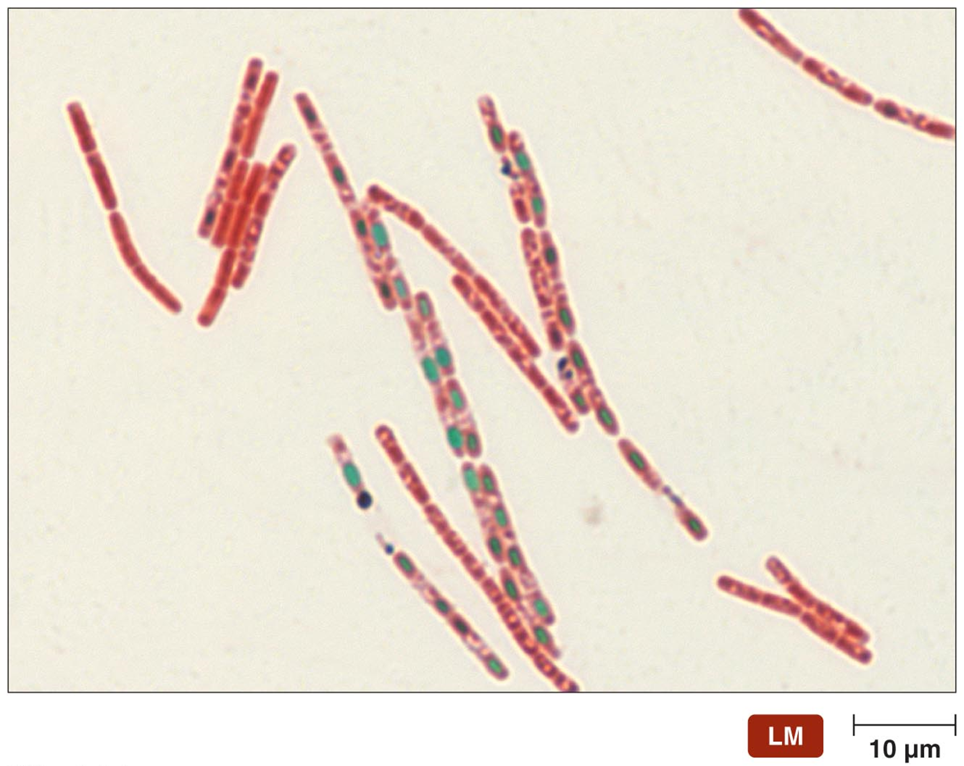
ENDOSPORE STAIN - differentiates cells with endospores
endospores found in genera Bacillus and Chostridium (both environmental)
heat is used to drive primary stain, malachite green, into the endospore
spore coat would not be penetrable by dye alone
5 min steam bath to force malachite green into endospore
counterstain with safranin → vegetative cells stained pink
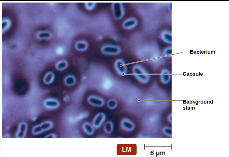
SPECIAL STAINS
negative (capsular) cell
stains the cell (and background) → capsule remains clear because it is negatively charges and repels the dye
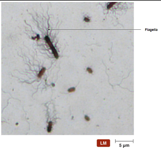
Flagellar
Structure normally not seen with light microscopy
Important for species identification
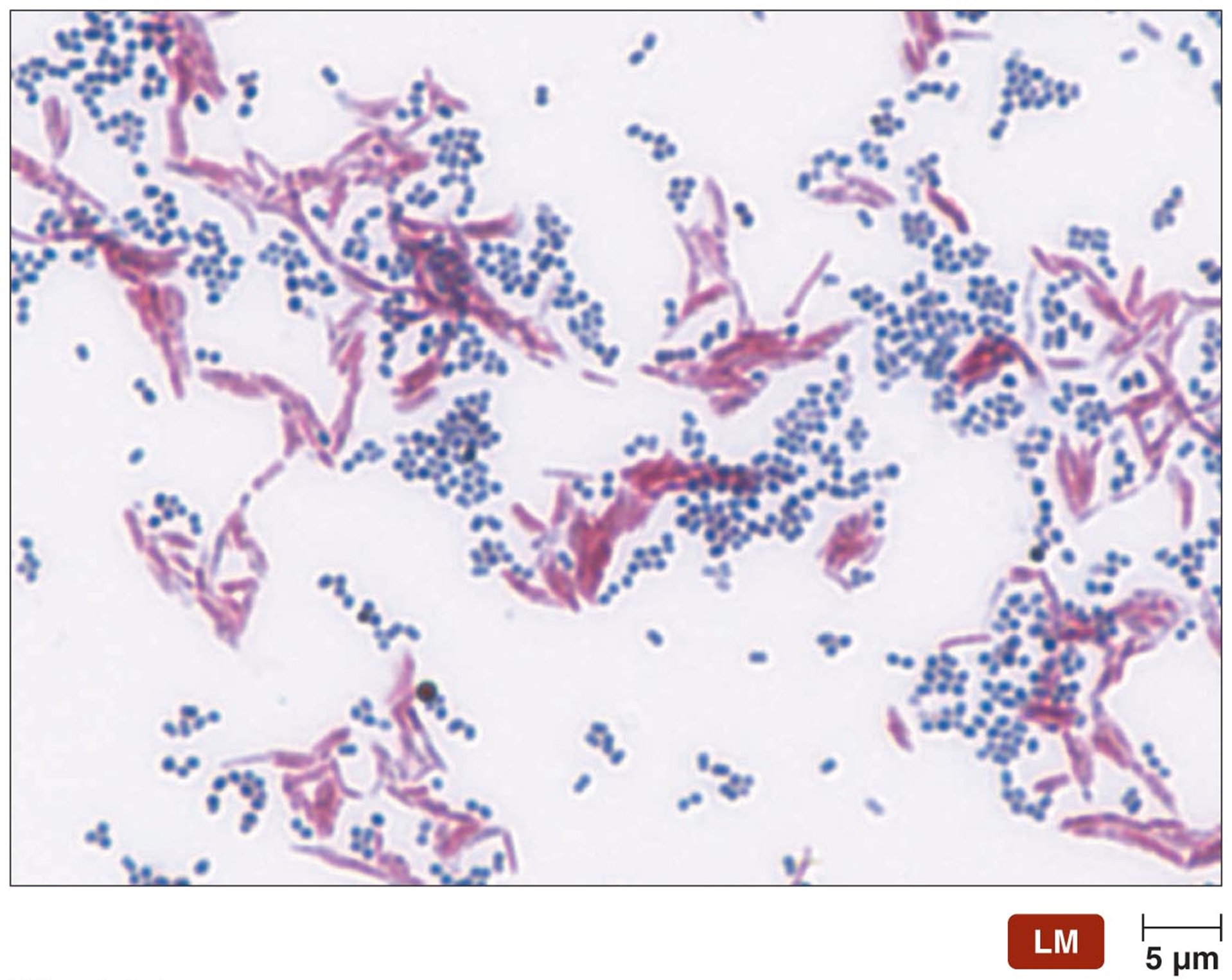
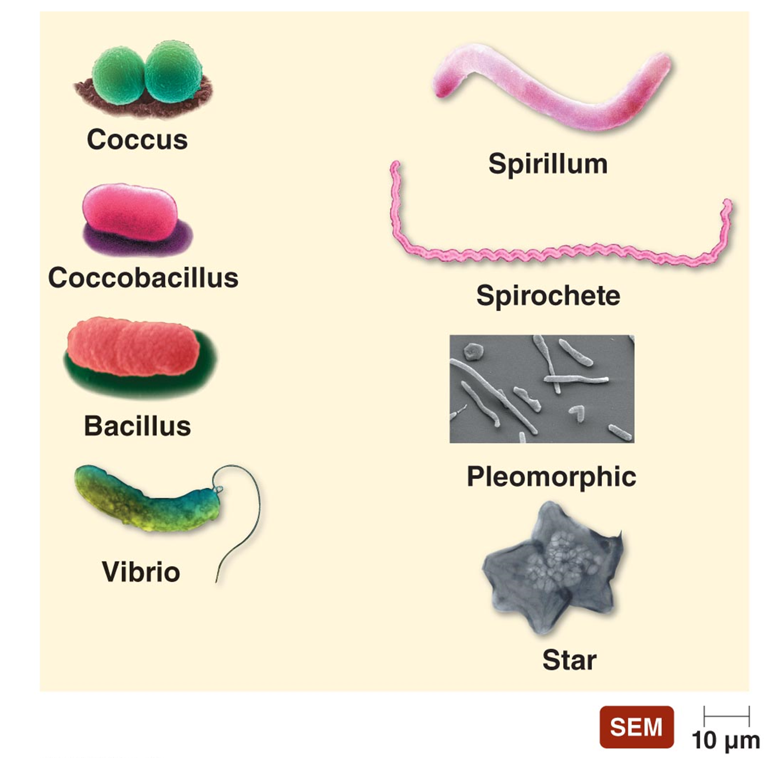
SHAPES OF BACTERIAL CELLS
coccus - roughly spherical
bacillus - rod shaped
spiral (motile bacteria)
spirlla - rigid
spirochetes - flexible
vibrios - curved rods “comma shaped”
Coccobacillus - intermediate between coccus and bacillus
Pleomorphic - Vary in shape or size
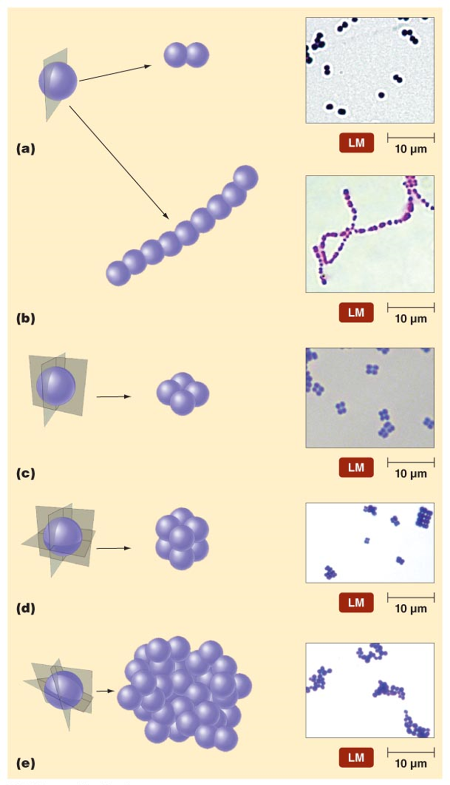
ARRANGEMENT OF PROKARYOTIC CELLS
Diplococci - two cocci that remain attached following binary fission
Streptococci - long chains of cocci
Tetrads (four)
Sarcinae - cuboidal packets
Staphylococci - grape-like bunches
**The above are similar in in bacilli - no tetrad, sarcinae or staph arrangement.
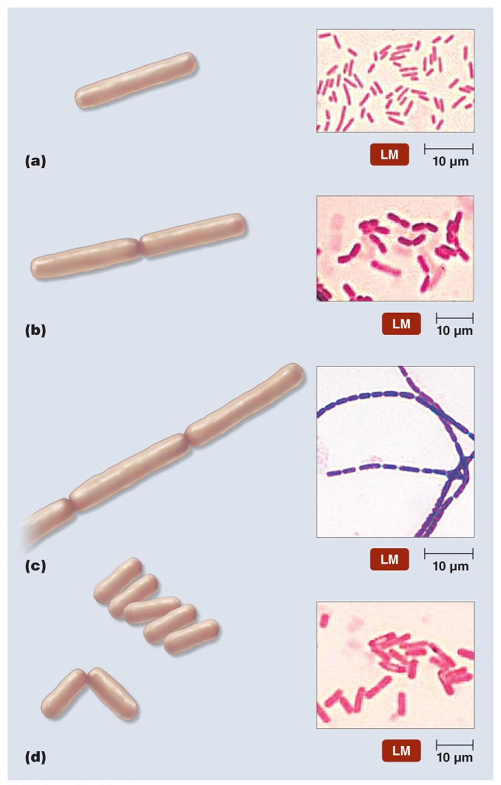
what is d?????
EUKARYOTIC CLASSIFICATION
protozoa, fungi, helminths, vectors
Protozoa - diverse, single-celled eukaryotic organisms primarily found in moist environments
***classifications
Parabasala: Lack mitochondria
ex. Trichomonas vaginalis: causes vaginal infections.
Diplomonadida: lack mitochondria, golgi bodies, and peroxisomes
ex. Giardia lamblia: causes gastrointestinal infections (giardiasis).
Kinetoplastids: have a single, large mitochondrion (kinetoplast) containing DNA.
ex. Trypanosomes: responsible for diseases such as African sleeping sickness and Chagas disease.
Ciliates: covered in cilia used for movement (move self or water over cell) and feeding.
ex. Balantidium coli: the only ciliate known to infect humans, causing intestinal infections.
Apicomplexans: Intracellular parasites capable of penetrating host cells;
examples:
Plasmodium: Causative agent of malaria.
Toxoplasma gondii: Causes toxoplasmosis, which can affect immunocompromised individuals and pregnant women.
Dinoflagellates: photosynthetic and important in marine ecosystems
Significance:
Red Tide: Can cause harmful algal blooms.
Neurotoxin Production: Some species produce toxins that can contaminate seafood, leading to poisoning.
Amoebozoa: characterized by lobe-shaped pseudopods for movement and feeding; lack shells.
Examples:
Naegleria: Can cause brain infections.
Acanthamoeba: Associated with eye infections.
Entamoeba histolytica: Causes amoebic dysentery.
FUNGI
Ascomycetes are the largest phylum of fungi
this phylum is defined by the formation of asci (singular: ascus), which are sac-like structures that contain ascospores
some species are beneficial, some species are pathogenic
HELMINTHS
Parasitic worms
Infectious agent usually the eggs or larvae
Diagnosis by presence of eggs or larvae in blood, feces or urine
ARACHNID AND INSECT VECTORS
Arachnids
Ticks - Rocky Mountain Spotted Fever, Lyme Disease
ticks have 6 and 8 leg life cycle stages
Fleas - Bubonic Plague
Lice – epidemic typhus
Flies - Leishmania, African Sleeping Sickness
Mosquitos - Malaria, Zika, Yellow Fever, West Nile, Dengue Fever
Kissing Bugs - Chagas’ Disease
VIRUSES
acellular; intracellular parasite
virion - the extracellular state of a virus; called virus when inside body
capsid - protective outside coat that surrounds viral genome
envelope - lipid membrane that surrounds some viruses, derived from the host cell's membrane during viral replication
VIRAL GENETIC MATERIAL
Double-stranded DNA (dsDNA)
Poxviridae, Herpesviridae
Single-stranded RNA (ssRNA)
Picornaviridae, Rhabdoviridae, Retroviridae
Single-stranded DNA (ssDNA)
Parvoviridae
Double-stranded RNA (dsRNA)
Reoviridae
LYTIC VS LYSOGENIC life cycle of viruses
lytic viruses
infects host cell, replicates, host cell is lysed and virus is released to adjacent cells
results in deaths of cells and rapid spread
lysogenic viruses
virus enters host cell, viral genome is replicated alongside the host’s DNA during cell division, allowing the virus to persist without causing immediate harm
***virus can remain dormant in lysogenic phase before transitioning into the lytic phase - all viruses will have a lytic phase
PRIONS
Proteinaceous Infectious Agent: Prions are misfolded proteins that can induce abnormal folding of normal proteins in the brain.
no nucleix acids → no genetic material
prion protein (PrP)
misfolds normal protein molecules and causing clumping throughout the brain
Impairs neuron function → displays as dementia
Holes appear in neural tissue