BioPsych Chapter 1
Basic Overview of Neurons
- Electrically charged
- ^^Synapse^^: the functional zone
- How neurons communicate
- Neurons are NOT connected continuously (like wires) but separated by functional space through which they communicate.
Parts of the Neuron
- ^^Soma^^ (aka the cell body or perikaryon)
- Stores genetic information and genes
- Neurites (aka the ^^Dendrites / Axon^^)
- Cellular fibers emerging from the soma
- ==Dendrites: “recieving”==
- ==Receives chemical info from other neurons==
- ==Axons: “output”==
- ==Relays info to other neurons==
- ^^Presyanptic Terminals^^ (aka boutons)
- ==Metabolism==
- Contains synaptic vesicles that contain ^^neurotransmitters^^ (neurochemicals essential for neuronal function)
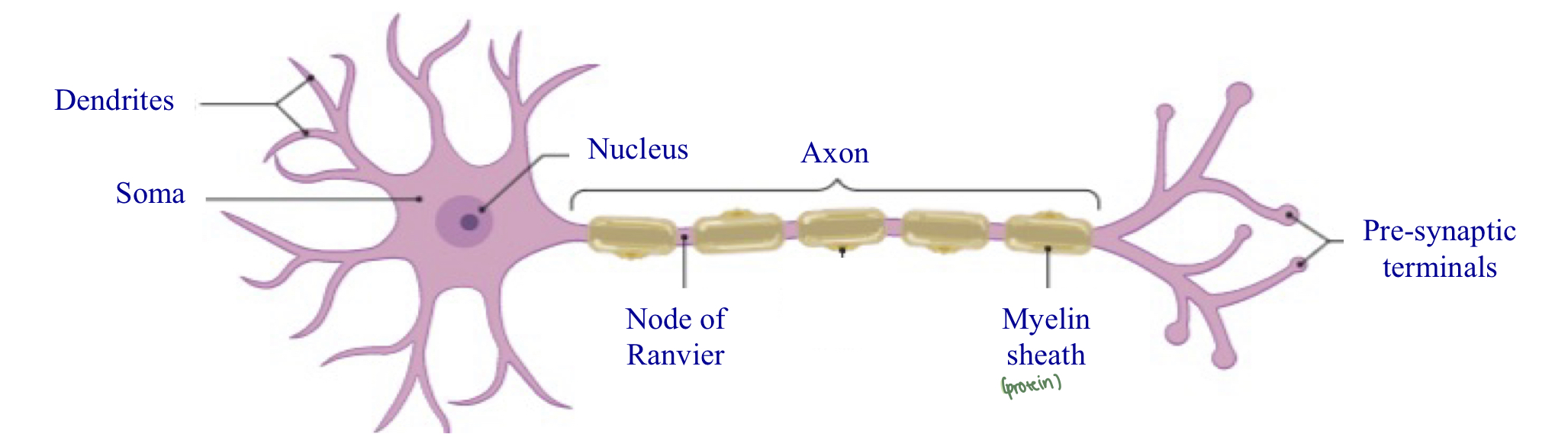
Myelin sheath is a protein
Neuronal Action at the Synapse
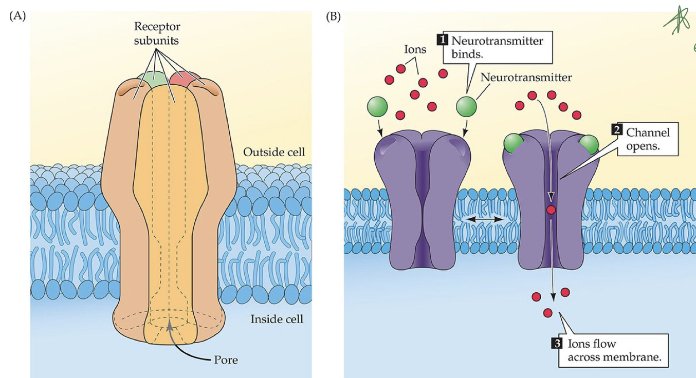
^^Neurotransmitter receptors:^^ specialized proteins in the membrane of post-synaptic neuron
- Neurotransmitter released from pre-synaptic terminal → binds to neurotransmitter receptor → opens pore for charged ions to enter/exit neuron → changes electrical charge of neuron
- ^^Inhibits:^^ cell body loses ions (cell becomes negatively charged)
- ^^Excites:^^ cell body gains ions (cell becomes positively charged)
- Example of Ionic neurotransmitter receptor type
- SSRIs and SSRNIs
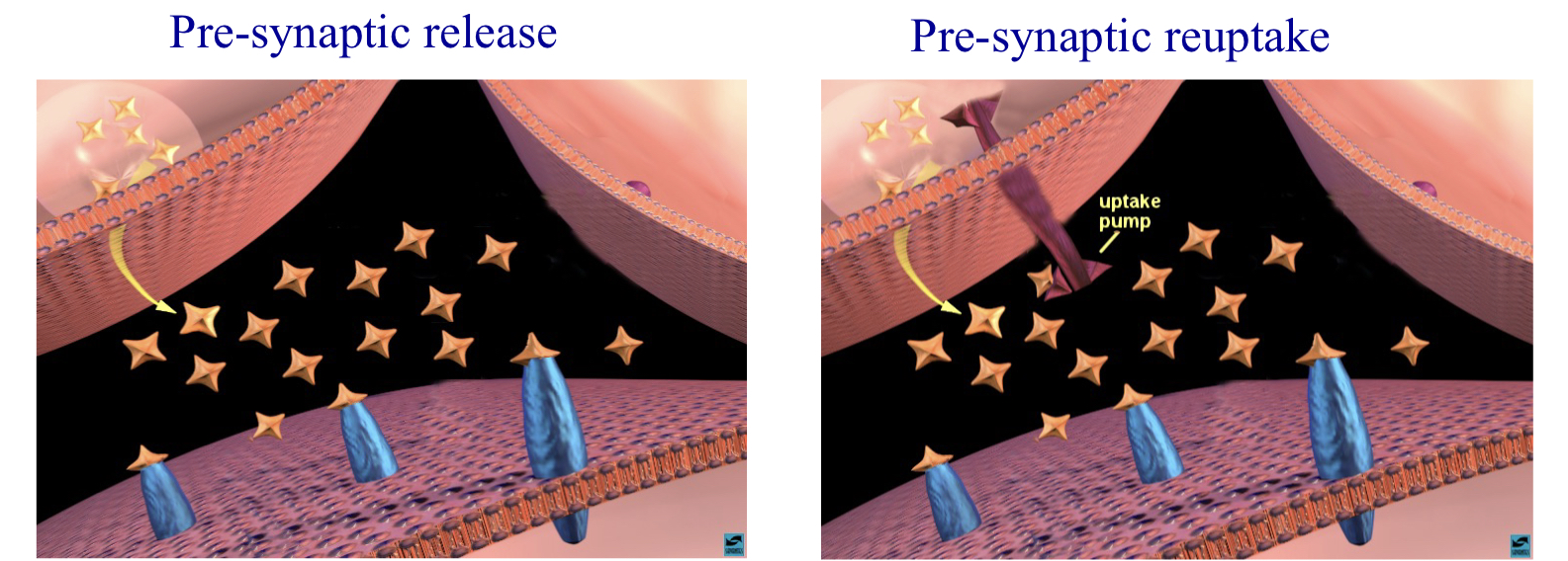
^^Neurotransmitter Re-uptake pumps:^^ specialized proteins on presynaptic membrane
- Bind and transport back in the pre-synaptic terminal
- Purpose: breaking down and packaging
- Example: SSRIs
Type of Neurons
How are they characterized?
- \ # of Neurites from soma
Classification
- Unipolar (One neurite)
- Bipolar (Two neurites)
- Multipolar (Three neurites)
![]()
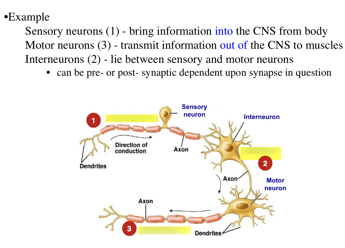
Neural Function
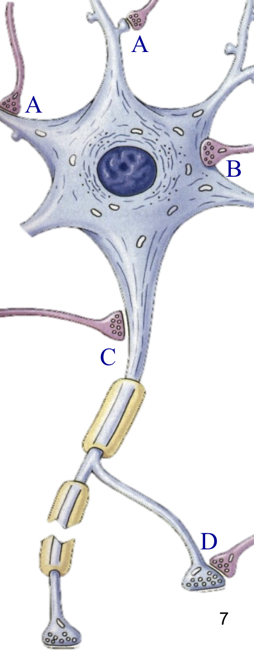
Original “Law of Dynamic Polarization” : Neuronal function from neuron structure (Ramon Cajal)
- Axo-dendritic connection/synapses (A)
- Info flows from dendrite → soma → axon → dendrite
*Revised “Law of Dynamic Polarization”: Info flows from presynaptic cell to post synaptic cell with respect to specific synapse \n *
- Axo-somatic synapses - Synapse on cell body (B)
- Axo-axonic synapses - Synapse at beginning of axon (C)
- Axo-synaptic synapses - presynaptic terminals connect w/ one another (D)
The Neuronal Membrane
Controls the neuronal function and separates the intracellular environment from the extracellular environment.
Polar/Nonpolar
Polar: have an electrical charge
- Water (+ charge on H, - charge on O)
- Like charges repel
Nonpolar: don’t have an electrical charge
- Organic molecules (contains carbon)
- Grease, oil, lipids(fats)
- Can’t interact with polar molecules (I.e. water); therefore not soluble
- Phospholipids: phosphate group acquired by nonpolar molecules WHEN COMBINED
Phospholipids
- The “head”: the phosphate group, charged (polar)
- Hydrophilic region
- The “tail”: the hydrocarbon group, not charged (nonpolar)
- Hydrophobic region
When together, this is a phospholipid.
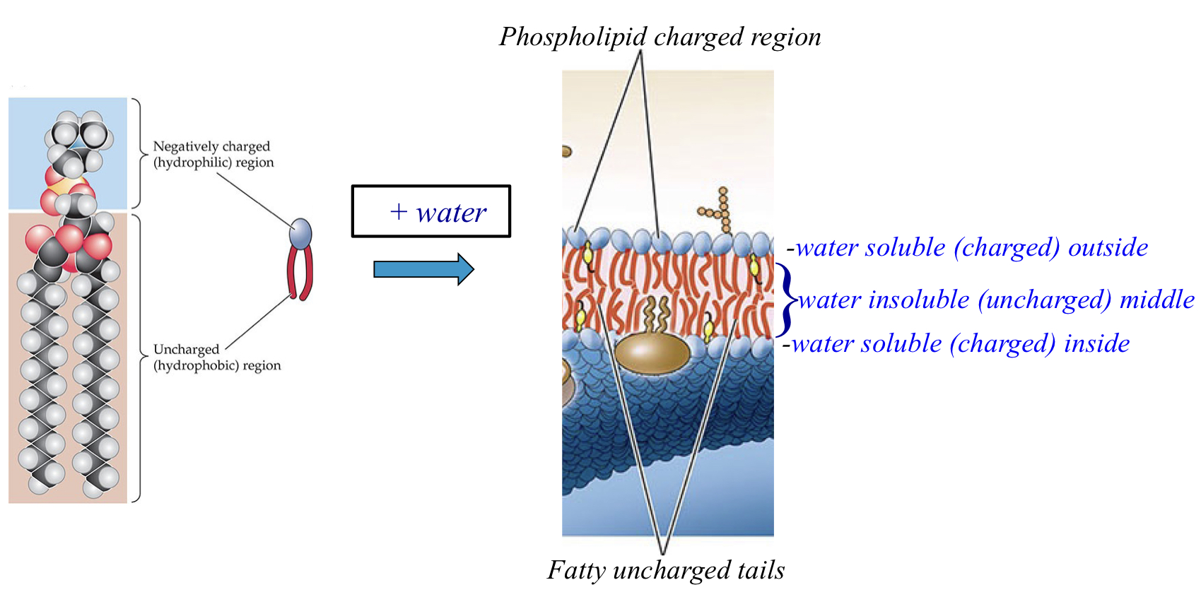
The cell membrane
- Made up of the phospholipids
- ^^Lipid bilayer^^
- Hydrocarbon group (tails) on the inside, phosphate group on the outside
- Inside and outside of the membrane is charged, and inside is uncharged.
- ^^Amphipathic^^
- Both hydrophobic and hydrophilic regions
Fluid Mosaic
- Bilayer’s hydrophobic interior stops charged ions from getting in and out of the cell, so,
- ^^Transmembrane proteins^^^^:^^ channels that allow ions to move in and out of the cell
- aka channels and pumps
- There are specified channels for each ion (sodium, hydrogen, chlorine, etc)
- thus, changing the charge of the cell
- These channels and pumps are why we call the membrane a fluid mosaic.
Genetics Overview
DNA (genes) is transcribed into RNA (genetic intermediary) that is translated into Protein (the functional molecule in a cell)
- DNA (Deoxyribonucleic acid)
- double-stranded genetic sequence
- synthesis of RNA
- RNA (ribonucleic acid)
- single-stranded copy of DNA
- ^^Transcription^^: production of an RNA copy of DNA
- occurs in the nucleus
- messenger RNA (mRNA) codes for specific amino acid sequence (protein)
- mRNA exists the nucleus and travels to the cytoplasm of cell.
- Protein
- mRNA is the synthesis of proteins
- Sequences of amino acids
- 3 amino acids make one protein
- amino acids from transcription(DNA to RNA) is then translated to a protein.
- Translation: assembly of amino acids in a specific sequence
Chemicals and Proteins Important for Neuronal Function
Chemicals
Neurotransmitters: Chemical molecules released from neurons that act as chemical signals between neurons
- Classical neurotransmitters: small chemical molecules
- Norephrine (Noradrenaline): Concentration
- Gaba: Calming
- Dopamine: Pleasure
- Glutamate: memory (excitation), most common
- Serotonin: mood (happiness)
- Acetylcholine: learning
- Peptide neurotransmitters: short peptides (small proteins)
- Endorphins
- Often works with classical neurotransmitters
- Can be 5 - 50 amino acids
Proteins
Ion channels/pumps
- proteins in the cell membrane that move ions (mainly salt [NaCl] and K] in and out of the cell
Neurotransmitter receptors
- proteins in the post-synaptic cells that bind to neurotransmitters released in the synapse
- The neurotransmitter binds to the receptor → opens a pore for charged ions (to enter/exit) → changes the charge post-synaptic neuron.
- Types of receptors
- ^^Metabotropic^^
- The neurotransmitter binds to receptor → activates protein (aka signaling molecule) → ion channel opens/closes
- ^^Ionotropic^^
- The neurotransmitter binds to ion channel → channel opens
- ion channel has its own neurotransmitter (ligand) bonding site
Types of Ligands for Neurotransmitter Receptors
Overview
- Continuum of Efficacy
- Neurotransmitters act by rapidly bind/activate receptors & then release and deactivate a neurotransmitter receptor
- Reuse after function is completed
- Neurotransmitters have
- ^^Affinity^^ (how fast and strong a ligand is)
- ^^Potency^^ (how biologically effective a ligand is once bonded)
- There are varying levels of both (high affinity and low potency)
- Properties of the receptors determine the properties of the transmitters
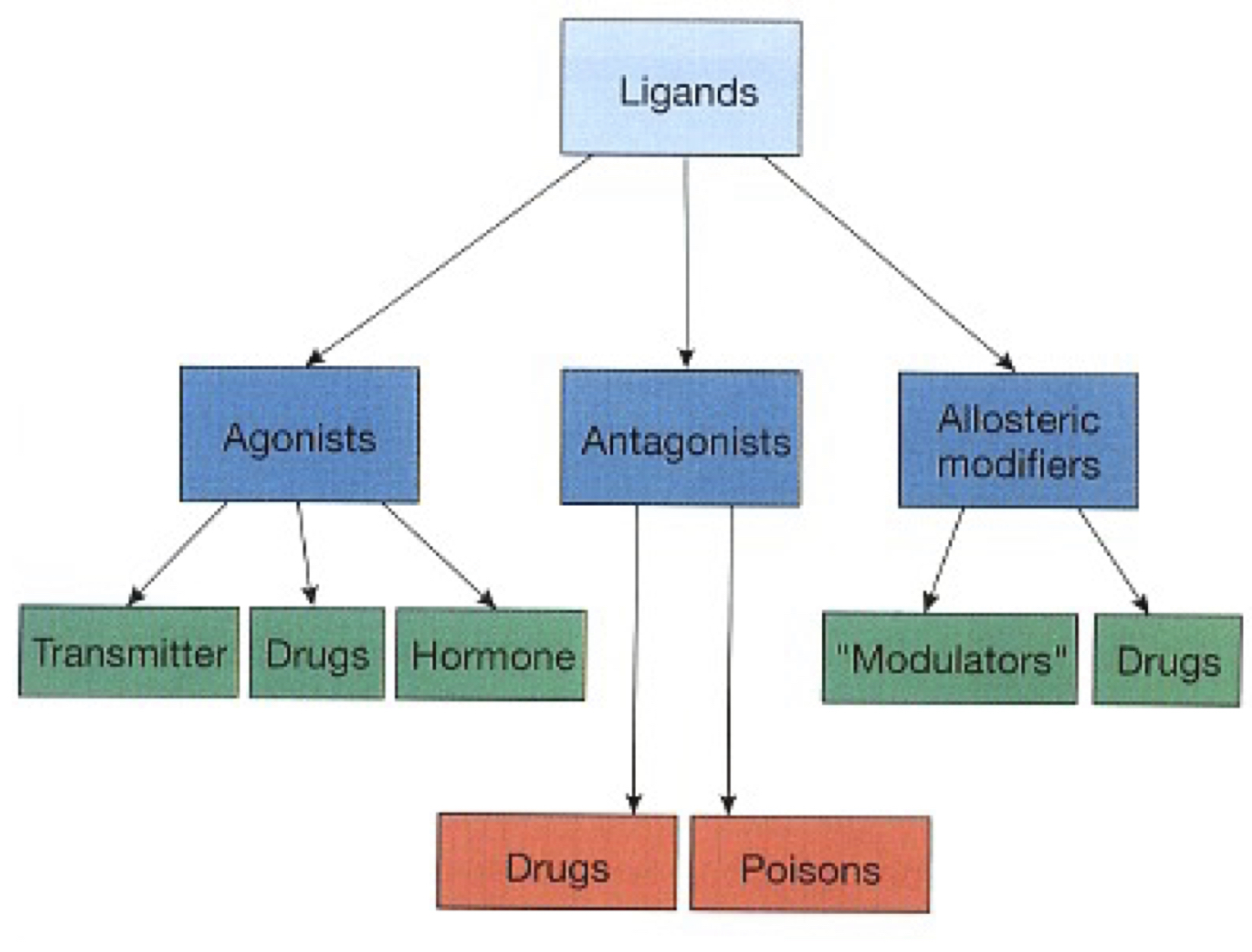
Types of Ligands
Remember: ligands are what bind to the receptors
^^Agonist^^
- A ligand that binds to a receptor and activates it biologically
- varying levels of affinity and potency
- Endogenous
^^Antagonist^^
- ligands that bind to a receptor and do not activate it biologically
- Typically have high affinity and zero potency
- So receptor is blocked from functioning
- all antagonists are exogenous: foreign substances
- Toxins and venoms
- Opioids
Allosteric modifiers
- helps naturally occurring ligand increase likelihood of receptor-ligand binding
- In humans, helps agonists
- Binds to a different location that agonist and antagonists
- Benzodiazepines
Excitation and inhibition are properties of the receptor, not the neurotransmitter/ligand
The Concept of Neuromodulation
Peptide transmitters
- Are often co-transmitters and are released with small chemical neurotransmitters such as dopamine/norepinephrine.
- These often act at allosteric sites
- Act as neuromodulators: a substance that binds to a receptor at a different location than the neurotransmitter itself
- Increases affinity of the receptor to bind to the neurotransmitter
Neurotransmitters
Acetylcholine
- Synthesized in pre-synaptic terminal by ^^cholineactyltransferase (ChAT)^^.
- Enzyme that transfers acetyal group to choline.
- Fuses acetate (from Acetyl-CoA) and choline together
- Packed in vesicle and sent out
To terminate post-synaptic ACh activity
- Desensitization: respectors become less responsive to presence of ACh
- Diffusion: ACh out of the synapse
- Breakdown: of transmitter molecules
- For ACh, use of AChE(acetylcholinesterase)
- Makes sure we don’t have too much Acetyl CoA in our system (to breathe)
- Drugs
- Physostigmine: naturally occurring drug that blocks AChE
- gets more Acetyl-CoA function
- Insecticides: manmade AChE blockers
Neuromuscular junction
- via nicotinic ACh receptors (stimulant)
Gamma-aminobutyric Acid (GABA)
Produces neural inhibition (most common)
- Synthesized from glutamate
- Glutamic acid decarboxylase (GAD) converts glutamate to GABA
GABA transaminase: recycles GABA back to glutamate for re-uptake and use
GABA A receptors
- Ionotropic receptors, Cl- channels
- cell becomes more negative
- at least 2 allosteric bonding sites
- when bound increases the ability of the receptor to bind to neurotransmitter
- Action seen in benzodiazepines (anti-anxiety) and barbiturates (depressants)
- doesn’t produce inhibition itself, but increase affinity for GABA receptor for GABA.
- increase ability of GABA a receptors to bind GABA
In the Brain
Majority of the synapses in the brain are GABA-containing
- Many use presynaptic inhibition (via axo-synaptic contacts)
- GABA blocks the synaptic terminal from releasing neurotransmitters
- GABA antagonists (receptor blockers) produce excitation
- blocking inhibition = excitatory effects
- can cause seizures
- Epilepsy: loss of GABA producing neurons
- treated with GABA simulating drugs
Glutamate
Produces neural excitation, ionotropic receptors.
- Associated with memory and learning, the big daddy
- Long term effects on cell function
Types of receptors
- Sodium receptors
- AMPA: most active
- Kainate
- Both cause excitation because of positive sodium ion.
- Sodium AND calcium receptors
- NMDA
- Excitation due to positive sodium and calcium ions.
Glycine
Amino acid for inhibition
- in spinal cord (used instead of GABA)
- Strychnine
- poison, glycine antagonist causing spinal seizures
Biogenic Amines
Have different amine groups(ring structure) attached
- Synthesized by tyrosine or tryptophan
Types of receptors
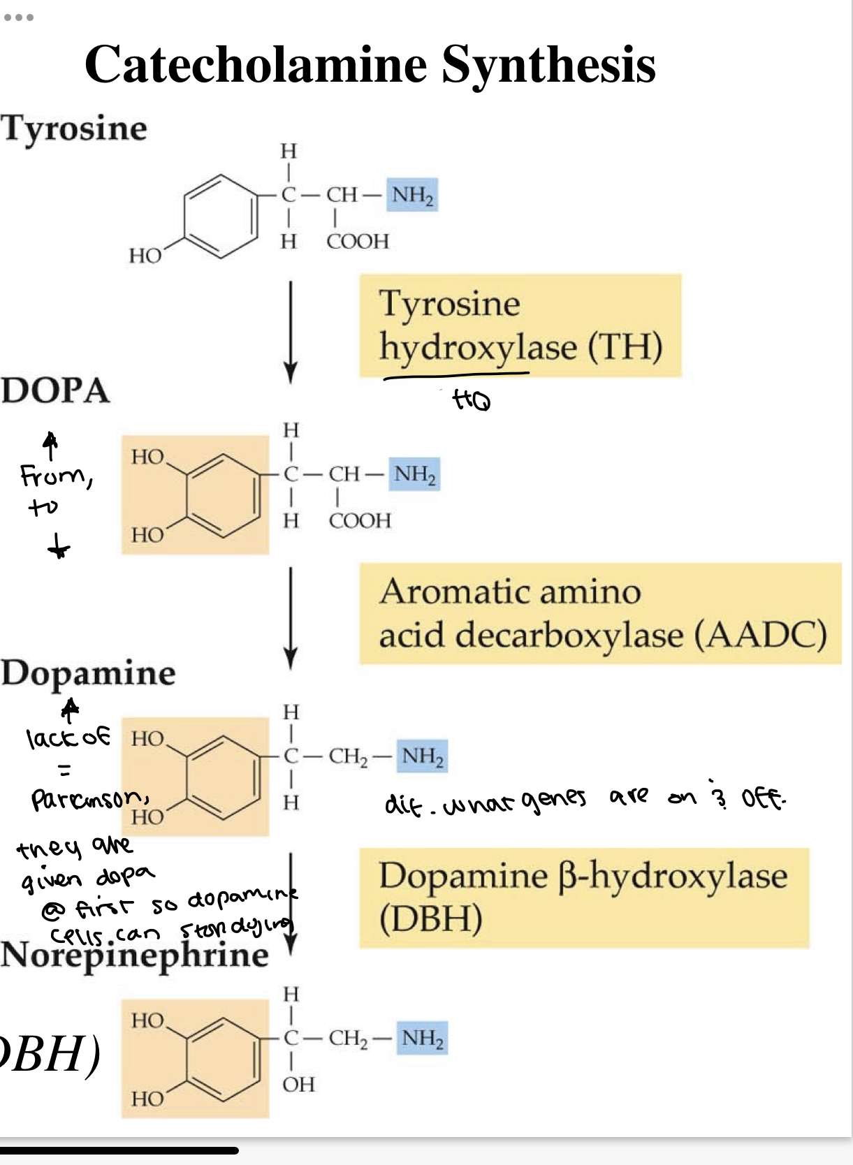
- Dopamine(DA) and Norepinephrine(NE)
- share core structure(catechol ring group) and nitrogen-containing side group(amine)
- Synthesis
- From Tryosine → DOPA (one hydroxl group to two) → Dopamine (Loss of COOH [carboxyl acid]) → Norepinephrine (adding oxygen to one hydrogen)
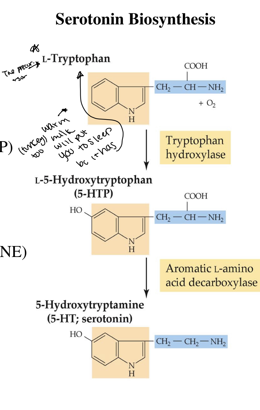
Seotonin
- Small molecule transmitter
- core structure (indole ring) and nitrogen containing side group (amine)
- Synthesis
- L-Tryptophan → L-5-Hydroxtryptophan aka 5-HTP (added HO group) → serotonin (loss of carboxyl acid)
Peptide Transmitters
A short chain of amino acids
Can act one of two ways
- Peptide transmitters: released in brain as neurochemical signal between neurons
- Peptide hormones: released from brain into the blood stream to act as neurochemicla signal between brain and body
How it is synthesized
- synthesized as large proteins to small “active” peptides →packaged into vesicles in cell body → ^^orhtograde axoplasmic transport^^ (sent down axon to synaptic terminal)
Once released, diffuse away from sunapse or broken down by enzymes (^^proteolysis^^)
Act at low concentrations for a long time
- Because they require a lot of energy (metabolically expensive)
- Therefore, they are called neuromodulators
Examples
- endorphins, enkephalins, oxytocin & vaspressin
Other important proteins
Structural proteins (actin, tubulin and elastin)
- Hold cells & keeps them In place.
- Determines the shape and movement of cell
Enzymes
- A catalyst that is a protein
- Creating products from building blocks
- Can be catabolic or anabolic!
The Active Neuron
Axoplasmic Transport
- Transport of proteins to distant location in the neuron.
- Moving between cell body and axon.
- Orthograde/anterograde transport
- Away from the cell body (to neutries)
- For release
- Retrograde transport
- Toward the cell body (from neurites)
- For degradation
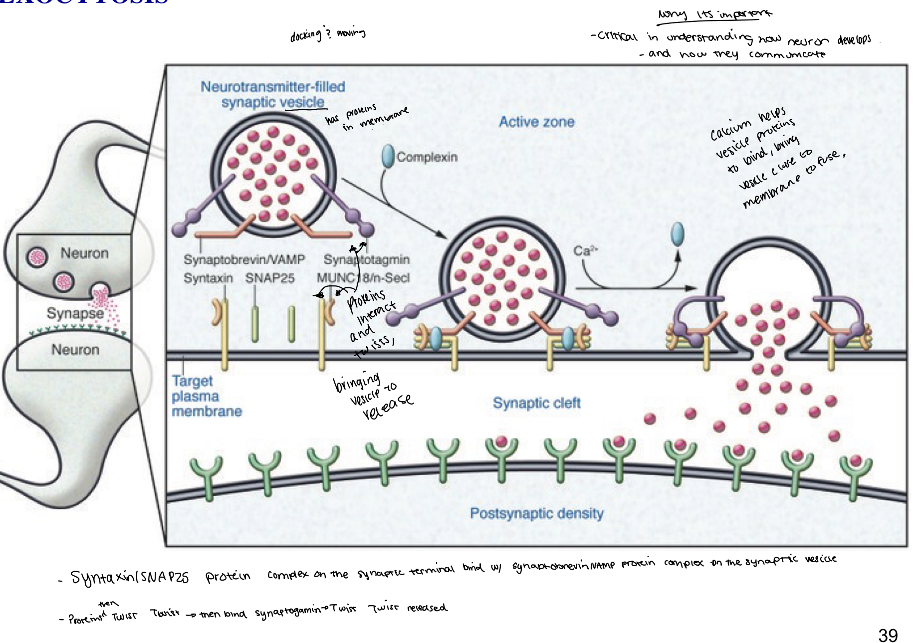
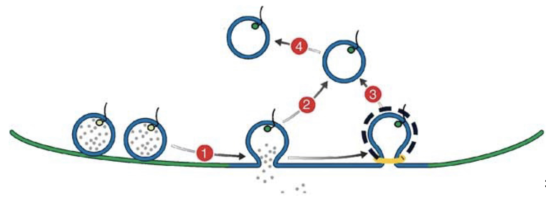
Neuronal Release
Exocytosis
- Fusion of the synaptic vesicle with the plasma membrane
- peptide transmitters of neurotransmitters are dumped into extracellular space (synapse)
- Fusing with terminal membrane and releasing its prescense
- Calcium helps vesicle proteins to bind with the membrane, bringing the vesicle close enough the fuse
Endocytosis
- Piece of the membrane that pinches back to form a new vesicle
- In order to maintain size
Neuronal Development/Aging
Neurons use the exocytosis and endocytosis to grow, develop and prune.
- Neuronal growth
- Neuron sends out neural processes
- axon growth
- exo > endo
- axon from one neuron grow and connect with another neuron to form synapse
- Neuronal pruning
- Neural processes (axons) withdraw
- exo > endo
- axon from one neuron withdraws connection with another
- Mature Synapse
- stable associations between neurons
- endo = exo
Chemoaffinity Hypothesis
- How neurites find their way through development
- Chemical signals (trophic factors) - proteins that help nerve cells develop and recognize each other are exchanged between potential synaptic partners
- Grow cone
- Tip of growing neuronal axon
- Withdrawl and approach cycles
- When correct synaptic partner is found
- filopodia flatten out
- presynaptic and post synaptic densities appear
Neuroplasticity
@ mature synapse, # of postsynaptic receptors in membrane can be increased (up-regulated) or decreased(down-regulated)
- Based on amount of neurotransmitter released and received by receptors on the postsynaptic cell.
- ^^Up regulation:^^ little neurotransmitters, lots of receptors
- presyn: decrease trophic influence
- postsyn: increase receptor number
- Problem with up-regulation: can cause hypersensitivity
- Decrease in neurotransmitter influence → starves post synp cell → post synp produces more receptors and becomes sensitive to the remaining neurotransmitter around it
- Example: phantom leg pain
- ^^Down regulation:^^ lots of neurotransmitters, not a lot of receptors
- presyn: increase in trophic influence(neurotransmitter)
- postsyn: decrease in receptor
- Problem with down-regulation: can cause desensitization
- Too much neurotransmitter can cause the post syn cell to feel overwhelmed → post synp cell decreases receptors and becomes less sensitive to the presence of neurotransmitters
- May or may not change back to normal
- Seen in drug addiction
- Too much, so I change
Regeneration
Schwann Cells
- Glial cells that myelinate neurons are
- Myelinate a single neuronal axon
- when axon is damaged, schwann cells form a guidance tube to guide the regnerating end of the axon to the target end of the axon.
- In PNS
- 1 cell body with one axon.
Oligodendrocyte
- Glial cells that myelinate neurons
- Myelinates multiple axons
- Axon is damaged → oligodendrocytes fail to respond → withdraws remaining support → axon degenerates and damage is permanent.
- There’s not enough space in the brain for other cells, which is why oligodendrocytes are used.
- In the CNS
- I.e. spinal cord injury
- 1 cell body with many axons