Biol 2030 lec exam two
functions of the circulatory system
- @@circularity system@@: consists of the heart, blood, and blood vessels
- @@cardiovascular system@@: refers to only the heart and blood vessels
- Functions of the circulatory system:
1. TRANSPORT- ^^O2^^, CO2, Nutrients, wastes, hormones, stem cells 2. PROTECTION- inflammation, limit the spread of infection, destroy cancer cells, neutralize toxins, and initiate clotting. 3. REGULATION- Fluid balance, stabilizes pH or ECF, and temperature control.
Components of blood
- adults have 4-6 L of blood
- Liquid connective tissue: cells and extracellular matrix
- @@Plasma@@: Matrix of blood, clear light yellow fluid
- Formed elements: blood cells (red, white, and platelets)
- Hematocrit- centrifuge blood to separate components
7 kinds of formed elements
1. @@Erythrocytes@@: Red blood cells
- @@Platelets@@: cell fragments from special cells in bone marrow
@@Leukocytes@@: White blood cells. Divided into two categories
* GRANULOCYTES (with granules)
* \
3. @@Neutrophils@@
* \
4. @@Eosinophils@@
* \
5. @@Basophils@@ \n
* AGRANULOCYTES ( Without granules)
* \
6. @@Lymphocytes@@
* \
7. @@Monocytes@@ \n 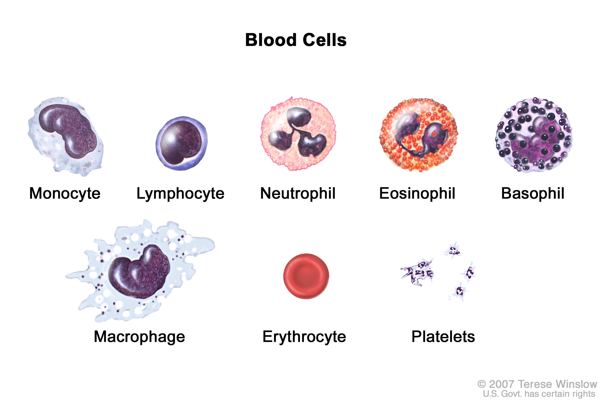
\
Blood Plasma
- @@Plasma@@- liquid portion of blood * serum: remaining fluid when blood clots and solifs are removed identical to plasma except for absence of fibrinogen (holds blood clots together)
- Three major categories of plasma proteins * ^^Albumins^^ : Smallest and most abundant; influences blood pressure, bloodflow, an fluid balance * ^^Globulins^^ (antibodies): provide immune functuons * ^^Fibrinogen^^: precursor of fibrin threads that help form blood clots
- dissolved O2, CO2, and Nitrogen electrolytes.
Blood Viscosity and Osmolarity
- @@Viscosity@@: Resistance of a fluid to flow, resulting from the cohesion of its particles * whole blood 4.5-5.5 times as viscous as water * Plasma is 2.0 times as viscous as water
- @@Osmolarity@@: total molarity of those dissolved particles that cannot pass through the blood vessel wall. (measure of solute inh a solution) -Water always diffuses from low to high * if to high, blood absorbs too much water, increasing the blood pressure * if to low, to much water stays in the tissue, blood pressure drops, edema occurs * optimum osmolarity is achieved by bodys regulation of sodium ions, proteins, and red blood cells
Starvation and plasma protein deficiency
- Hypoprotenimia: deficiency of plasma proteins * Extreame starvation * liver or kidney disease * severe burns
- Kwashiorkor: children with severe protein deficiency * Thin arms * Distended abdomen
How blood is produced
- Adult production of 400 billion platelets, 100-200 billion RBC, and 10 billion WBCs every day
- @@Hemopoiesis@@: production of blood
- @@Hemopoietic tissues@@ produce blood cells * Yolk sac produces stem cells for first blood cells * liver stops producing blood cells at birth * splen remains involved with lymphocyte production
- Red bonme marrow produces all seven formed elements
- @@colony- forming unit@@- specialized stem cells only producing one class of formed element of blood
- @@Myeloid Hemopoiesis@@- blood formation in the bone marrow
- @@Lymphoid Hemopoiesis@@- blood formation in the lymphatic organs
Erythrocytes
- Two principle functions * carry O2 from lungs to tissues * Pick up CO2 from tissues and bring to lungs
*inssuificient EBC can cause death in minutes due to lac of oxygen to tiussues
- disk shaped cell with thick rim * ^^Lack of nucleus^^: no mitosis * ^^lack of mitocondria^^: anarobic resperitaion * ^^full of hemoglobin^^ to carry O2
- Gas transport: major function
- O2 must bind to hemogloben
- @@Carbonic anhydrase@@ (CAH) in cytoplasm * * produces carbonic acid from CO2 and water * important role in gas transport and pH balance
Hemoglobin
- four protein chains
- four Heme groups each carrying one O2 * nonprotein moiety that binds O2 to Fe at its center * *1 hemoghloben can bind 4 O2
- @@Men have more RBC and Hemogloblen@@ * women have a menstrual cycle and more body fat
Erythrocyte production
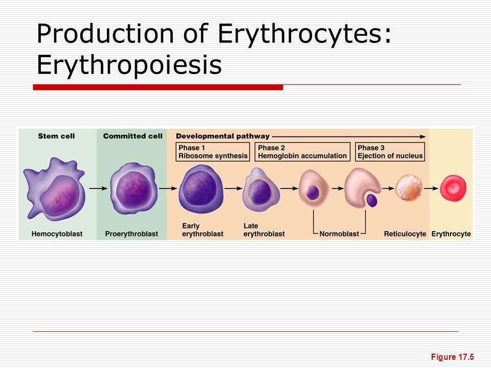
- Erythropoiesis (EPO)- RBC production * EPO made buy kidneys * The average lifespan of RBC is 120 days * development takes 3-5 days
- Production
1. Pluripotent stem cell 2. Erythrocyte colony-forming unit (committed cell) * has receptors for EPO from kidneys 3. precursor cells * Erythroblasts multiply and synthesize hemoglobin * Reticulocyte discards nucleus to make room for hemoglobin 4. mature cell (Erythrocyte)
\
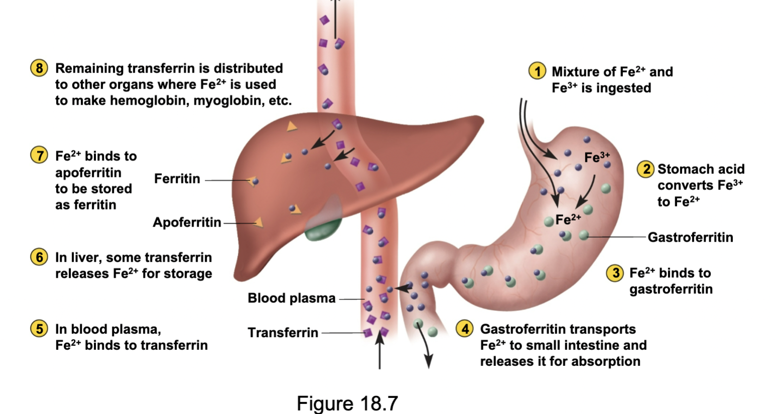
Iron Metabolism
Erythrocyte homeostasis
1. hypoxemia is sensed by the liver and kidneys
- secretion of EPO from kidneys
- stimulation of red bone marrow
- accelerated erythropoiesis (formation of red blood cells)
- increased RBC count
- increased O2 transport
- negative feedback control * Dropped RBC causes Hypox (‘emia’ means blood) * Kidney production of preproprotein stimulates bone marrow
*erythropoiesis (EPO) is RBC production * RBC increases in 3-4 days
- Stimuli for increasing EPO * Low levels of O2 * high altitude * increase exercise * loss of lung tissue in emphysema
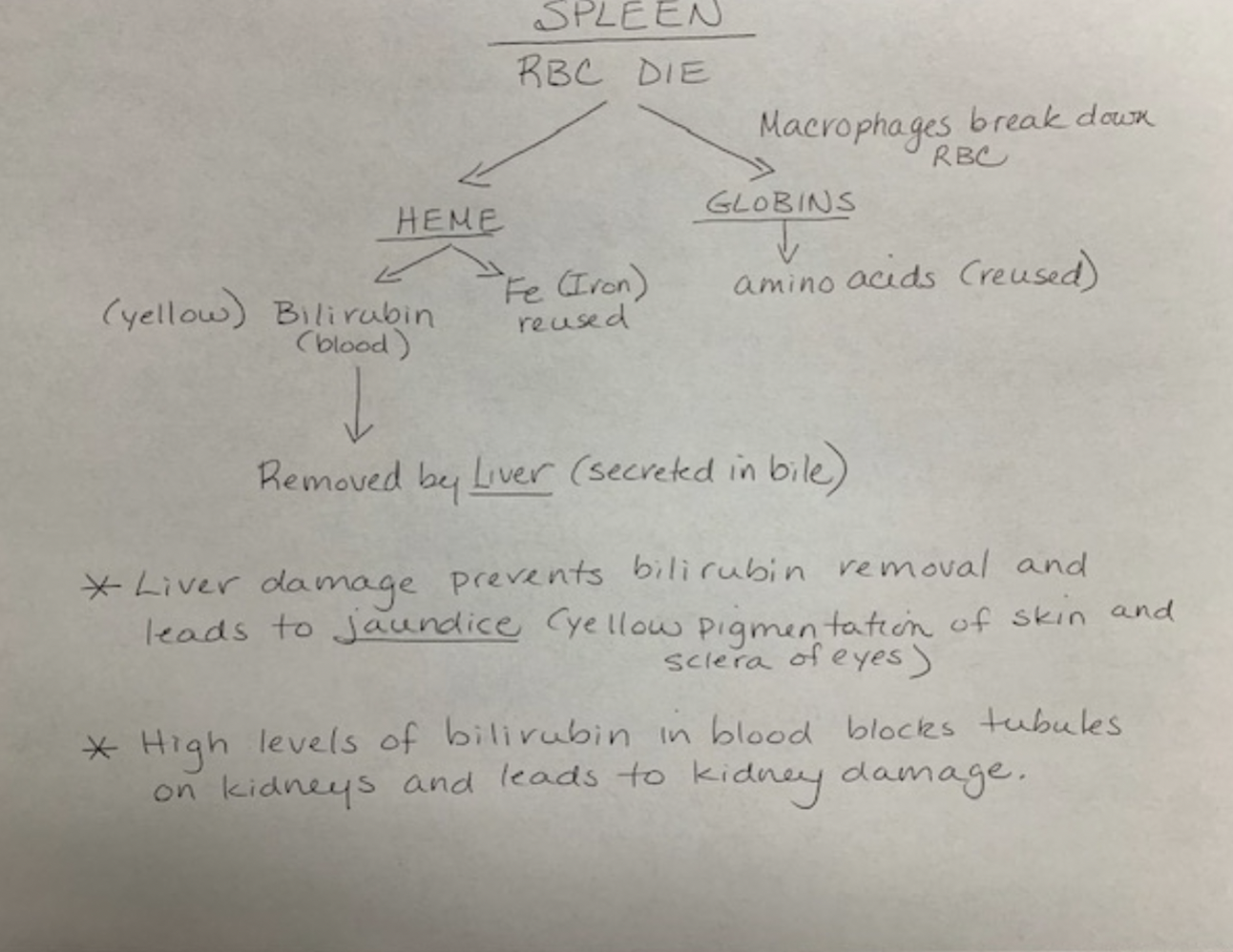
Erythrocyte death and disposal
- RBC rupture (Hemolysis) in narrow channels of spleen and liver
- macrophages in the spleen (spleen considered RBC graveyard) * Digests membrane bits * separates heme from globin
Erythrocyte disorders
- @@polycythemia (High RBC count)@@ * primary polycythemia * cancer of erythropoietin cell line in red bone marrow * High RBC count high as ^^11 million^^ RBC * Secondary polycythemia * from dehydration, emphysema, high altitude, or physical conditioning * RBC up to ^^8 million^^ * Dangers of polycythemia * increased blood volume, pressure, viscosity * can lead to embolism, stroke, or heart failure
- @@Anemia@@ * inadequate EPO or hemoglobin synthesis * kidney failure and insufficient erythropoietin * Hemorrhagic anemias from bleeding * Hemolytic amenia from RBC destruction * Dangers * Tissue hypoxia and necrosis * pt lethargic, shortness of breath with exertion, necrosis to the brain, heart, or kidney * blood osmolarity is low, producing tissue edema * blood viscosity is low, cardiac failure
- @@Sickle cell disease@@ * hereditary defect in mostly African descent (due to malaria) * differed only on the 6th amino acid on the bata chain * full carriers usually don’t survive without medical help * can lead to kidney failure, heart failure, stroke, joint pain, or paralysis
Blood typing
- Agglutinogens are RBC antigens
- Called antigen A and B
- Ditermined by glycolipids on RBC surface
- antibodies are called agglutinins
I is recessive
A is dominant
B is dominant
| blood type | antigens | Antibodies |
|---|---|---|
| O | none | A and B |
| A | A | B |
| B | B | A |
| AB | A and B | none |
| blood type | donate to | receive from |
|---|---|---|
| O | O,A,B,AB | O |
| A | A and AB | O and A |
| B | B and AB | O an B |
| AB | AB | A, B, AB, O |
- RH group: Rh+ is if you have the D antigen
- Rh- people can only receive from other Rh- people but Rh+ can receive from Rh- and Rh+
Hemolytic disease of the newborn
Can occurr if Rh- mother has formed antibodies and is pregnant with a second Rh+
- prevention * RhoGAM given to prevent Rh- women
WBC form and function
- least abundant formed element
- protects against infectious microorganisms and other pathogens
- conspicuous neculus
- retaint their organelles for protein synthesis
- granules
Types of leukocytes
- granulocytes * Nutrophils - aggressively antibacterial * nurtophilia- rise in number of nutrophils in response to bacterial infection * Eosinphils- releases enzyme to destroy large parasites * increased numbers in parasitic infections, collagen disease, allergies, and diseases of the spleen * Basophils- secreasts histamine (speeds flow o blood to an injured area) and Heparin ( promotes mobility of other WBCs in the area. * increased numbers in chickenpox, sinitis, diabeties.
- Agranulocytes * Lymphocytes- Distroy cells ( cancer, foreign, and infected cells) * coordinate actions to other immune cells * secrete antibodies to provide immune memory * increased number in diverse infections and immune responses * monocytes- leaves blood stream to transform into macrophages * increased numbers in viral infections and inflammation
Leukocyte life history
- leukopoiesis- production of white blood cells
- Hemopoiteic stem cells differeate into: * myeoblasts- from nutrophils, eosinophils, basophils * monoblasts- from monocytes * lymphoblasts- gove rise to all forms of lymphocytes
- red bone marrow stores and releases monocytes and granulocytes
Leukocyte life cycle
- circulating WBC do not stay in blood stream * granulocytes leave in 8 hours and live 5 days longer * monocytes leave in20 hours, transform into macrophages, and live several years * lymphocytes provide long term immunity (decades) being continuously recycled from blood to tissue fluid to luymph back to blood.
Lekocyte disorders
- lueokpenia- low WBC count, below 5,000 WBCs * causes: radiation, poisons, infectious diseases * effects: elevated risks of infection
- Leukpcytosis: high WBC count, above 10,000 * causes: infection, allergy, diseases * differential WBC count: identifies what percentage of total WBC consists of each type of leukocyte.
- leukemia: cancer of hematopoietic tissue usually producing a very high number of circulating leukocytes * myeloid leukemia: uncontrolled granulocyte production * Lymphoid lukema: uncontrolled lymphocyte or monocyte production
## Platelets
Small fragments of megakaryocyte
- platelet function: * secrete vasoconstriction that help reduce blood loss * stick together to form platelet plugs ( collegon is found under skin layer and is stickey) * secrete procoagulants to promote clotting * chemically attract nuetrophils and monocytes to sites of inflammation * secrete growth factors that stimulate mitosis to repair blood vessels
- Platelet production: * Thrombopoiesis: Stem cells that becomnme megakaryoblasts * Megakaryoblasts: repeatedly replicate DNA without dividing * \
## Platelets and hemostasis
- Hemostasis: the cessation of bleeding * hemorrhage: excessive bleeding
- Three homeostacic mechanisms * vascular spams * platelet plug formation * coagulation ( blood clotting)
- vascular spasm- prompt constriction of a broken vessel. * causes: pain receptors * most immediate protection against blood loss
- platelet plug formation- plsayelet psudeopods stick to damaged vessels and other psudeoods contract * collagen is directly under endotheial tissue and is sticky. platelets stick to collagen.
- Coagulation- clotting. conversion of fibrinogen into fibrin threads framework together * procoagulants (clotting factors)- usually produced by the liver * extrenisic pathway- factors released by damaged tissues begin to cascade ( activate one factor and it will activate the next) * intrinsic pathway Factors found in blood begin to cascade
Caogulation of intrinsic and extrensic pathways
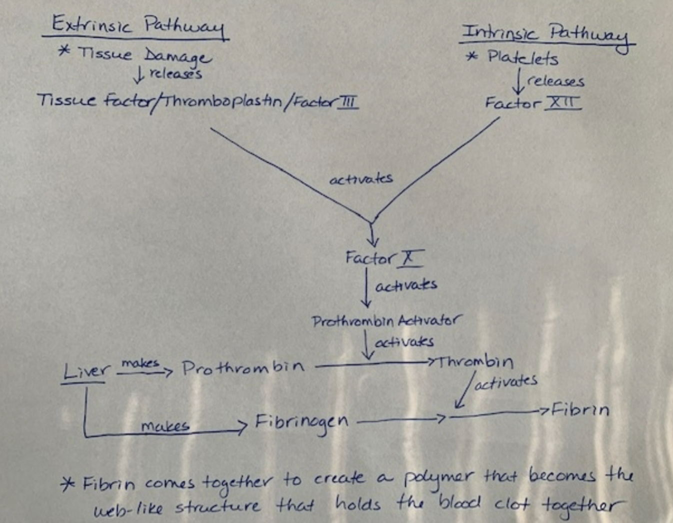
The fate of blood clots
- clot retraction occurs within 30 minutes
- fibrinolysis- dissolution of a clot * @@plasminogen turns into plasma which dissolves fibrin that breaks up clot@@
- Thre prevention of inappropriate clotting is caused by a prostacyclin coated endothelium
Clotting disorders
- Hemophilia- deficiencies in one factor or another in coagulation * usually missing Factor 8/VIII * Physical exertion causes bleeding and pain
- Hematomas- blood colt in the tissues outside of the vessel
- Thrombosis- blood clotting inside a blood vessel
- embolus- anything that can travel in the blood and block blood vessels
Blood vessel anatomy
- arteries- caqrry blood away from the heart
- veins- carry blood to the heart \
- capillaries- connects smallest arteries to smallest veins
- vessel walls * Tunica interna- inner most layer, lines blood vessels and exposed to blood * Endthelium- simple squamous epithelium overlaying basement membrane. normally repels blood cells and platelets * Tunica Media- middle layer that consists of smooth muscle * regulates diameter of the blood vesssel * Tunica Externa- outermost layer * provides passage for small nerves, lymphatic vessels
## Arteries
- classified by size * conducting (elastic or large) arteries * biggest arteries ( aorta, common corotid, pulmonary trunk,..) * Distributing (muscular or medium) arteries * distributes blood to specific organs * Resistance (small) arteries * Arterioles: smallest arteries * control amount of blood to various organs
Aneurysm
Weak point in artery or heart wall
- forms thin- walled, bulging sac and may rupture at any time
- dissecting aneruysm * blood acculumulates between tunics of artery and separates them.
Atrial sense organ
- sensory structures in walls of major blood vessels that monitor blood pressure and chemistry * carotid sinuses: baroreceptors monoter blood pressure and found in internal carotid * carotid bodies: chemoreceptors, stableize pH
\
\