SHARC Nervous tissues
What are the components of nervous tissue? - Neurons - send and receive signals
Neuroglia (glial cells) - support and protect neurons
What are the parts of a neuron? What is the function of each part?
Large nucleus and nucleolus - nucleolus makes ribosomes
Neurotransmitters (proteins) - chemical messengers
Mitochondria - ATP
RER and ribosomes - make proteins for neurotransmitters
Cytoskeleton - microtubules for neuronal communication
Nissl bodies - dense areas of RER and free ribosomes
Dendrites - highly branched 80/90% of neuron surface area - receive information
Axon hillock - signal integration (translates signal via changes in ion concentration)
What are the 3 general functions of the nervous system?
Sensory: translating sensory information (touch, vision, hearing, blood pressure change)
Integration: using the brain to translate signals
Motor: moving skeletal muscle (part of response)
What are the organs of the CNS?
Spinal cord, brain
What are the functions of the CENTRAL Nervous system?
Sensory data: from inside and outside the body
Motor commands: controlling activity of peripheral organs (ex: skeletal muscles)
Higher functions of brain: intelligence, memory, learning, emotion
Functional divisions of the nervous system:
Input - the sensory nervous system detects stimuli and transmits information from receptors -> CNS
Somatic sensory: consciously perceived input from receptors
Visceral sensory: unconsciously perceived input from receptors of blood vessels and internal organs
Output: motor nervous system initiates and transmits information from CNS -> effectors
Somatic motor: voluntarily controlled output (ex: skeletal muscle)
Autonomic motor: involuntary output (ex: cardiac muscle, smooth muscle, glands)
Functional divisions of the peripheral nervous system:
afferent/sensory division: periphery to CNS (bottom to top)
efferent/motor division: CNS to the periphery (top to bottom)
Functions of receptors of the afferent division? Examples of receptors in the body?
Detect changes or respond to stimuli (mainly proteins that change shape in response to stimuli)
Neurons and specialized cells, complex sensory organs (eyes, ears)
Functions of the effectors of the afferent division? Examples of effectors in the body?
Respond to efferent signals
Cells, organs
What are the differences between a sensory neuron and a motor neuron? Sensory neurons are receptors detecting changes/responding to stimuli. Motor neurons are effectors responding to signals from the central nervous system.
Neurons can be classified in a number of ways- what is each type of neuron discussed in class and what are defining characteristics/functions/examples/locations.
By structure:
Unipolar neuron - sensory. Detects touch, pressure, and temperature. (cell body extending off axon). Singular process branching off cell body like a T shape.
Bipolar neuron - relays information for olfactory epithelium and the retina (cell body in center. Two processes extending from cell body)
Multipolar neuron - interneurons of CNS, all skeletal muscle motor neurons (multiple processes extending out from cell body).
By function:
Sensory - monitor internal environment and effects of external environment on body (incl. Corpuscles, nociceptor, Merkel cell)
Interneurons - only located in the CNS; between sensory and motor neurons; higher functions (memory, planning, learning)
What is a neurotransmitter in general terms, what does it cross and what is its function?
A chemical messenger (usually proteins). Neurotransmitters are released by the presynaptic cell to cross the gap junction; they are chemical messengers of an action potential.
What are the categories of neuroglia cells in both the CNS and PNS and what are their
primary functions?
CNS:
Ependymal cells - secrete cerebrospinal fluid into spinal cord and brain ventricles. Monitors CSF and contains stem cells.
Astrocytes - form blood brain barrier, repair damaged neural tissue, guide fetal brain development
Microglia - clean up cellular debris, waste, protect against pathogens
Oligodendrocytes - form myelin sheaths
PNS:
Satellite cells - surround ganglia, regulate environment around neuron, maintain nerve health
Schwann cells - form myelin sheath around axons
What does myelination do to conduction speed? Why? Myelination increases the speed of the action potential by insulating the electric signal, preventing leakage of ions.
What are the differences between gray and white matter? White matter contains many myelinated neurons, gray matter contains few myelinated neurons and forms the outer layer of the brain (cerebral cortex).
What is an action potential? What channels are involved? An action potential is an electrical signal that travels long distances on the axon. Only voltage gated type channels are involved.
What is saltatory conduction? Conduction of action potential through myelinated axons. This is a “jumping signal” where movement is very fast.
What is a resting potential? Why do you see a certain charge inside the cell (think ions,etc.). Draw a basic image of a cell and the ions, etc involved.
Resting potential is the neuron’s membrane charge when the cell is at rest/not conducting action potential. It is -70 mV. This is caused by Na K ATPase pumping sodium out and potassium in the cell, and also because there are many more potassium leak channels than sodium leak channels allowing more potassium to leave the cell. Negatively charged phosphates and proteins are also stuck inside the cell.
Na+, K+ ATPase. What does it do?
Transport protein that uses active facilitated transport (ion-pump) to move potassium and sodium against their concentration gradients. It uses the energy from ADP and phosphate separating to move 3 Na out and 2 K in.
Explain the difference in AP conduction between myelinated and unmyelinated neurons?Unmyelinated neurons conduct AP much slower and neurons must be close together for AP to conduct. Myelinated neurons conduct AP much faster because ions are prevented from leaking out, so the signal essentially jumps between muelin sheaths.
Define depolarization, hyperpolarization. Which ions can be involved? Depolarization is when cells are made more positive. This can occur by increasing cell permeability to Na+ (enters) and decreasing permeability to K+ (trap inside)
What is the CNS and how is it protected? The central nervous system consists of the spinal cord and brain; responsible for integrating and processing nervous information. Protected by cranial bones, cranial meminges, cerebrospinal fluid, and the tight junctions of blood brain barrier.
What happens during an action potential? Steps involved?
Graded potential reaches threshold at axon hillock (minimum electric voltage to achieve action potential)
Depolarization: Gated sodium channels open, allowing sodium to rush in. Membrane potential becomes more positive.
Action potential reaches peak. Gated Na channels close.
Repolarization: Gated K channels open, letting K rush out. Membrane potential becomes more negative.
Hyperpolarization: Membrane potential dips below resting potential.
Gated K channels close, return to resting potential.
Why is the inactivation of the voltage gated Na+ channel important?
Closing of the channels allows the neuron to return to membrane potential so it is ready for the next action potential. If the channels remained open, the cell would become too positive.
What is a graded potential? What channels can be involved? Temporary changes in charge (aka deviations from resting potential of -70 mV). They occur the most in the dendrites and cell body. Leak channels, voltage gated channels, ligant gated channels, or mechanically gated channels may be involved.
What can you find at Nissle bodies? Nissl bodies are dense areas of rough ER and ribosomes.
Where can you find bipolar neurons? What about the other neuron types? Unipolar neurons are sensory neurons located in the skin, joints, muscles, and organs. Bipolar neurons are found in olfactory epithelium, inner ear, and retina (special senses). Multipolar neurons are the most common; CNS and skeletal muscle motor neurons.
What are the three layers (in order) of the meninges? What are their functions/specific
Components?
Dura mater (most external) - external tough, dense irregular tissue layer. Contains venous sinuses to drain blood from brain.
Arachnoid mater - web of collagen and elastic fibers; cerebrospinal fluid flows under the arachnoid mater in the subarachnoid space
Pia mater (most internal) - highly vascularized and tightly adhered to brain
What are the functions of CSF? How is it produced? Liquid protecting brain and spinal cord against chemical/physical injury; carries oxygen and glucose from the blood to nervous tissue cells. Produced by the choroid plexuses (capillaries).
What is the blood brain barrier? What general type of substances can and which cannot
pass through the BBB?
Blood brain barrier protects brain cells from harmful substances and pathogens by serving as a selective barrier to prevent passage of many substances from the blood into the brain.
The general type of substance that can pass through the BBB is hydrophobic and lipid-soluble substances.
The general type of substance that can’t pass through the BBB is ions, certain waste products in the blood, and certain drugs.
What are the lobes of the brain? Can you describe general functions for each lobe?
Frontal lobe - reasoning, planning, parts of speech, emotions, and problem solving
Parietal lobe - orientation, recognition, perception of stimuli, equilibriu, (inner ear)
Temporal lobe - perception/recognition of auditory stimuli (hearing), memory, speech
Occipital lobe - visual processing
Insula - awareness, emotional response, empathy, taste
What is the major function of the thalamus? Epithalamus?
Thalamus is the main entry route of sensory information into the cerebral cortex and translates data. The epithalamus contains the pineal gland that regulates sleep (secrete melatonin) and habenular nuclei responsible for emotional response to odor.
What are the major functions of the hypothalamus & pituitary?
Hypothalamus is the body’s thermostat (regulate temperature) and is the master gland of homeostasis by regulating all that occurs in the pituitary gland. The pituitary gland releases hormones that regulate growth and metabolism.
What are the structures in the diencephalon?
All of the “thalamuses” — Thalamus, hypothalamus, epithalamus
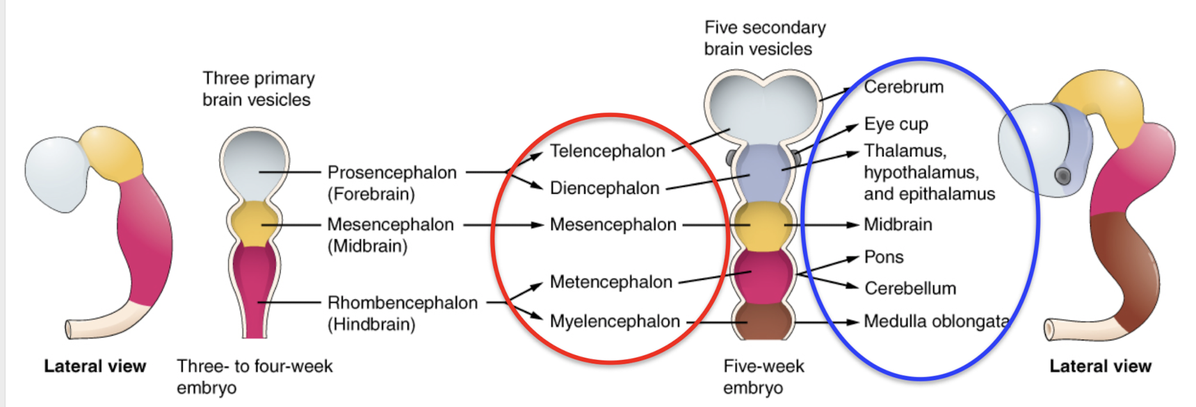
How does the CNS form? Be specific. Week 3-4, etc. What do the primary and secondary
vesicles become?
Week 3-4: Three primary vesicles — Prosencephalon (forebrain), mesencephalon (midbrain), rhombencephalon (hindbrain)
Primary vesicles further divide:
Prosencephalon (forebrain) divides into telencephalon, diencephalon
Mesencephalon (midbrain) stays as mesencephalon (does not divide)
Rhombencephalon (hindbrain) divides into metencephalon, myelencephalon
Five week embryo:
Telencephalon becomes cerebrum
Diencephalon forms eye cup and all the thalamuses (Thalamus, hypothalamus, epithalamus)
Mesencephalon forms midbrain
Metencephalon divides into pons and cerebellum
Myelencephalon becomes medulla oblongata
What is the “limbic system”?
Organs include hypothalamus, hippocampus, amygdala. Involved in memory, learning, emotion, and behavior. Located just under the cerebrum on both sides of thalamus.
What do the amygdala and hippocampus regulate?
Amygdala translates perception, so it is involved in emotion (especially FEAR) and sorts memories based on emotional perception. Hippocampus converts short term memory into long term memory. If the hippocampus is damaged, new memories are not formed.
What do the basal nuclei do? Reticular formation?
Basal nuclei regulate and organize movement; regulates unconscious movements (involuntary movements (such as laugh and walk)
Reticular formation - extends slightly into the diencephalon and the spinal cord. Reticular formation receives and processes various types of stimuli and controls the sleep-wake cycle.
What is the function of the pons? Medulla oblongata?
Pons:
Sensory role in swallowing, tasting
secreting saliva and tears
controlling respiration
Medulla oblongata:
“Cardiorespiratory center”
Heart rate, respiratory rate, swallowing, coughing, vomiting, sneezing, hiccuping
What is the function of the cerebellum?
Coordinates skeletal muscle contraction (basic movement), maintains muscle tone, posture, and balance.