

IGCSE Biology
The Nature and Variety of Living Organisms
KLO 1.2: describe the common features shown by eukaryotic organisms: plants, animals, fungi, protoctists.
Eukaryotic = organisms that contain a nucleus, their cells contain nucleus surrounded by a membrane along with other membrane bound organelles.
They can be multicellular or unicellular, must have a nucleus with a distinct membrane
Plants
Are multicellular organisms consisting of many different types of cells
Plants carry out the process of photosynthesis through its chloroplast, this is called autotroph which is when an organism makes its own food, and it feeds on this.
Stores carbohydrates (energy) as starch or sucrose
Starch is a complex carbohydrate/sugar stored within various cells
Contains cell walls made of cellulose
Examples: Herbaceous Legumes, Cereal, Flower Plants
Contains Extra Organelles Than Animal Cells
Structure | Function |
Cell Wall |
|
Chloroplast |
|
Permanent Vacuole |
|
Animals
Are multicellular organisms
Cells do not contain cell walls, hence cells can change shape
Cells do not contain chloroplast, hence are unable to carry out photosynthesis
Feed on organic material/substance made by other living things: heterotroph
Stores carbohydrates as glycogen, commonly stored in liver of most animals
Has nervous coordination
Are able to move from place to place
Examples: Mammals, Insects, Amphibian, Reptiles
Contains Organelles In Both Animal and Plant Cells
Structure | Function |
Nucleus |
|
Cytoplasm |
|
Cell Membrane |
|
Ribosomes |
|
Mitochondria |
|
Fungi
Usually multicellular but some are unicellular (yeasts)
These organisms can not carry out photosynthesis as they do not contain chloroplasts
Bodies are usually organised into networks called mycelium
make of thread like structure known as hyphae which contain many nuclei
Contain fruiting body of mushroom/toadstool
Cells have cell walls made of chitin
Feed by secreting extracellular digestive enzymes onto food material, usually decaying organic matter, then absorbing digested molecules - Saprotrophic nutrition
Some are parasitic and feed in living material
Stores carbohydrates as glycogen
Do not have nervous coordination
Examples: Moulds, Mushroom, Yeasts
Protoctists
Diverse group of organisms that don’t belong to other eukaryotic kingdoms
Mainly extremely microscopic and single celled but some aggregate into large forms (colonies/chains of cells)
Some have features that make them look like animal cells, protozoa
E.g Plasmodium - pathogenic protist that causes malaria
Some have plant cell like feature, algae
E.g Chlorella
Meaning some protoctista photosynthesise and some feed on organic material made by other organisms
Examples: Amoeba, Paramecium, Plasmodium, Chlorella
(Should be able to recognize, draw, and interpret images of cells, so practice drawing & labelling in revision)
KLO 1.3: describe the common features shown by eukaryotic organisms such as bacteria
Prokaryotic = organisms are always unicellular and do not contain nucleus, instead nuclear material of prokaryotic cells are found in found in the cytoplasm
Bacteria
Microscopic unicellular organisms that are the largest group of prokaryotes
Contain cell walls made of peptidoglycan, cell membranes, cytoplasm, and plasmids ( small circular loops of DNA that contain genes)
lack a nucleus, but contains a nucleoid (circular chromosome of DNA)
Does not have a mitochondria and other membrane bound organelles like eukaryotic cells
Some water bound bacteria contain flagellums, a tail like projection allowing flagella movement
Some can carry out photosynthesis (even without chloroplast as they can posses chlorophyll and enzymes needed to synthesise sugars from carbon dioxide
Examples:
Lactobacillus: a rod shaped bacterium used in the production of yoghourt from milk
Pneumococcus - a spherical bacteria that acts as the pathogen causing pneumonia
Eukaryotic Cells & Prokaryotic Cells
Component | Eukaryotes | Prokaryotes |
Cell Membrane | Yes | Yes |
Cytoplasm | Yes | Yes |
Genetic Material | Yes - in the nucleus | Yes - in cytoplasm |
Nucleus | Yes | No |
Cell Wall | Some | Yes - made of peptidoglycan |
KLO 1.4: understand the term pathogen and know that pathogens may include fungi, bacteria, protoctista, and viruses
Pathogens = any microorganisms that causes disease in other organisms
Not all species of bacteria, protoctists, fungi, and pathogens
But all viruses are pathogens as they can only exist by living inside living cells of other organisms or by using these cells to create more viruses
Pathogenic fungi, Pathogenic bacteria, Pathogenic Protoctista
Virus
Are not alive as they do not carry out the 8 life’s processes
Are parasitic and can only reproduce in living cells by taking over host cell’s metabolic pathways to make multiple copies of itself
Share biological characteristics
Wide variety of shapes and sizes
Microscopic particles that are much smaller than bacteria
Infect every type of living organism
Has no cellular structure
Contain a protein coat called capsid
Contains 1 type of nucleic acid, either DNA or RNA
Contains a viral envelope, similar to the cell membrane
Examples:
Tobacco Mosaic Virus (TMV): Causes discoloration on leaves of tobacco plants by preventing the formation of chloroplast
HIV Virus: Causes Aids
Influenza Virus: Causes flue
Ecology and the Environment
KLO 4.1: understand the terms population, community, habitat, ecosystem
Organism/Individual
Individual living things, all have same basic needs
Population
A group of organisms of the same species living in the same place at the same time
Community
All of the different populations living in the same area at the same time
In a community, species depend on other species for food, shelter, pollination, seed dispersal, etc
If 1 is removed, it impact the whole community, this shows interdependence
Habitat
The place where an organism lives
Ecosystem
All the biotic and abiotic factors in a community that interact with each other in an area at the same time
Biotic factors include all the living component
Plants, animals, etc
Abiotic factors include all the non living components
Light intensity, mineral/nutrient concentration, water, etc
Ecosystems can vary greatly in size and scale
An ecosystem can be as small as a garden pond and as large as the whole of antarctica
KLO 4.2: practical: investigate population size of an organism in 2 different areas using quadrat
Ecology is a branch of biology that studies the distribution and abundance of species along with interactions between species and species interactions within abiotic environment
Ecologist study these interactions by investigation ecosystems
1 Piece of equipment that might be used to investigate population size is a quadrat
Quadrat
Square frames made of wood/wire that can be a variety of size (0.2 m2 to 1 m2
Are placed on the ground and organism within are recorded
Plant species are commonly studied using quadrats to identify abundance
Abundance can be measured by recording
Number of an individual species: total number of individuals of a single species
Species richness: total number of different species
Percentage cover: approximate percentage of quadrat area in which an individual species covers (used when it is difficult to count individually)
Investing population size in 2 different areas using quadrats
Method
Use 2 tape measures to lay out survey area (ie 10 m by 10 m) in chosen habitat
Use random number generator to create a set or coordinates to place 1st quadrat (ie. (4,5) -> 4 m along x axis and 5 m along y axis)
Count number of individual of chosen species (plant) that are found in quadrat
Repeat step 2 and 3 to a total of 10 times and take average of results for accuracy
Move survey area number 2 and repeat steps 2 - 4
Calculate population of species using: estimated population = total area/ area sample x total number of species countred
Ie. survey area 1 = 100(total area was 10 m x 10 m) / 10 (each quadrat square was 10 ) x 21 (total number of individuals counted)
Results
Once averages are calculated, abundance of study species in each area can be calculated
Species abundance is affected by
Biotic Factors like: competition, predator-prey relationships, interactions with other organisms
Abiotic factors like: light intensity, mineral availability, water availability, pH, temperature, salinity
Limitation
It can be easy to miss individuals in quadrat, especially is covered by leaves
Use pencil/stick to move leaves out of the way
Identifying species may be tricky
Use a species key to identify species
Apply CORMS
Change —----> What is being changed in the investigation (Independent Variable)
Organism —----> A control relating to the organism being used
Repeat —----> Repeats must be carried out for reliable results
Measurement 1 —----> How will you measure the measurement (Dependent Variable)
Measurement 2 —----> What time scale is used (Dependant Variable)
Same —----> What will be controlled/ kept the same in the investigation/experiment (Control Variable)
Independent variable – the variable that is altered during a scientific experiment.
Dependent variable – the variable being tested or measured during a scientific experiment.
Controlled variable – a variable that is kept the same during a scientific experiment. Any change in a controlled variable would invalidate the results.
In this Experiment
C —----> changing the study area in which data was collected
O—----> count the same species of organism in each quadrat
R —----> repeated a total of 10 times for reliable results
M1 —----> counting the the total number of individual from designated species found across quadrat
M2 —----> isn’t relevant in this scenario
S —----> control size of quadrat, random way quadrats are placed on the study area and the day the results were collected
KLO 4.5: understand how biotic and abiotic factors affect the population size and distribution of organisms
Biotic = Living
Abiotic = Non - Living
Biotic Factors | Abiotic Factors |
|
Terrestrial Extra factors
Aquatic Extra Factors
|
Note: when answering questions regarding charts/graphs, should always give not only analysis but specific examples of where it is happening in the chart and why it is connected to and contributes to the point
KLO 4.6: understand different trophic level names including producers, primary, secondary, tertiary consumers, and decomposer
Trophic Levels
Describes feeding relationships between organisms
Energy flows from sun into producers in the form of light (sunlight)
Produces convert light energy into chemical energy, this chemical energy gets passed on through the trophic levels
Eventually all energy is transferred to the environment as being passed on is lost or used up by the organism
Is a non cyclic process as it is “lost” to the environment
Is a direct contrast to chemical elements an organism
Producers: produce their own organic nutrients through energy from sunlights, organism that makes its own energy rich food compounds
Primary Consumer: Feed on producers/Consumer producers
Secondary Consumer: Feed/Consume primary consumers
Tertiary,Quaternary, quinnary …. Apex predator/top carnivores
Decomposers: Organisms that break down/consume dead materials helping recycle it
KLO 4.10: describe stages in the carbon cycle including respiration, photosynthesis, decomposition, combustion
Carbon Cycle
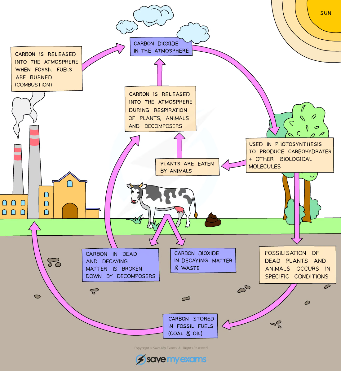
Carbon is taken out of the atmosphere in the form of carbon dioxide by plants to be used in photosynthesis
Passed onto animals and microorganisms by feeding/consuming
Returned/Released back into the atmosphere in the form of carbon dioxide by plant, animals, and decomposers through respiration
If animals and plants die in conditions where decomposing organisms aren’t present, carbon in their body can be converted over millions of year of significant pressure, fossilising into fossil fuels
When fossil fuels are burned, carbon combines with oxygen and carbon dioxide is released
Increased use of fossil fuels contributes to the increase CO2 content in the atmosphere
Stages
Photosynthesis: “fixes” carbon atoms into organic compounds like starch and glucose
Respiration: produces inorganic CO2 from organic compound (mainly carbohydrates) as they are broken down to release energy
Decomposition: Carbon in dead decaying matter is broken down and once in decomposer, is respired into carbon reserves
Combustion: releases carbon dioxide into atmosphere as fossil fuels are burned
KLO 4.16: understand biological consequences of water by sewage
Bioaccumulation
Concentration of a substance, ie pesticides/herbicides, in an organism by not breaking down. Building up/accumulating into the tissues of the organism over time
Biomagnification
Increase in concentration of toxic substances as it moves through the food chain
Water Pollution by Sewage
Aerobic bacteria in the water, polluted by the sewage, use up the majority of the dissolved oxygen in the water as it decomposes the organic material, reduction in oxygen levels leads to death of larger aquatic organisms
Only species like anaerobic bacteria are able to survive
Water becomes more oxygenated again as it moves towards and mixes with cleaner waters.
Untreated sewage contains pathogenic bacteria which is a danger to human health
KLO 4.17: understand biological consequences of eutrophication caused by leached minerals from fertilisers
Eutrophication
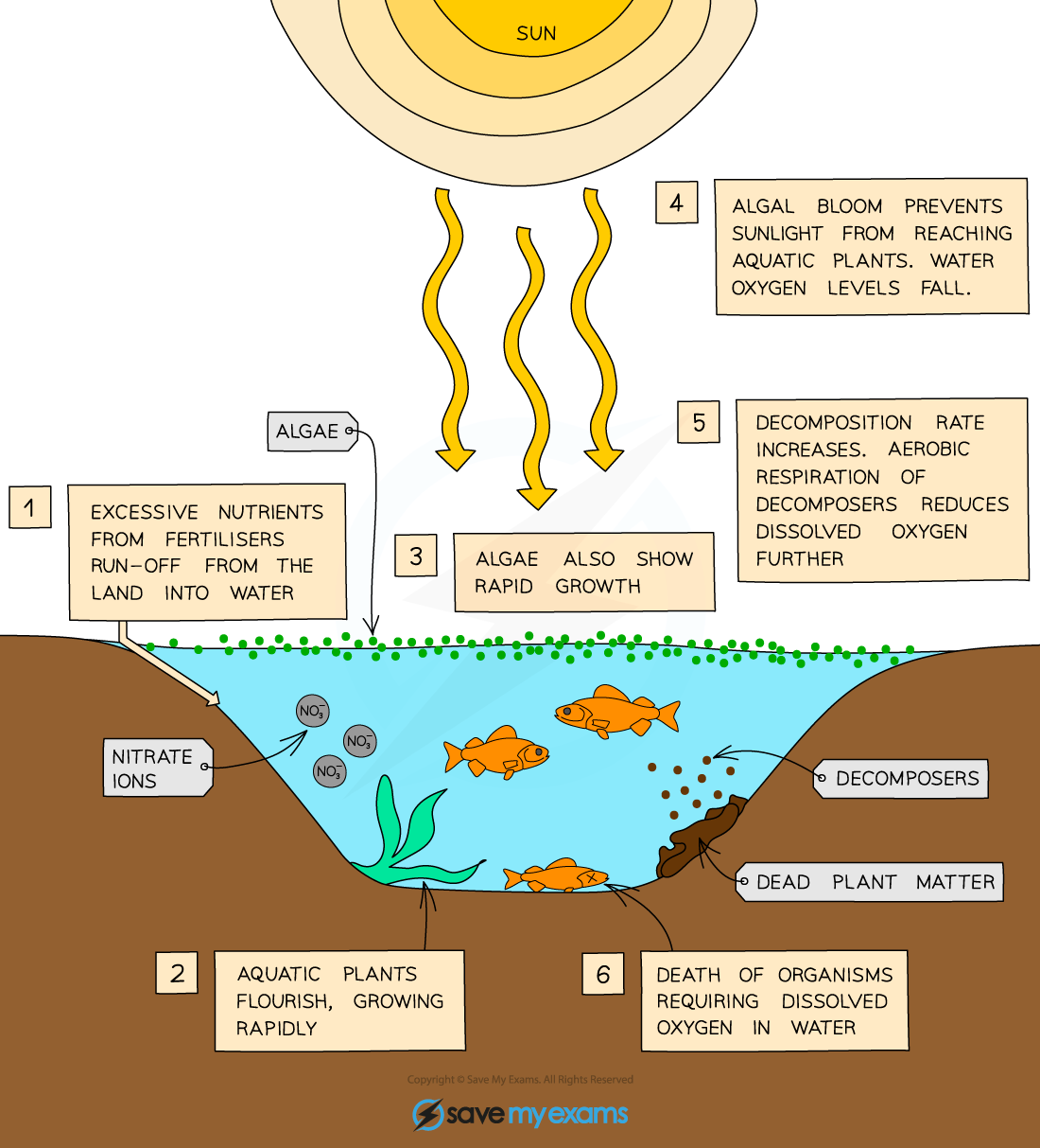
Leaching: process of nutrients removed from soil as excess water passes through
Eutrophication
Excess minerals/nutrients enter a water body in 2 ways
Artificial nitrate/phosphate fertilisers-causes algal blooms
Sewage - increased decomposing done by decomposing bacteria leads to decreasing levels of dissolved oxygen
Common misconception but sewage does not cause algal blooms
Structure & Function in Living Things
KLO 2.1: describe levels of organisation in organisms: organelle, cell, tissue, organs, organ systems
In a multicellular organism…
Organelle
A component within a cell that carries out a specific task
Cell
Basic functional and structural unit in a living organism
Tissues
A group of cells of similar structure working together to perform a particular function
Organ
Made from groups of tissues working together to perform a particular function
Organ Systems
Several different organs with related functions working together to perform body functions within the organisms
Examples
Plant: chloroplast - palisade cell - epidermis mesophyll - leaf - shoot system
Animal/Human: mitochondria - muscle cell - muscle - heart - circulatory system
KLO 2.2: describe cell structures including nucleus, cytoplasm, cell membrane, cell wall, mitochondria, ribosomes, vacuole
Cell structure
Cells are separated from surrounding environment by cell membrane
Within cell membranes is the cytoplasm
Eukaryotic cells have organelles contained within their cytoplasm
Organelles are subcellular compartments where specific processes take place within the cell
Following organelles are present
Nucleus
Mitochondria
Ribosomes
Plant cells contain a few additional organelles
Cell walls
Chloroplasts
permanent vacuole
KLO 2.3: describe the functions of nucleus, cytoplasm, cell membrane, cell wall, mitochondria, ribosomes, vacuole
Organelle Functions
Structure | Function |
Nucleus |
|
Cytoplasm |
|
Cell Membrane |
|
Ribosomes |
|
Mitochondria |
|
Chloroplast (Plant cell only) |
|
Cell Wall (Plant Cell Only) Bacteria and Fungi have it but from different materials |
|
Permanent Vacuole (Plant Cell Only) |
|
KLO 2.4: know the similarities and differences between plant and animal cell structures
KLO 2.7 identify the chemical elements present in carbohydrates, proteins and lipids (fats and oils)
Most of the molecules in living organisms fall into three categories: carbohydrates, proteins and lipids
These all contain carbon and so are described as organic molecules
Chemical Elements
Biological Molecules | Chemical Elements |
Carbohydrates | Carbon, Hydrogen, Oxygen |
Protein |
|
Lipids | Carbon, Hydrogen, Oxygen |
KLO 2.8 describe the structure of carbohydrates, proteins and lipids as large molecules made up from smaller basic units: starch and glycogen from simple sugars, protein from amino acids, and lipid from fatty acids and glycerol
Carbohydrates
Molecules made of carbon (carbo) hydrogen (hydr) oxygen (ate)
Used by the body for respiration to release energy
Humans get most of their carbohydrates in the form of starch
Potatoes, rice, pasta
Starch is a large insoluble carbohydrate molecule made by plants
Animals store carbohydrates as glycogen
Insoluble: Large carbohydrates like starch need to be broken down by enzymes in digestion to release simple sugars needed for respirations. Ie. glucose
Glycosidic bonds join/bond simple monomer sugars.
A monosaccharide is a simple sugar e.g. glucose (C6H12O6) or fructose
Glucose molecules contain lots of energy which can be released in respiration by breaking the bonds between the carbon atoms
Glucose = hexagon simple sugars bonded together with glycosidic bonds into carbohydrate molecules
Fructose = pentagon simple sugars bonded together with glycosidic bonds into carbohydrate molecules
A disaccharide is made when two monosaccharides join together
Maltose is formed from two glucose molecules
Sucrose is formed from one glucose and one fructose molecule
A polysaccharide/polypeptide is formed when lots of monosaccharides join together
Starch, glycogen or cellulose are all formed when lots of glucose molecules join together
Polysaccharides are insoluble and therefore useful as storage molecules
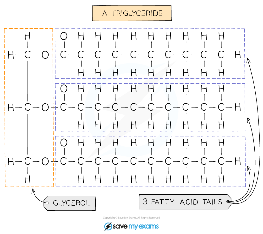
Lipids
Molecules made of carbon, hydrogen, oxygen
Lipids are divided into fats (solids at room temperature) and oils (liquids at room temperature)
Food high in animal fats: meat, butter, cheese, milk, eggs, fish
Foods high in plant oils: sunflower oil, olive oil, rapeseed oil, margarine
Uses
Makes cell membrane
Provides insulation
Protects organs
Stores energy
Most fats (lipids) are made up of triglycerides
One glycerol molecule chemically bonded by ester bonds to three fatty acid chains
The fatty acids vary in size and structures
Bonded by ester bonds
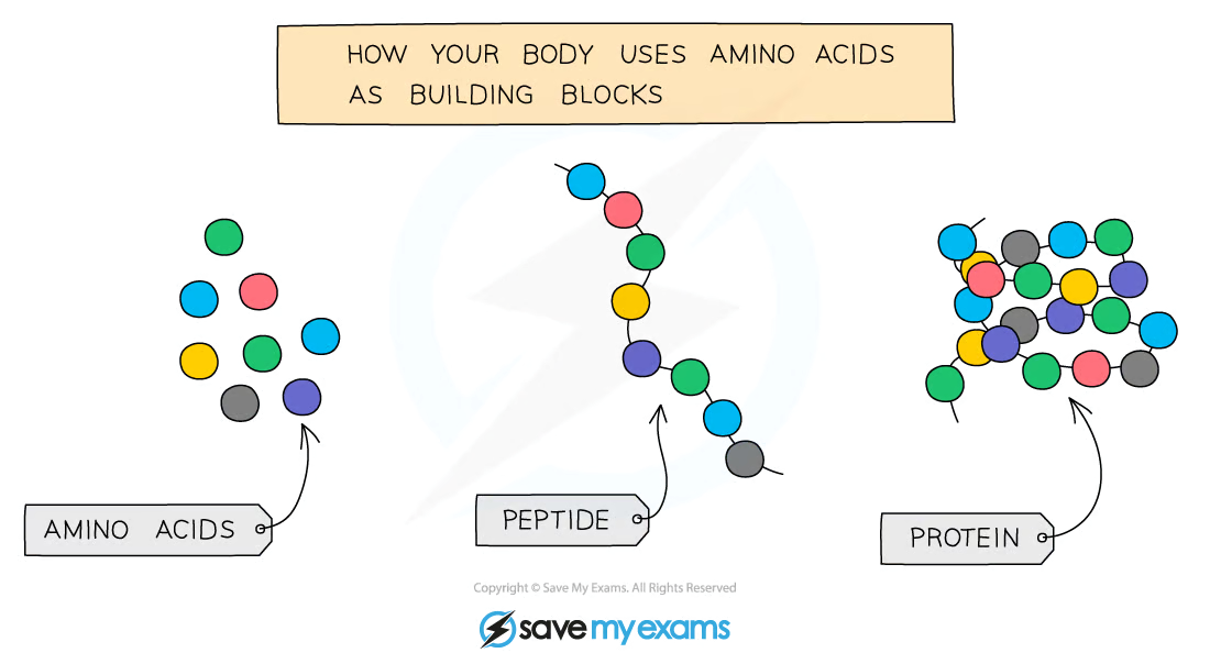
Proteins
Molecules made of carbon, hydrogen, oxygen, and nitrogen
All cells contain protein
Needed for tissue growth and repair
Animal foods high in protein
Meat
Fish
Eggs
Cheese
Plants generally contain less protein that animals, but some have more
Beans
Peas
Nuts
Proteins are large molecules made up of amino acids
20 different types of amino acids used to make up 1000s of different proteins
Different proteins have different amino acid sequences resulting in them being different shapes
Even a small difference in the amino acid sequence will result in a completely different protein being formed
The different sequences of amino acids cause the polypeptide chains to fold in different ways and this gives rise to the different shapes of proteins
In this way, every protein has a unique 3-D shape that enables it to carry out its function
The shape of a protein determines its function
Peptide bonds are used to bond amino acids
KLO 2.9 practical: investigate food samples for the presence of glucose, starch, protein and fat
There are various chemical tests which can detect which molecules are present
Before you can carry out any of the food tests described below, you may need to prepare a food sample first (especially for solid foods to be tested)
To do this:
Break up the food using a pestle and mortar
Transfer to a test tube and add distilled water
Mix the food with the water by stirring with a glass rod
Filter the mixture using a funnel and filter paper, collecting the solution
Proceed with the food tests
Iodine - Starch
Add a small amount of food sample into a test tube
Add a few drops of iodine solution to the food sample
A sample containing starch will show a colour change from orange-brown to blue-black
Benedict’s Solutions - Glucose (a reducing sugar)
Take a food sample and add it into a test tube
Add Benedict's solution to the sample solution in a test tube
Heat the tube in a hot water bath for 5 minutes
Remove from heat and observe the colour
A positive test will show a colour change from blue to orange to brick red
Biuret Solution - Protein
Take a food sample (must be in liquid form) and add it into a test tube
Add drops of Biuret solution to the food sample
A positive test will show a colour change from blue to violet / purple
Emulsion Test - Lipids
Take a food sample and add it into a test tube
Add a few drops of ethanol to the food solution
Shake the test tube with a bung on the top and leave for one minute
Pour the ethanol into another test tube of water
If the solution turns cloudy, the food contains lipids. This cloudiness is called an emulsion.
Results Table
Food Test | Colour At Start | Positive Result | Negative Result |
Iodine for Starch | Orange - Brown | Blue - Black | Orange - Brown (no change) |
Benedicts for Sugar | Light Blue | Green to Brick Red | Light Blue (no change) |
Ethanol for Lipids | Colourless | Cloudy Emulsion | Colourless (no change) |
Biurets for Protein | Blue | Lilac - Purple | Blue (no change) |
CORMS Evaluation
C: Changing the type of food in the sample
O: isn’t relevant in this experiment
R: Repeat the investigation several times for each food sample to ensure a reliable result
M1: presence of the specific biological molecule in each food type by noting the colour change
M2: ....after testing with each specific testing agent
S: control the volume of each testing agent used, the quantity of the food sample, the concentration of the testing agents, the temperature of the water bath for the Benedict's test.
Tips
When describing food tests in exam answers, make sure you give the starting colour of the solution and the colour it changes to for a positive result.
KLO 2.10 understand the role of enzymes as biological catalysts in metabolic reactions
Biological Catalysts
Biological Catalyst: A substance that increase the rate of a chemical reaction without being changed or consumer in the reaction
Enzymes
proteins that act as biological catalysts to speed up the rate of a chemical reaction without being changed or used up in the reaction
Amino acid chains are folded, each enzyme is folded in a different shape help by peptide bonds between amino acids
They are biological because they are made in living cells
Necessary to all living organisms as they maintain reaction speeds of all metabolic reactions at a rate that can sustain life
Metabolic Reaction: All the chemical or mechanical reactions that happen within a cell or organism to keep it alive
Ie. if we did not produce digestive enzymes, it would take around 2 - 3 weeks to digest one meal; with enzymes, it takes around 4 hours
Activation energy: amount of energy needed for a reaction
Lowering activation energy means not as much energy is needed as before. Reactions can happen sooner as energy for reaction is smaller
Controls all reactions that happens in a cell
How they Work
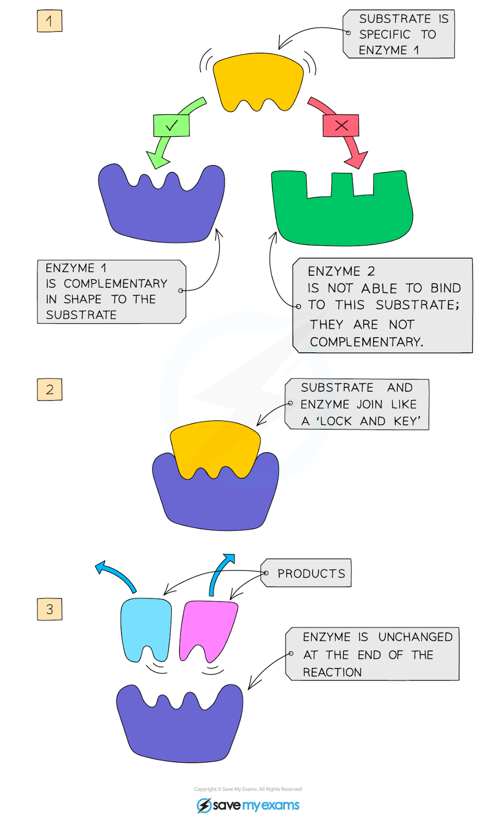
Active Site: Part of the enzyme that has the same shape as the substrate
Substrate: Molecule enzyme wants to change, correctly fits into the active site
Specific to one particular substrate(s) as the active site of the enzyme, where the substrate attaches, is a unique shape to the substrate
When the substrate moves into the enzyme’s active site they become known as the enzyme-substrate complex
After the reaction has occurred, the products leave the enzyme’s active site as they no longer fit it and it is free to take up another substrate
Enzymes and substrates randomly move about in solution
When an enzyme and its complementary substrate randomly collide an enzyme-substrate complex forms, and the reaction occurs
A product (or products) forms from the substrate(s) which are then released from the active site. The enzyme is unchanged and will go on to catalyse further reactions
KLO 2.11 understand how temperature changes can affect enzyme function, including changes to the shape of active site
Temperature
Enzymes are proteins and have a specific shape, determined by the amino acids that make the enzyme and held in place by bonds
This is extremely important around the active site as the specific shape is what ensures the substrate will fit into the active site and enable the reaction to proceed
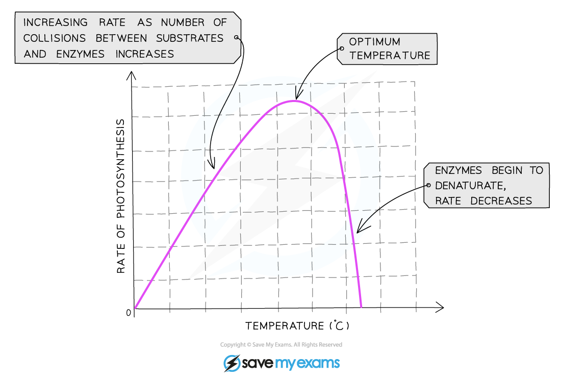
Enzymes work fastest at their ‘optimum temperature’
Optimum temperature: temperature in which the maximum number of successful collisions occur
In the human body, the optimum temperature is 37⁰C
Increasing the temperature towards the optimum increases the activity of enzymes as the more kinetic energy the molecules have the faster they move and the number of collisions with the substrate molecules increases, leading to a faster rate of reaction
This means that low temperatures do not denature enzymes, they just make them work more slowly due to a lack of kinetic energy
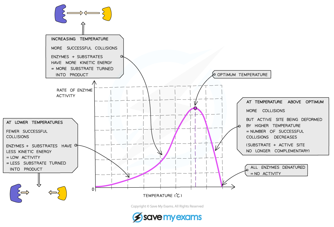
Heating to high temperatures (beyond the optimum) will break the bonds that hold the enzyme together and it will lose its shape
This is known as denaturation
Substrates cannot fit into denatured enzymes as the shape of their active site has been lost
Denaturation is irreversible - once enzymes are denatured they cannot regain their proper shape and activity will stop

KLO 2.12 practical: investigate how enzyme activity can be affected by changes in temperature
Equipement
Amylase
Starch
Iodine
Thermometer
Small testing tubes
Beaker filled with water
Dropper
Stopwatch
Spotting tile
Method
Place around 5 cm cubed of starch into a testing tube and 1 cm cubed of amylase in a testing tube into a water bath of around 22 degrees celsius
Add drops of iodine into the spotting tile
Pour amylase into the starch tube
Immediately take an put a couple of drops in one tile
Take one minute intervals to add drops to the next tile till the tile remains the same colour as the amylase has broken down the starch
Heat up the water bath to around 47 degrees celsius
Mix together a new amount of starch and amylase
Immediately take an put a couple of drops in one tile
Take one minute intervals to add drops to the next tile until the tile remains the same colour as the amylase has broken down the starch, should be quicker as amylase is heated
Use various temperatures to identify the reactions
Result
Amylase is an enzyme which breaks down starch
The quicker the reaction is completed, the faster the enzyme is working
This investigation shows:
At the optimum temperature, the iodine stopped turning blue-black the fastest
This is because the enzyme is working at its fastest rate and has digested all the starch
At colder temperatures (below optimum), the iodine took a longer time to stop turning blue-black
This is because the amylase enzyme is working slowly due to low kinetic energy and few collisions between the amylase and the starch
At hotter temperatures (above optimum) the iodine turned blue-black throughout the whole investigation
This is because the amylase enzyme has become denatured and so can no longer bind with the starch or break it down
CORMMS Evaluation
C: Changing the temperature of each round, impacting rate of reaction
O: isn’t relevant in this experiment
R: Repeat the investigation several times for each temperature to ensure a reliable result
M1: measure the time taken for the reaction
M2: the iodine to remain the same colour
S: control the concentration and volume of starch solution, iodine and amylase used in the investigation
Tips
Describing and explaining experimental results for enzyme experiments is a common type of exam question so make sure you understand what is happening and can relate this to changes in the active site of the enzyme when it has denatured, or if it is a low temperature, relate it to the amount of kinetic energy the molecules have.
KLO 2.13 understand how enzyme function can be affected by changes in pH altering the active site
pH
An enzyme is denatured once it is placed beyond its optimum pH, the shape get denatured
Acids or alkali interfere with the enzyme’s peptide bonds holding the enzyme together
The optimum pH for most enzymes is 7
Some enzymes that are produced in acidic conditions, such as the stomach, have a lower optimum pH (pH 2)
Some that are produced in alkaline conditions, such as the duodenum, have a higher optimum pH (pH 8 or 9)
If the pH is too high or too low, the bonds that hold the amino acid chain together to make up the protein can be disrupted/destroyed
This will change the shape of the active site, so the substrate can no longer fit into it, reducing the rate of activity
Moving too far away from the optimum pH will cause the enzyme to denature and activity will stop

Tip:
Remember the terminology when writing about enzymes is very important. Make sure you refer to an enzyme becoming 'denatured' not 'dying'.Being able to describe AND explain the effect of each environmental condition on enzyme action is key.Practise describing and explaining using the graphs and then check your descriptions against your notes.
KLO 2.15 understand the processes of diffusion, osmosis and active transport by which substances move into and out of cells
In order for cells to carry out the chemical reactions it needs to, substance must enter and leave the cell. This happens in the following 3 ways
Diffusion
Osmosis
Active Transport
Diffusion
Diffusion is the movement of molecules from a region of its higher concentration to a region of its lower concentration
Concentration gradient: high to low concentration
Diffusion happens when a substance is more concentrated in one area than another. This difference is concentration is the concentration gradient
Molecules move randomly due to kinetic energy, however net movement follows the concentration gradient.
Diffusion in living organisms
For living cells, the principle of the movement down a concentration gradient is the same, but the cell is surrounded by a cell membrane, which can restrict the free movement of the molecules
The cell membrane is a partially permeable membrane - this means it allows some molecules to cross easily, but others with difficulty or not at all
The simplest sort of selection is based on the size of the molecules (i.e. smaller molecules can diffuse across the membrane but larger molecules cannot)
Diffusion helps living organisms to:
Obtain many of their requirements
Get rid of many of their waste products
Carry out gas exchange for respiration
Tip:
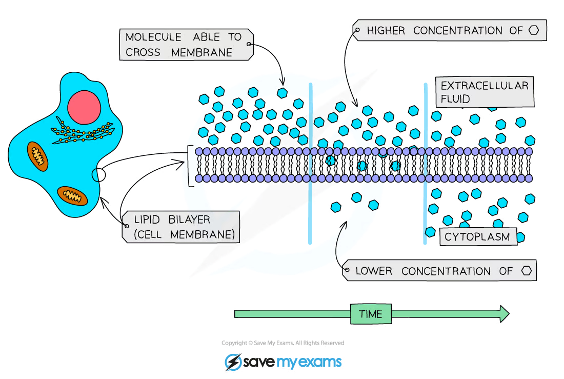
Remember that diffusion is a passive process, so when it occurs in a living organism, the cells of that organism do not provide the particles involved with energy to diffuse. The particles that are moving about randomly have their own kinetic energy.
Osmosis
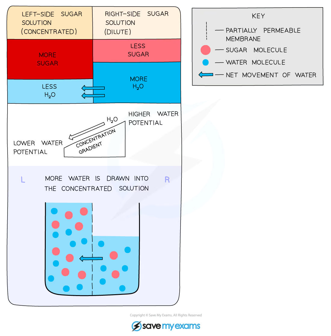
Movement of water from a dilute solution to a more concentrated one across the partially permeable cell membrane
Water moves from an area of high water potential to an area of low water potential
Partially permeable: this membrane has pores through which very small molecules, including water, can pass, but not larger molecules
Osmosis is the net movement of water molecules from a region of higher water potential (dilute solution) to a region of lower water potential (concentrated solution), through a partially permeable membrane
All cells are surrounded by a cell membrane which is partially permeable
Water can move in and out of cells by osmosis
In doing this, water is moving down its concentration gradient
The cell membrane is partially permeable which means it allows small molecules (like water) through but not larger molecules (like solute molecules)
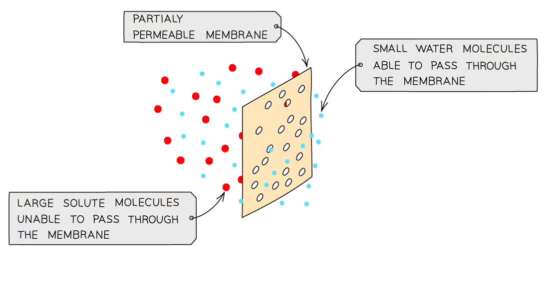
a dilute solution has a high water potential (the right-hand side of the diagram below) and a concentrated solution has a low water potential (the left-hand side of the diagram below)
Osmosis in Animal Cells
Animal cells lose and gain water as a result of osmosis
As animal cells do not have a supporting cell wall, the results of osmosis can be severe
If an animal cell is placed into a strong sugar solution (with a lower water potential than the cell), it will lose water by osmosis and become crenated (shrivelled up)
If an animal cell is placed into distilled water (with a higher water potential than the cell), it will gain water by osmosis as it has no cell wall to create turgor pressure
It will continue to gain water until the cell membrane is stretched too far and it bursts
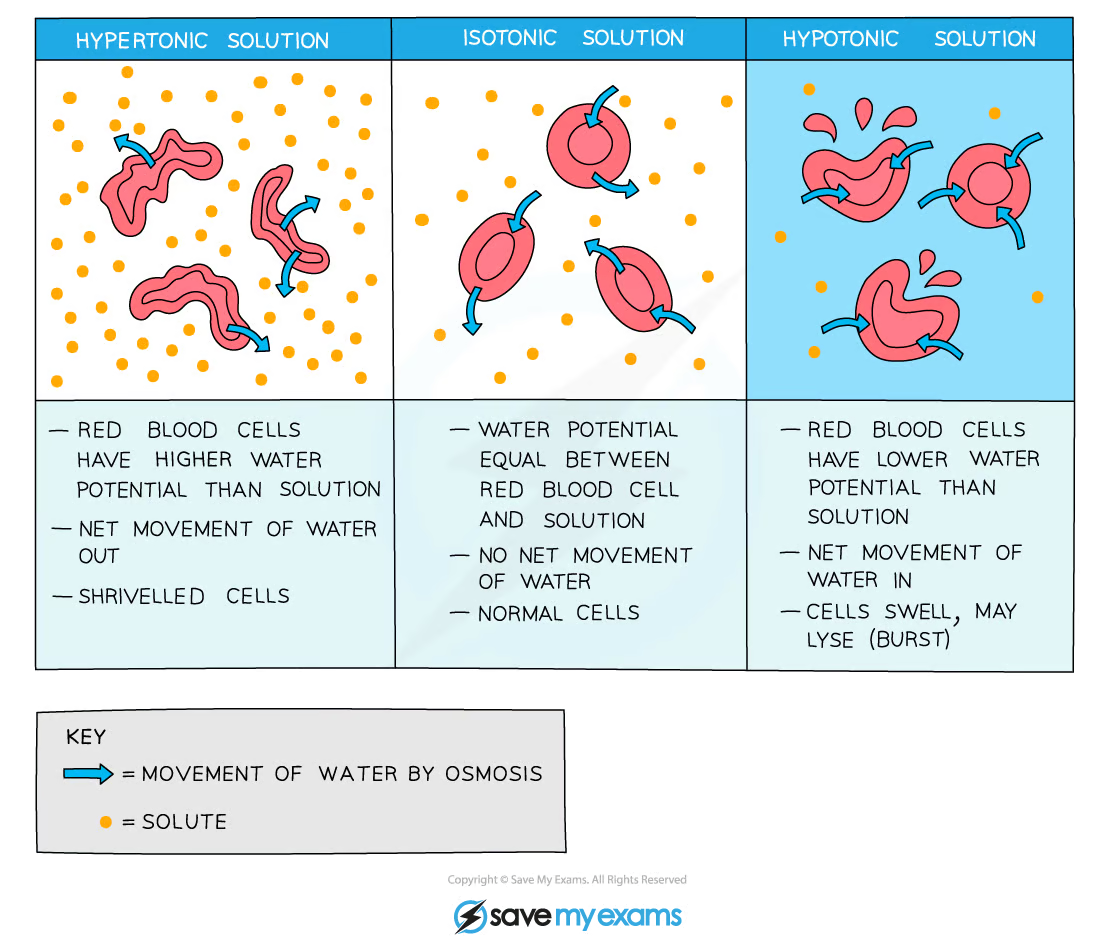
It is important that osmosis is carefully controlled in organisms to avoid damage to cells through lysis
the disintegration of a cell by rupture of the cell wall or membrane
The human body is adapted to maintain the optimum osmotic balance using processes such as sweating or increasing and decreasing urine concentration
This is all part of osmoregulation
Osmosis in Plant Cells
Once a plant cell is placed in pure water, water enters the cell through osmosis and the cell becomes turgid
Plant cells lose or gain water as a result of osmosis
As plant cells have a supporting cell wall, they are protected from cell lysis
If a plant cell is placed into a strong sugar solution (with a lower water potential than the cell), it will lose water by osmosis
The vacuole gets smaller and the cell membrane shrivels away from the cell wall
It becomes flaccid or plasmolysed (shrivelled up)
If a plant cell is placed into distilled water (with a higher water potential than the cell), it will gain water by osmosis
The vacuole gets bigger, pushing the cell membrane against the cell wall
The plant cell is described as being turgid or as containing a high turgor pressure (the pressure of the cytoplasm pushing against the cell wall)
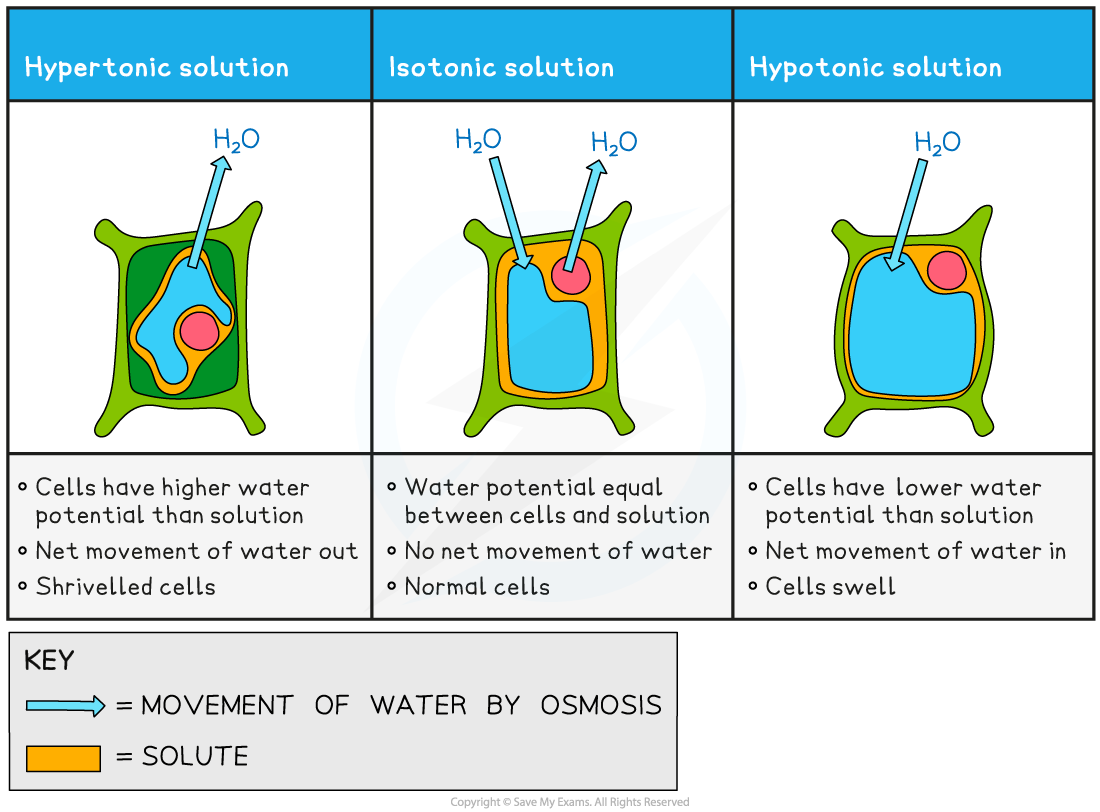
Water entering the cell by osmosis makes the cell rigid and firm
This is important for plants as the effect of all the cells in a plant being firm is to provide support and strength for the plant - making the plant stand upright with its leaves held out to catch sunlight
If plants do not receive enough water the cells cannot remain rigid and firm (turgid) and the plant wilts
Tips:
Osmosis refers only to the movement of water molecules, so if in an exam you are talking about the movement of water, make sure you mention osmosis as this will often earn you a mark. The best explanations to do with osmosis will refer to water potential, so if you are aiming for a 7, 8 or 9 you will need to understand the concept and use it in your explanations.
Active Transport
The movement of particles through a cell membrane from a region of lower concentration to a region of higher concentration using energy from respiration
Energy is needed because particles are being moved against a concentration gradient, in the opposite direction from which they would naturally move (by diffusion)
Active transport across the cell membrane involves protein carrier molecules embedded in the cell membrane
Movement of molecules from a low concentration to a high concentration against the concentration gradient
Energy is required for movement to occur, energy from respiration
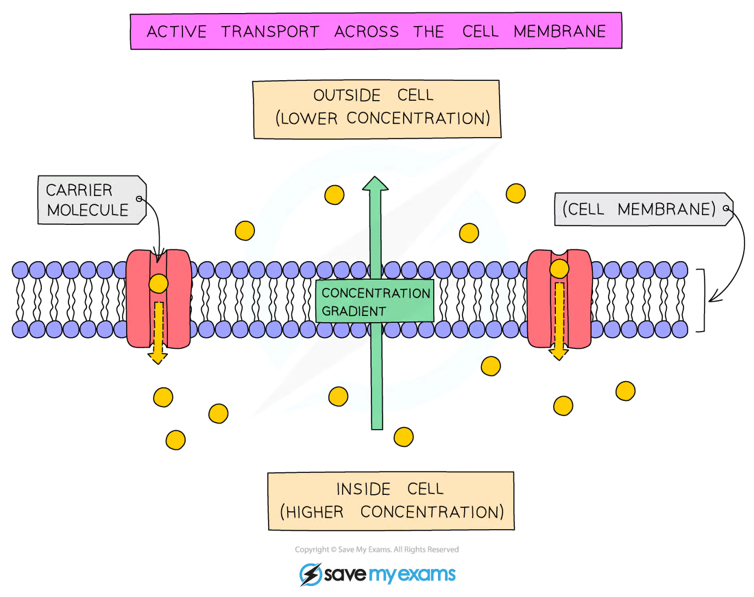
Molecules here are being transported against the concentration gradient, from a region of lower concentration (outside the cell) to a region of higher concentration (inside the cell)
Active Transport in Animals
Food molecules (such as the sugar glucose) can be absorbed across the wall of the small intestine by diffusion, but this is dependent on a concentration gradient existing between the lumen of the intestine and the bloodstream
Active transport allows molecules such as glucose to be transported into the bloodstream from the lumen of the small intestine (the gut) when the concentration of sugar molecules in the blood is higher
The active uptake of glucose by epithelial cells in kidney tubules in the kidney nephron allows for the reabsorption of glucose back into the blood so that none is lost in the urine
Sugar molecules are used in respiration to release energy for cells to function
Active Transport in Plants
Root hair cells lining the surface of plant roots need to move minerals such as magnesium ions from a region of lower concentration (the very dilute solution of minerals in the soil surrounding the roots) to a region of higher concentration (inside the cytoplasm of the cell)
Mineral ions are needed by plants to function
Magnesium ions are required to make chlorophyll
Nitrate ions are needed to make amino acids for protein synthesis (and subsequently growth)
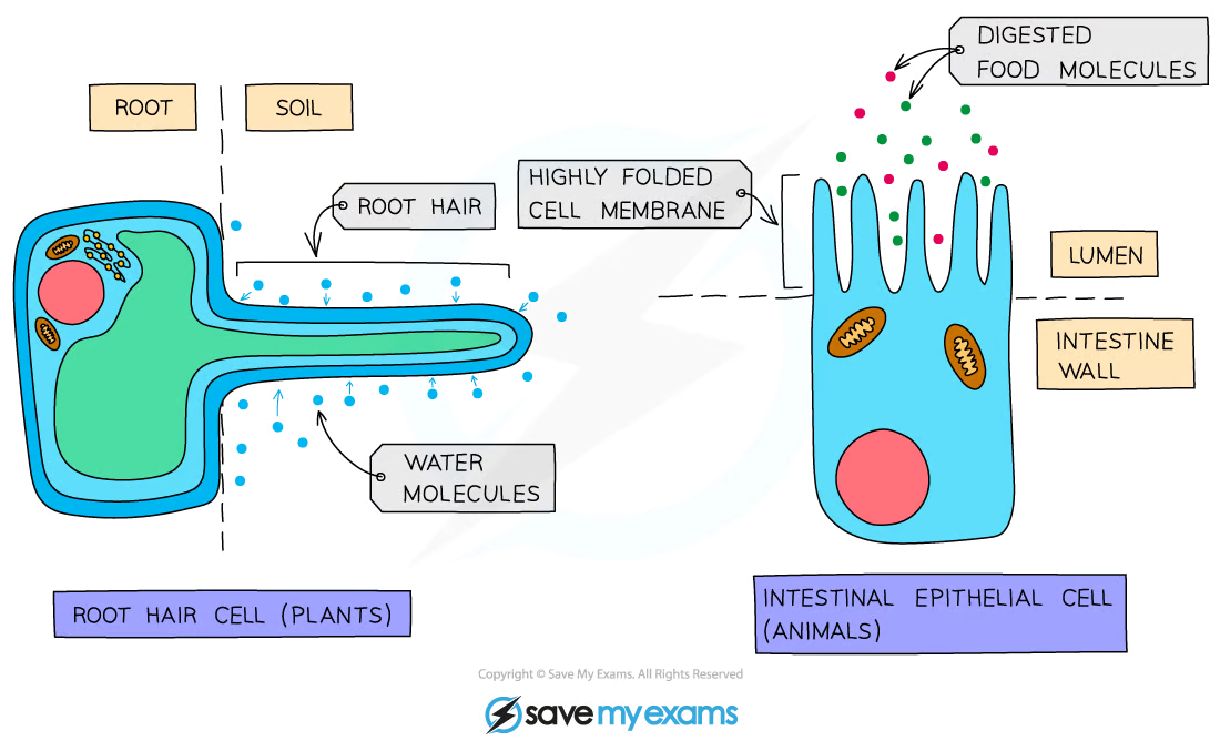
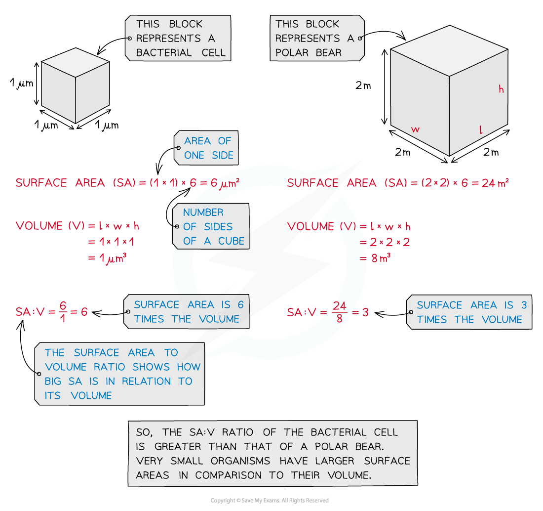
KLO 2.16 understand how factors affect the rate of movement of substances into and out of cells, including the effects of surface area to volume ratio, distance, temperature and concentration gradient
Factors influencing Diffusion
Surface area to volume ratio
The bigger a cell or structure is, the smaller its surface area to volume ratio is, slowing down the rate at which substances can move across its surface
Many cells which are adapted for diffusion have increased surface area in some way
e.g. root hair cells in plants (which absorb water and mineral ions) and cells lining the ileum in animals (which absorb the products of digestion)
You should be able to calculate and compare surface area to volume ratios
Diffusion distance
The smaller the distance molecules have to travel the faster transport will occur
This is why blood capillaries and alveoli have walls which are only one cell thick, ensure the rate of diffusion across them is as fast as possible
Temperature
The higher the temperature, the faster molecules move as they have more kinetic energy (movement)
This results in more collisions against the cell membrane and therefore a faster rate of movement across them
Concentration gradient
The greater the difference in concentration on either side of the membrane, the faster movement across it will occur, faster overall diffusion
This is because on the side with the higher concentration, more random collisions against the membrane will occur
Tips:
You should have carried out investigations into the factors that influence the rate of diffusion and as so should be able to use the information above to explain experimental results in an exam. You should also be able to plan and carry out an experiment which can investigate the effect of one of these factors.
KLO 2.17 practical: investigate diffusion and osmosis using living and non-living systems
Equipment/Apparatus
Potatoes
Knife
Sucrose solutions (from 0 Mol/dm3 to 1 mol/dm3)
Test tubes
Balance
Paper towels
Ruler
Test tube rack
Method
Make up a dilution series of sugar solutions in the range of 0.2M – 1.0M as follows:
Test-tube Number | Sugar solution (cm3) | Distilled water (cm3) | Concentration of solution in moles/L |
1 | 10 | 0 | 1.0 |
2 | 8 | 2 | 0.8 |
3 | 6 | 4 | 0.6 |
4 | 4 | 6 | 0.4 |
5 | 2 | 8 | 0.2 |
6 | 0 | 10 | - |
Set up 6 labelled test tubes with 10cm3 of each of the sucrose solutions
Using the knife and ruler, cut 6 equally-sized cylinders of potato
Blot each one with a paper towel and weigh on the balance to the nearest 0.1g
Put 1 piece into each concentration of sucrose solution
After 40 mins, remove them, blot with paper towels and reweigh them
Calculate the percentage change in mass.
Plot a graph of the percentage change in mass against the concentration of the sugar solution
Results
The potato cylinder in the distilled water will have increased its mass the most as there is a greater concentration gradient in this tube between the distilled water (high water potential) and the potato cells (lower water potential)
This means more water molecules will move into the potato cells by osmosis, pushing the cell membrane against the cell wall and so increasing the turgor pressure in the cells which makes them turgid - the potato cylinders will feel hard
The potato cylinder in the strongest sucrose concentration will have decreased its mass the most as there is a greater concentration gradient in this tube between the potato cells (higher water potential) and the sucrose solution (lower water potential)
This means more water molecules will move out of the potato cells by osmosis, making them flaccid and decreasing the mass of the cylinder - the potato cylinders will feel floppy
If looked at underneath the microscope, cells from this potato cylinder might be plasmolysed, meaning the cell membrane has pulled away from the cell wall
CORRMS Evaluation
C: Mass of the potato cylinders and the concentration of sucrose in the solutions
O: The potato cylinders will all be taken from the same potato or potatoes of the same age
R: We will repeat the investigation several times to ensure our results are reliable
M1: difference in mass of potato cylinder before and after sugar solution
M2: potatoes are left for 40 mins in sugar solution
S: control the volume of sucrose solution, size/dimensions of potatoes, type of potatoes, amount of time left in the solution.
Tips:
Questions involving osmosis experiments are common and you should be able to use your knowledge of these processes to explain the results.Don’t worry if it is an experiment you haven’t done – simply figure out where the higher concentration of water molecules is – this is the solution with the higher water potential - and explain which way the molecules move due to the differences in water potential.
KLO 2.18 understand the process of photosynthesis and its importance in the conversion of light energy to chemical energy
Photosynthesis
Photosynthesis happens in the chloroplast of a plant cell
Chloroplasts contain chlorophyll which traps energy from the sun and uses it to turn water and carbon dioxide into glucose for the plant
Photosynthesis is an endothermic reaction in which energy from sunlight is transferred to the chloroplasts in green plants
Energy from sunlight is absorbed by chlorophyll, a green pigment found inside chloroplasts
Green plants use this energy to make the carbohydrate glucose from the raw materials carbon dioxide and water
At the same time, oxygen is made and released as a waste product
Photosynthesis can be defined as the process by which plants manufacture carbohydrates from raw materials using energy from light
Plants are
Autotrophs – they can make complex molecules (glucose) from simple molecules (carbon dioxide and water)
Producers - they can make their own food and so are the first organism at the start of all food chains
The products of photosynthesis
Plants use the glucose they make as a source of energy in respiration
They can also use it to
Produce starch for storage
Synthesise lipids for an energy source in seeds
To form cellulose to make cell walls
Produce amino acids (used to make proteins) when combined with nitrogen and other mineral ions absorbed by roots
Stored in fruit as sucrose
Tips:
If asked for the raw materials required for photosynthesis, the answer is carbon dioxide and water.Although required for the reaction to take place, light energy is not a substance and therefore cannot be a raw material.
KLO 2.19 know the word equation and the balanced chemical symbol equation for photosynthesis
Word Equation
light
Carbon dioxide + water —----------> Glucose + Oxygen
Chlorophyll
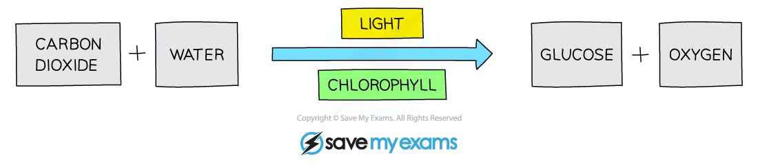
Carbon Dioxide
Diffuses into leaves through stomata
Water
Taken up by the roots and transported through the xylem into the leaves
Glucose
Used to make substance needed in the plant
Used for respiration
Oxygen
Diffuses out of the leaf through the stomata
Used in respiration
Balanced Chemical Equation
light
6CO2 + 6H20 —----------> C6H1206 + 6O2
Chlorophyll
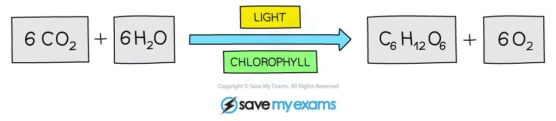
Six carbon dioxide molecules combine with six water molecules to make one glucose molecule and six oxygen molecules
Tips:
The photosynthesis equation is the exact reverse of the aerobic respiration equation so if you have learned one you also know the other one! You will usually get more marks for providing the balanced chemical equation than the word equation.
KLO 2.20 understand how varying carbon dioxide concentration, light intensity and temperature affect the rate of photosynthesis
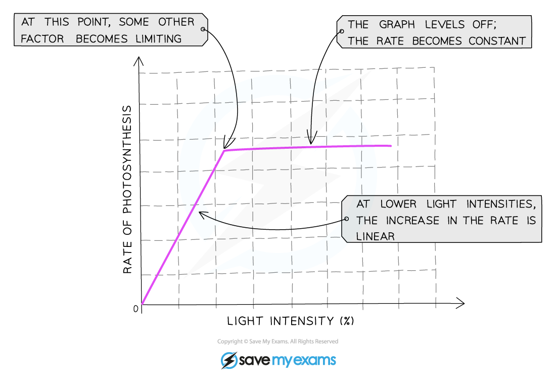
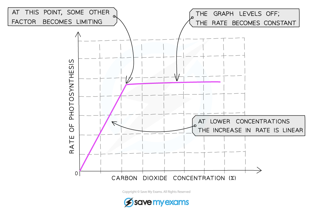
Plants do not have unlimited supplies of their raw materials so their rate of photosynthesis is limited by whatever factor is the lowest at that time
So a limiting factor can be defined as something present in the environment in such short supply that it restricts life processes
There are three main factors that limit the rate of photosynthesis:
Temperature
Light intensity
Carbon dioxide concentration
Although water is necessary for photosynthesis, it is not considered a limiting factor as the amount needed is relatively small compared to the amount of water transpired from a plant so there is hardly ever a situation where there is not enough water for photosynthesis
The number of chloroplasts or the amount of chlorophyll in the chloroplasts can also affect the rate of photosynthesis
Temperature
The temperature of the environment affects how much kinetic energy all particles have – so temperature affects the speed at which carbon dioxide and water move through a plant
The lower the temperature, the less kinetic energy particles have, resulting in fewer successful collisions occurring over a period of time
Increasing temperature increases the kinetic energy of particles, increasing the likelihood of collisions between reactants and enzymes which results in the formation of products
At higher temperatures, however, enzymes that control the processes of photosynthesis can be denatured (where the active site changes shape and is no longer complementary to its substrate) – this reduces the overall rate of photosynthesis
Light Intensity
The intensity of the light available to the plant will affect the amount of energy that it has to carry out photosynthesis
The more light a plant receives, the faster the rate of photosynthesis
This trend will continue until some other factor required for photosynthesis prevents the rate from increasing further because it is now in short supply
Carbon dioxide concentration
Carbon dioxide is one of the raw materials required for photosynthesis
This means the more carbon dioxide that is present, the faster the reaction can occur
This trend will continue until some other factor required for photosynthesis prevents the rate from increasing further because it is now in short supply
Chlorophyll
The number of chloroplasts (as they contain the pigment chlorophyll which absorbs light energy for photosynthesis) will affect the rate of photosynthesis
The more chloroplasts a plant has, the faster the rate of photosynthesis
The amount of chlorophyll can be affected by:
Diseases (such as tobacco mosaic virus)
Lack of nutrients (such as magnesium)
Loss of leaves (fewer leaves means fewer chloroplasts)
Tips:
Interpreting graphs of limiting factors can be confusing for many students, but it’s quite simple. In the section of the graph where the rate is increasing (the line is going up), the limiting factor is whatever the label on the x axis (the bottom axis) of the graph is. In the section of the graph where the rate is not increasing (the line is horizontal), the limiting factor will be something other than what is on the x axis – choose from temperature, light intensity or carbon dioxide concentration.
KLO 2.21 describe the structure of the leaf and explain how it is adapted for photosynthesis
Plant leaves have complex structures with layers of different tissues containing specially adapted cells
Leaf Structures
Structure | Description |
|
|
|
|
|
|
|
|
|
|
|
|
|
|
|
|
|
|
|
|
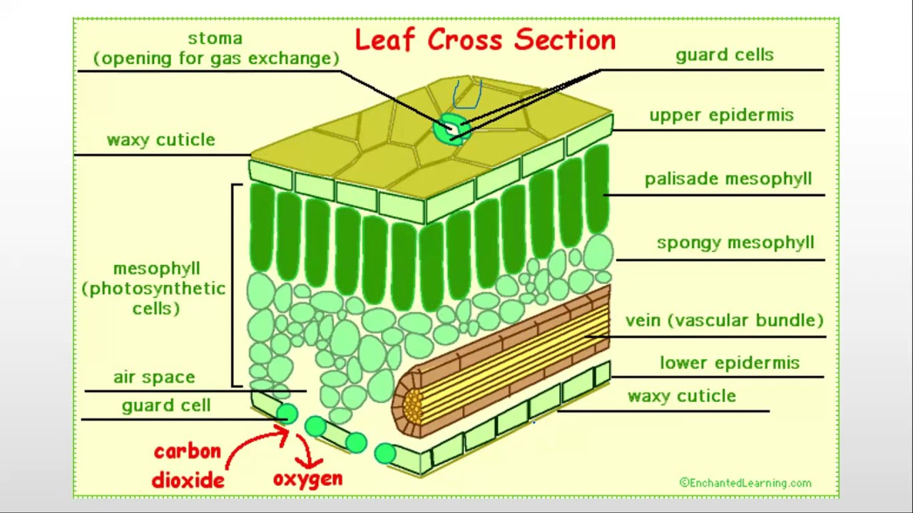
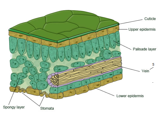
What we typically think of a plant cell is actually the palisade mesophyll cell
Leaf’s Adaptation to Photosynthesis
The specialised cells in leaves have adaptive features which allow them to carry out a particular function in the plant
Feature | Adaption |
Large Surface Area (leaf) | Increased surface area allows better diffusion of carbon dioxide and absorption of light for photosynthesis |
Thin | Allows carbon dioxide to diffuse to palisade mesophyll cells quickly |
Chlorophyll | Absorbs light energy so that photosynthesis can take place |
Network of Veins | Allows the transport of
|
Stomata | Allows carbon dioxide to diffuse into the leaf and oxygen to diffuse out |
Epidermis is Thin and Transparent | Allows more light to reach the palisade cells |
Thin Cuticle made of Wax | Protects the leaf without blocking the sunlight |
Palisade cell layer at the top of the leaf | Maximises absorption of light as it will hit chloroplast in the cells directly |
Spongy Layer | Air spaces allow carbon dioxide to diffuse through the leaf, increasing the surface area |
Vascular Bundles | Thick cell walls of the tissue in the bundles help to support the stem and leaf |
KLO 2.22 understand that plants require mineral ions for growth, and that magnesium ions are needed for chlorophyll and nitrate ions are needed for amino acids
Mineral Ions
Photosynthesis provides a source of carbohydrates, but plants contain and require many other types of biological molecule; such as proteins, lipids and nucleic acid (DNA)
As plants do not eat, they need to make these substances themselves
Carbohydrates contain the elements carbon, hydrogen and oxygen but proteins, for example, contain nitrogen as well (and certain amino acids contain other elements too)
Two fundamental mineral ions required by plants are nitrogen and magnesium, without a source of these elements, plants cannot photosynthesise or grow properly
Plants obtain these elements in the form of mineral ions actively absorbed from the soil by root hair cells
‘Mineral’ is a term used to describe any naturally occurring inorganic substance
Obtaining Minerals:
Moved into root cells in a water solution.
Often in law concentration (soil) compared to high concentration of root hair cell
Minerals need to be actively transported against concentration gradient
This is quite energy consuming
Mineral Ion | Function | Deficiency |
Magnesium |
| Causing yellowing between the veins of the leaves (chlorosis) and growth to slow down |
Nitrate |
| Causes stunted growth and older leaves turning yellow and dying of leaves |
KLO 2.23 practical: investigate photosynthesis, showing the evolution of oxygen from a water plant, the production of starch and the requirements of light, carbon dioxide and chlorophyll
There are some key experiments that you should know about in this topic.
Show that starch is produced in photosynthesis
Demonstrate that oxygen is given off by a water plant in photosynthesis
You need to be able to adapt these experiments to prove the requirement for light, chlorophyll and carbon dioxide.
Plants make glucose during photosynthesis and store it in their cells as starch!
You can test a leaf with iodine to show it contains starch.
Investigating Light & Photosynthesis
Although plants synthesise glucose during photosynthesis, their leaves cannot be tested for its presence as the glucose produced is quickly used up, converted into other substances and transported or stored as starch.
Starch is stored in the chloroplasts where photosynthesis occurs so testing a leaf for starch is a reliable indicator of which parts of the leaf are photosynthesising
Equipment/Apparatus
Beakers
Leaf tissue
Bunsen burner
Tripod
Gauze platform
Prongs
Iodine solution
White tile
Apron
Safety goggles
Gloves
Method 1
First remove a leaf from the plant.
Place it in a beaker of boiling water for 30 seconds to kill it.
This kills the tissue and breaks down the cell walls
Then place it in a boiling tube of ethanol, inside the beaker of boiling water for 5 to 10 minutes, and make sure the Bunsen flame is switched off.
This removes the green pigment called chlorophyll from the leaf so you can see the results clearly.
Remove the leaf after a minute using forceps and rinse with cold water under the tap.
This is done to soften the leaf tissue after being in ethanol
Spread the leaf out on a white tile and cover/drip it with iodine solution
Iodine will turn blue black if starch is present.
Method 2
Destarch the plant by placing it in a dark cupboard for 24 hours
This ensures that any starch already present in the leaves will be used up and will not affect the results of the experiment
Following de-starching, partially cover a leaf of the plant with aluminium foil and place the plant in sunlight for a day
Remove the covered leaf and test for starch using iodine using the method above
Results
The leaf from method 1 will turn black/blue, a positive iodine test because there is starch present
The leaf from method 2 will not turn black/blue, a negative iodine tests, it contains no starch
This is because the leaf has been destarched.
It has been left in a dark cupboard for a few days, the plant has not been able to photosynthesise and has therefore used up its store of starch for energy instead.
Some leaves are variegated, this means they have some green and some white parts, if tested for starch
The white parts will just turn yellow/brown, the same colour as the iodine as there is no starch made here due to the absence of chlorophyll
The parts of the leaf with chlorophyll turn blue/black as starch has been produced here by photosynthesis
This means that chlorophyll is needed for photosynthesis to occur
In a green leaf, the entire leaf will turn blue-black as photosynthesis is occurring in all areas of the leaf
The area of the leaf that didn’t receive light will remain orange-brown as it did not receive any sunlight and could not photosynthesise, while the area exposed to sunlight will turn blue-black
This proves that light is necessary for photosynthesis and the production of starch
CORRMS
C: appearance of light on the leaves
O: leaves taken from same plant or plants of similar age
R: We will repeat the investigation several times to ensure our results are reliable
M1: observe the colour change on the leaf when iodine is applied
M2: leaves are boiled in ethanol for 5 - 10 minutes
S: control the temperature of the room, the volume of water, the volume of ethanol, the size of the flame
Investigating Carbon Dioxide & Photosynthesis
A iodine starch test can be used to determine carbon dioxide
The soda lime absorbs CO2 so if you leave the plant in the sealed bell jar for a while it will stop photosynthesising and use up its starch reserves for energy.
Therefore if you tested a leaf for starch it wont give a positive result.
Photosynthesis proof: CO2 is required
CORRMS
C - We are changing whether there is carbon dioxide or no carbon dioxide
O - The leaves will be taken from the same plant or same species, age and size of plant
R - We will repeat the investigation several times to ensure our results are reliable
M1 - We will observe the colour change of the leaf when iodine is applied
M2 - ...after a while
S - We will control the temperature of the room and the light intensity
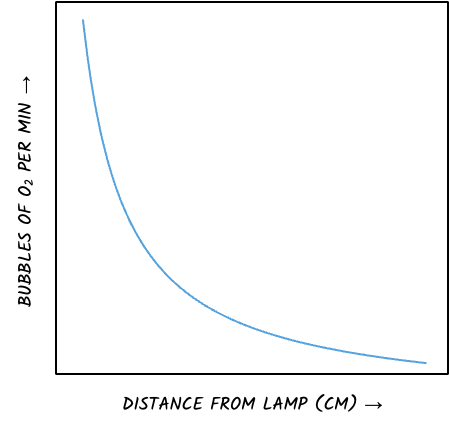
Investigating Oxygen Gas & Photosynthesis
You can use an aquatic plant such as Elodea (pondweed) to measure the rate of photosynthesis under various conditions.
As the plant photosynthesises it produces oxygen gas as bubbles.
You can measure the volume produced in a given time to calculate the rate.
You could also count the number of bubbles in a given time
You could change the distance the lamp is from the plant to investigate the effect of light intensity on the rate of photosynthesis
Or sodium hydrogen-carbonate can be added to increase the CO2 in the water, or the temperature of the water could be changed.

KLO 2.24 understand that a balanced diet should include appropriate proportions of carbohydrate, protein, lipid, vitamins, minerals, water and dietary fibre
Balanced Diet
Why is Food needed
It supplies us with fuel for energy
Helps fight disease
Keeps bodies healthy
Provides materials for growth and repair of tissues
Balanced Diet
A balanced diet consists of all of the food groups in the correct proportions
The necessary key food groups are:
Carbohydrates
Proteins
Lipids
Dietary Fibre
Vitamins
Minerals (mineral ions)
Water
Malnutrition
Having an unbalanced diet can lead to malnutrition
Malnutrition can cause a variety of different health problems in humans
Type | Cause | Effect |
Starvation | Taking in less energy than is used (over a long period of time) | Body starts to break down its energy stores.
|
Coronary Heart Disease | Diet too high in saturated fats and cholesterol | Fat deposits build up in the arteries supplying the heart, reducing blood flow to the heart muscle cells which do not work properly due to lack of oxygen. Can lead to heart attacks and death |
Constipation | Lack of fibre in the diet | Food lacks bulk for muscles to push it through the alimentary canal. Risks of diseases like bowel cancer is increased |
Obesity | Taking in more energy than is used | Extra energy is stored as fat
|
KLO 2.25 identify the sources and describe the functions of carbohydrate, protein, lipid (fats and oils), vitamins A, C and D, the mineral ions calcium and iron, water and dietary fibre as components of the diet
Sources & Functions of Dietary Elements
Dietary Element | Function | Sources |
Carbohydrate |
|
|
Protein |
|
|
Lipid |
|
|
Dietary Fibre |
|
|
Vitamins |
|
|
Minerals |
|
|
Water |
|
|
Vitamins & Minerals
Vitamin/Mineral | Function | Sources | Deficiency Causes … |
Vitamin A | Used to make a chemical/pigment in the retina that maintains the retina and allows vision |
| Cornea damage/night blindness |
Vitamin D | Helps bones absorb calcium and phosphate, meaning it is also important for growing strong bones and teeth |
| Ricket & Osteoporosis Poor Teeth |
Vitamin C | Stick cell lining surfaces together to make connective tissues Forms an essential part of collagen protein - makes up hair, skin, gums, bones |
| Scurvy |
Calcium | Needed to make teeth and bones. Involved in the clotting of blood |
| Osteoporosis - weakened bones |
Iron | Used to make parts of haemoglobin, pigment in red blood cells which transports oxygen around the body |
| Iron deficiency/Anaemia |
KLO 2.26 understand how energy requirements vary with activity levels, age and pregnancy
Variations in Energy Requirements
The nutritional requirements for individuals will vary throughout their lifetime
An individual will still require the same key food groups, but in different quantities depending on a number of factors such as age, height, sex, activity levels, pregnancy and breastfeeding
Factors | Dietary Needs |
Age |
|
Activity Levels |
|
Pregnancy |
|
Breastfeeding |
|
Sex |
|
KLO 2.27 describe the structure and function of the human alimentary canal, including the mouth, oesophagus, stomach, small intestine (duodenum and ileum), large intestine (colon and rectum) and pancreas
Digestion
The chemical and mechanical breakdown of food where relatively large insoluble molecules are broken into smaller soluble molecules that can be absorbed into the bloodstream
The digestive system is an example of an organ system in which several organs work together to digest and absorb food
Digestion is a process in which relatively large, insoluble molecules in food (such as starch, proteins) are broken down into smaller, soluble molecules that can be absorbed into the bloodstream and delivered to cells in the body
These small soluble molecules (such as glucose and amino acids) are used either to provide cells with energy (via respiration), or with materials with which they can build other molecules to grow, repair and function
The human digestive system is made up of the organs that form the alimentary canal and accessory organs
The alimentary canal is the channel or passage through which food flows through the body, starting at the mouth and ending at the anus
Digestion occurs within the alimentary canal
Accessory organs produce substances that are needed for digestion to occur (such as enzymes and bile) but food does not pass directly through these organs
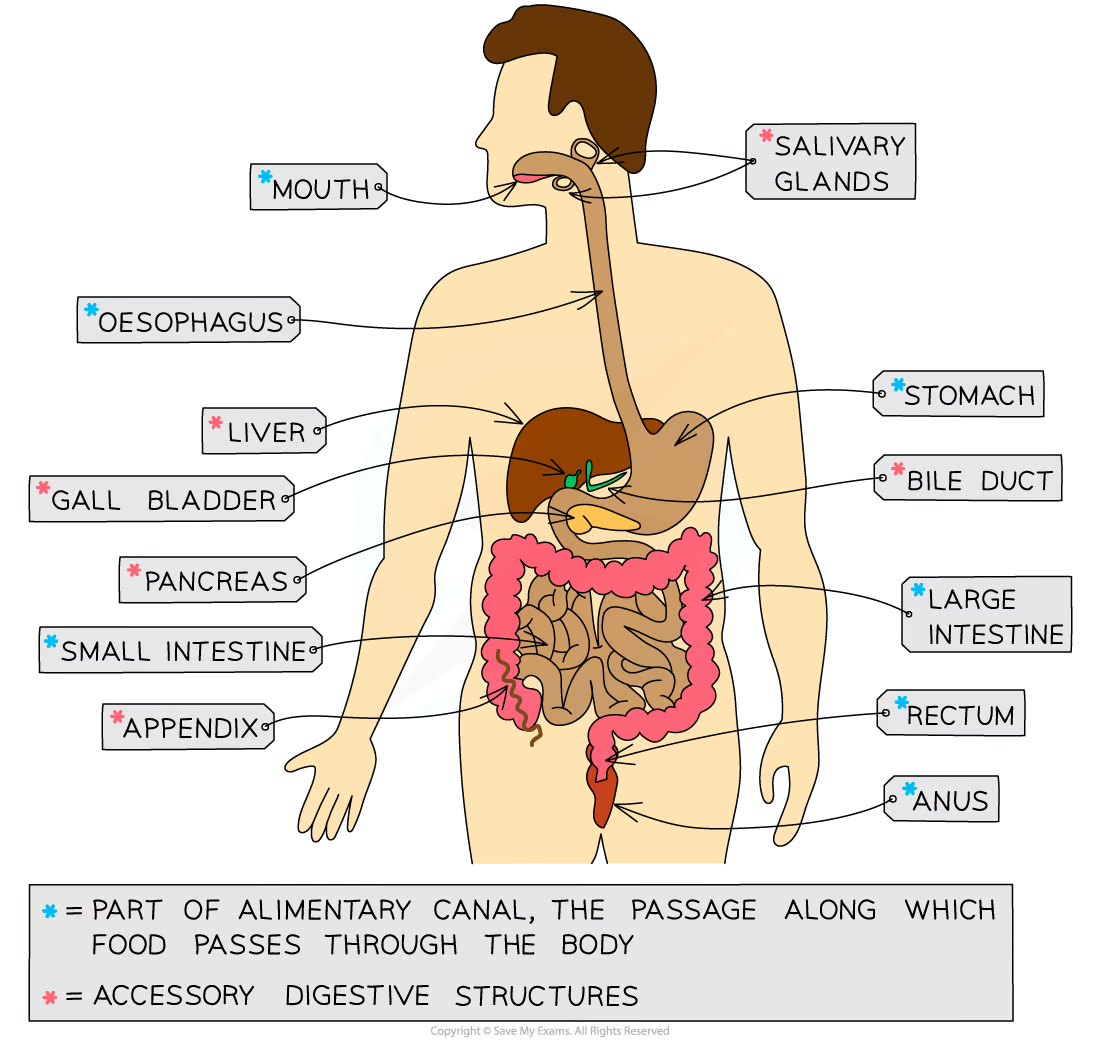
Digestive System
Structure | Function |
Mouth Ingestion |
|
Oesophagus Ingestion |
|
Stomach pH of 2 Chemical & Mechanical Digestion |
|
Duodenum pH of 8-9, slightly alkaline Chemical Digestion |
|
Ileum pH of 8-9, slightly alkaline Absorption |
|
Large Intestine Egestion |
|
Pancreas |
|
Liver |
|
Gallbladder |
|
The Stages of Food Breakdown
Ingestion - the taking in of substances, e.g. food and drink, into the body through the mouth
Mechanical digestion - the breakdown of food into smaller pieces without chemical change to the food molecules
Chemical digestion - the breakdown of large, insoluble molecules into small, soluble molecules
Absorption - the movement of small food molecules and ions through the wall of the intestine into the blood
Assimilation - the movement of digested food molecules into the cells of the body where they are used, becoming part of the cells
Egestion - the passing out of food that has not been digested or absorbed (as faeces) through the anus
KLO 2.28 understand how food is moved through the gut by peristalsis
Peristalsis
Peristalsis is a mechanism that helps moves food along the alimentary canal
Muscles in oesophagus create waves of contraction forcing movement
In the stomach, it is churned into a less solid form called chyme
Peristalsis is controlled by circular and longitudinal muscles
Circular muscles contract to reduce the diameter of the lumen of the oesophagus or small intestine
Longitudinal muscles contract to reduce the length of that section the oesophagus or the small intestine
Mucus is produced to continually lubricate the food mass and reduce friction
Dietary fibre provides the roughage required for the muscles to push against during peristalsis
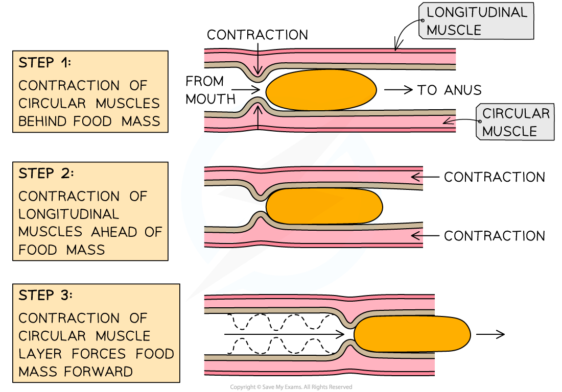
Circular and longitudinal muscles in the alimentary canal contract rhythmically to move the partially digested food mass along in a wave-like action
KLO 2.29 understand the role of digestive enzymes, including the digestion of starch to glucose by amylase and maltase, the digestion of proteins to amino acids by proteases and the digestion of lipids to fatty acids and glycerol by lipases
Digestive Enzymes
Food is partially digested mechanically (by chewing, churning and emulsification) in order to break large pieces of food into smaller pieces of food which increases the surface area for enzymes to work on
Digestion mainly takes place chemically, where bonds holding the large molecules together are broken to make smaller and smaller molecules
Chemical digestion is controlled by enzymes which are produced in different areas of the digestive system
Enzymes are biological catalysts – they speed up chemical reactions without themselves being used up or changed in the reaction
There are three main types of digestive enzymes – carbohydrases, proteases and lipases
Carbohydrases
Carbohydrases are enzymes that break down carbohydrates to simple sugars such as glucose
Amylase is a carbohydrase which is made in the salivary glands, the pancreas and the small intestine
Amylase breaks down starch into maltose
Maltase then breaks down maltose into glucose
Enzyme | Source of Enzyme | Breaks Down | Product |
Amylase | Salivary Glands & Pancreas | Starch | Maltose |
Maltese | Small Intestine | Maltose | Glucose |
Proteases
Proteases are a group of enzymes that break down proteins into amino acids
Pepsin is an enzyme made in the stomach which breaks down proteins into smaller polypeptide chains
Proteases made in the pancreas and small intestine break the peptides into amino acids
Enzyme | Source of Enzyme | Breaks Down | Product |
Pepsin | Stomach | Proteins | Polypeptide Chains |
Trypsin | Pancreas, Small Intestine | Polypeptide Chains | Amino Acids |
Lipases
Lipases are enzymes that break down lipids (fats) to glycerol and fatty acids
Lipase enzymes are produced in the pancreas and secreted into the small intestine
Enzyme | Source of Enzyme | Breaks Down | Product |
Lipase | Pancreas | Lipids | Glycerol & Fatty Acids |
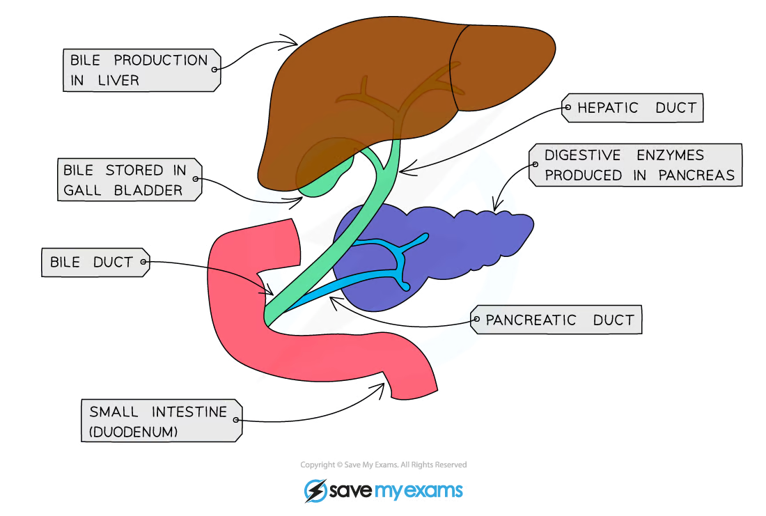
Note: The pancreas is an accessory organ in the digestive system. Food does not pass directly through it, but it has a key role in producing digestive enzymes as well as the hormones that regulate blood sugar (insulin and glucagon).
KLO 2.30 understand that bile is produced by the liver and stored in the gallbladder
Bile
Bile is an alkaline substance produced by cells in the liver
Before being released into the small intestine bile is stored in the gallbladder
Is Not an Enzyme
KLO 2.31 understand the role of bile in neutralising stomach acid and emulsifying lipids
Role of Bile
Neutralising the hydrochloric acid from the stomach
The alkaline properties of bile allow for this to occur
This neutralisation is essential as enzymes in the small intestine have a higher (more alkaline) optimum pH than those in the stomach
Breaking apart large drops of fat into smaller ones (and so increasing their surface area)
This is known as emulsification
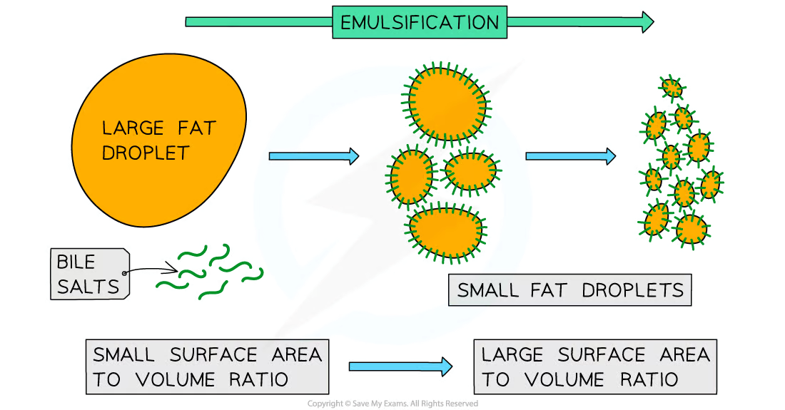
Bile salts break large lipid droplets into smaller ones with a larger surface area
NOTE: Emulsification is the equivalent of tearing a large piece of paper into smaller pieces of paper.This is an example of mechanical digestion, not chemical digestion – breaking something into smaller pieces does not break bonds or change the chemical structure of the molecules which make it up, which is the definition of chemical digestion.
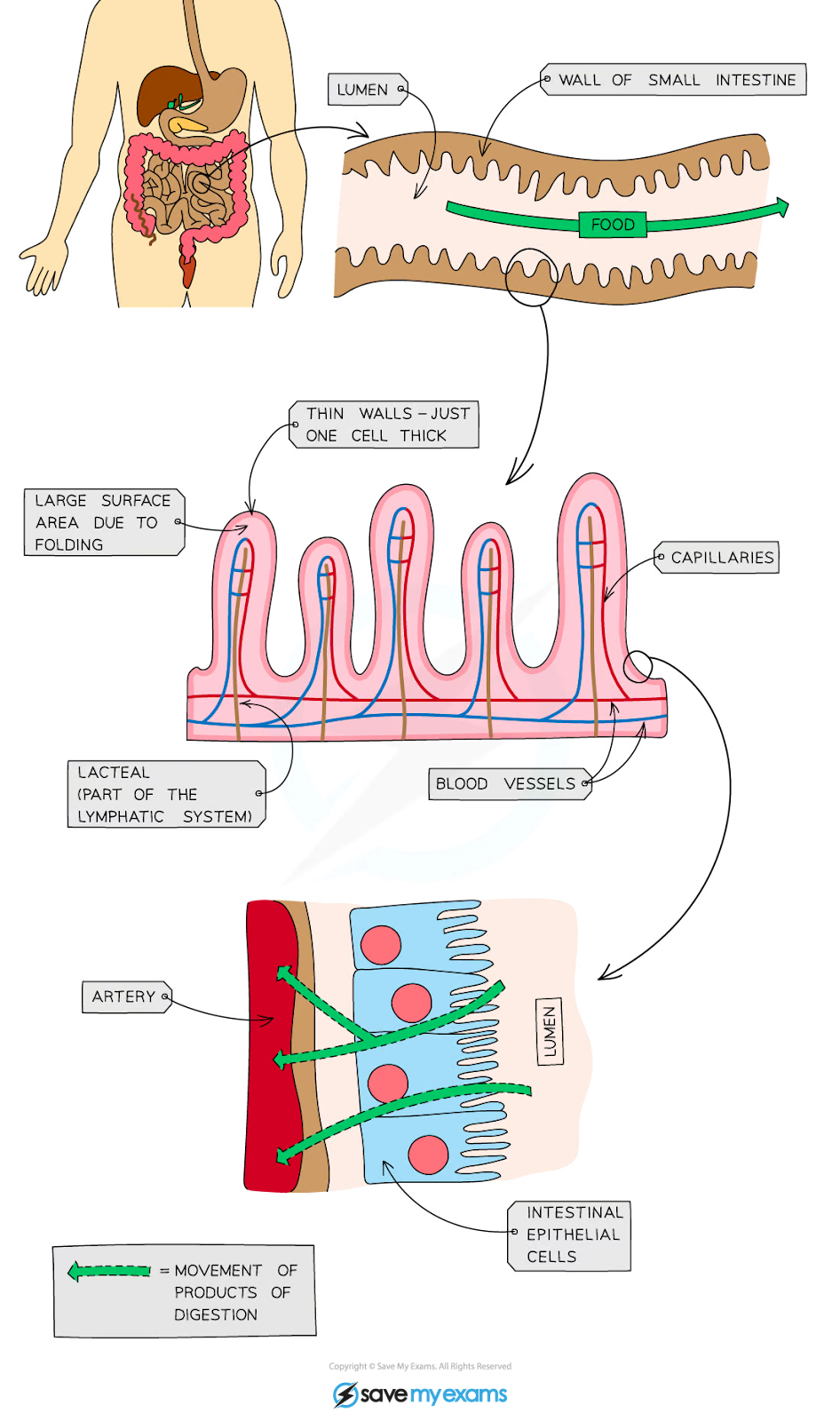
KLO 2.32 understand how the small intestine is adapted for absorption, including the structure of a villus
Adaptations of Small intestine
The small intestine is adapted for absorption as it is very long and has a highly folded surface with millions of villi (tiny, finger-like projections)
These adaptations massively increase the surface area of the small intestine, allowing absorption to take place faster and more efficiently
Peristalsis helps by mixing together food and enzymes and by keeping things moving along the alimentary canal
Villi of Small intestine
Villi have several specific adaptations which allow for the rapid absorption of substances
A large surface area
Microvilli on the surface of the villus further increase the surface available for absorption
A short diffusion distance
The wall of a villus is only one cell thick of intestinal epithelial cells
A steep concentration gradient
The villi are well supplied with a network of blood capillaries that transport glucose and amino acids away from the small intestine in the blood
A lacteal (lymph vessel) runs through the centre of the villus to transport fatty acids and glycerol away from the small intestine in the lymph
Enzymes produced in the walls of the villi assist with chemical digestion
The movement of villi helps to move food along and mix it with the enzymes present
NOTE: The way in which the structure of a villus is related to its function comes up frequently in exam questions so it is worth ensuring you have learned these adaptations and how they influence the rate of absorption.