Topic 19- Musculoskeletal System
Outcomes:
Comprehend the Levels of Muscle Structure
Sequence the Process of Muscle Contraction
Hypothesize and Diagnose Variability in Contraction
Compare and contast muscle types
I. Vertebrate Skeletal System
A. Introduction to Muscle
Function: Enables voluntary movement; stimulated by neurons and attached to bones via tendons.
Abundance: Most abundant tissue in the body.
Origin: Derived from mesoderm, which also forms:
Cardiac muscle
Smooth muscle (gut)
Kidney cells
Red blood cells
Structure:
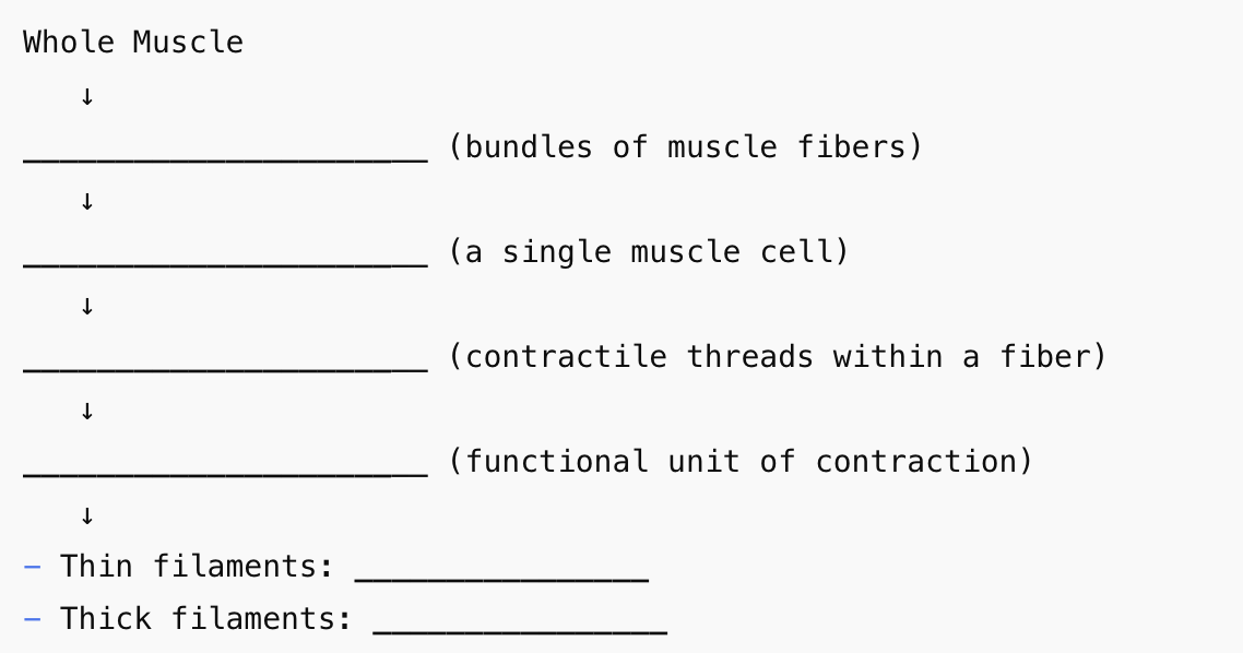
Muscle fiber = muscle cell
Multinucleated (high protein output)
Elongates rather than divides (no cytokinesis)
Bundled together to form whole muscles
Myofibrils
Long, contractile fibers inside each muscle fiber
Made of repeating units called sarcomeres
T-Tubules (Transverse Tubules)
Invaginations of the sarcolemma (plasma membrane)
Carry action potentials deep into the cell
Ensure simultaneous contraction throughout the fiber
Sarcoplasmic Reticulum (SR)
Network surrounding myofibrils
Stores and releases Ca²⁺ to initiate contraction
B. Muscle Fibers
Myofibrils:
Run the length of the muscle fiber
Composed of thick and thin filaments:
Thin Filaments:
Two strands of actin
Covered by tropomyosin, anchored by troponin
Tropomyosin blocks myosin binding sites at rest
Thick Filaments:
Made of ~350 myosin molecules
Myosin heads have ATPase activity to drive contraction
C. Sarcomere
Definition: Functional unit of contraction in muscle fibers
Structure:
Z lines: Boundary of each sarcomere; anchor thin filaments
M line: Middle; anchors thick filaments
Overlap: Region where thick and thin filaments interact
Contraction:
Sarcomeres shorten (filaments slide past each other)
Z lines move closer, filament lengths remain constant
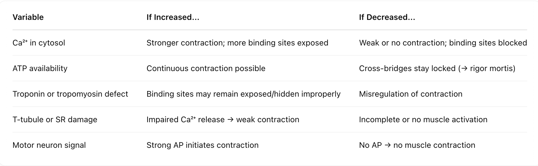
Greater overlap = stronger contraction
D. Sliding Filament Model
1. At Rest
Tropomyosin blocks actin binding sites
No Ca²⁺ → no contraction
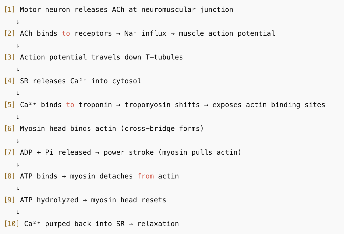
2. Neural Signal Initiation
Motor neuron releases acetylcholine (ACh) at neuromuscular junction
ACh binds to receptors → Na⁺ influx → muscle action potential
3. Calcium Release
AP travels through T-tubules
SR releases Ca²⁺ into cytosol
4. Exposure of Active Site
Ca²⁺ binds troponin → conformational change
Tropomyosin moves → exposes myosin-binding sites on actin
5. Cross-Bridge Cycle
Cross-Bridge Formation:
Energized myosin head binds actin (ADP + Pi attached)
Power Stroke:
ADP + Pi released → myosin pulls actin toward M line
Cross-Bridge Detachment:
New ATP binds → myosin releases actin
Reactivation of Myosin Head:
ATP hydrolyzed → myosin returns to high-energy (cocked) state
Cycle repeats as long as ATP and Ca²⁺ are available
6. Relaxation
Ca²⁺ is pumped back into SR
Tropomyosin re-covers actin
Muscle fiber returns to resting length
E. Energy Use in Muscle
Muscle contraction is ATP-intensive
ATP is not stored long-term, must be regenerated continuously:
Creatine Phosphate:
Transfers phosphate to ADP → rapid ATP regeneration
Glycogen:
Stored in muscle → glucose → ATP
Cellular Respiration:
Oxygen + glucose → long-term ATP production
F. Rigor Mortis
Definition: Post-death muscle stiffening
Cause:
No ATP → myosin heads cannot detach from actin
Ca²⁺ stays in cytosol → sustained contraction
Cross-bridges remain locked
II. Other Muscle Types
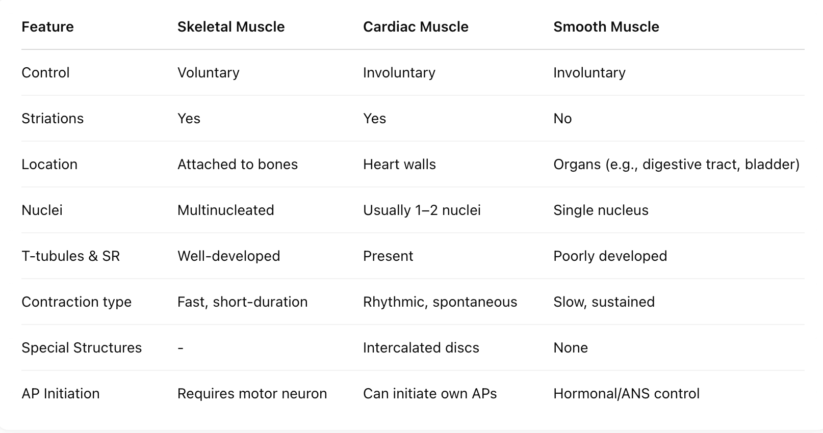
A. Cardiac Muscle
Location: Heart walls only
Function: Rhythmic, coordinated contraction → pumps blood
Spontaneous Activity: Contains pacemaker cells
Intercalated Discs:
Specialized junctions between cells
Allow ion flow (Na⁺, Ca²⁺) → synchronized contractions
Autonomous Rhythm:
Heart beats independently of nervous input
Can keep beating outside the body (e.g., during transplants)
B. Smooth Muscle
Location: Digestive tract, bladder, blood vessels, reproductive organs
Structure:
Involuntary, non-striated, not attached to bones
No sarcomeres, no T-tubules, underdeveloped SR
Contraction:
Slow and sustained
Regulated by hormones and autonomic nervous system
Found in peristalsis, blood flow regulation, and organ function