25
Chapter 25 Blood and Lymphatic System Infections
Circulation of blood and Lymph
Supplies nutrients and o2 to cells, removes waste
Systemic system – carried throughout the body
suffix (emia means in the blood)
Doesn’t always imply disease
Can become transiently bacteremic after brushing teeth
Sepsis- blood poisoning is from circulation agent or toxins can cause b/p to drop, leading to septic shock
Lymphatic (lymphatic vessels)
Infection of limb may result in visible red streaking, from infection sited called- lymphangitis.
Blood, lymph carries leukocytes, antimicrobial proteins (antibodies, complement, lysosome, an interferon) inflammatory response may cause clotting
Spleen
Fist sized organ behind the stomach containing two tissue types
Red Pulp (multiple blood filled passageways)
Clean blood by filtration, can produce new blood cells,
reverse of monocytes
White pulp (lymphoid tissue)
Contains B and T lymphocytes
Lymph nodes and spleen
Blood vascular diseases
Acute bacteria endocarditis- starts suddenly with fever.
Usually cause by virulent species
Staphylococcus aureus
Streptococcus pneumoniae
Quickly destroy heart valve- forms abscesses in muscle that leads to
Heart failure
Subacute endocarditis
Usually caused by organism with little virulence, proceeds slowly
Less likely to be fatal.
Sepsis is caused by both gram – and positive also other infectious agents.
Signs and symptoms
Fatigue, slight fever, typically ill gradually, decreased energy of weeks/months.
Stroke/heart failure (life threatening complication)
Causative agent
endotoxin gram -
Staphylococcus aureus
Streptococcus epidermis
Pathogenesis
Normal microbiota- dental procedures, teeth brushing, and trauma
Microbes adhere to heart tissue, trapped in blood cloth- disformed in heart valve – an infection of one or more heart valves or other inner surfaces of the heart (endocardium)
Multiply creating biofilm
Bacterial continually wash off into circulation which
Infected colt (septic emboli) can break off
Blocking blood vessel, leading to tissue death
Can also weaken/ balloon out forming (aneurysm)
High levels antibodies
Immune complex
Kidney---- glomerulonephritis
Also in skin, eye and other structures -trigger inflammatory response
Hypersensitivity type #3
Treatment and prevention
Antibacterial meds
Artificial valve implants replaced
Sepsis/ septic shock
Symptoms – violent shaking, chills, fever, rapid breathing, anxiety,
*** if septic shock occurs urine output decreases, respiratory and pulse increase
Arms and legs produce a cool dusky color
Causative agent
Gram – bacteria- most fatal cases involve.
Contains lipopolysaccharides- endotoxins.
Epidemiology
Healthcare associated
Rate of increase
Longer lifespan, antibiotic suppression, immunosuppressive meds and use of medical equipment
Pathogenesis
Tissue or infection gram -
compromised boy defense
cause - Medical treatment surgery catheters medications
out of control inflammatory response
Pattern recognition receptors (neutrophils)
overwhelming response cause cytokine storm
blood flow down to organs leads to shock
lungs— toxin and fluids increase oxygen exchange decrease (hypoxemia)
DIC- clotting due to disseminated intravascular coagulation
organ failure due to o2 lack, hemmorage
shortage of clotting protiens
Endotoxin forms gram (-) bacteria, released into the bloodstream
Macrophages activated (shock impaired O2 exchange)
Cytokines released (proteins)
Decreased muscle tone of heart and arteries
Fever
Increased adhesiveness of neutrophils
Increased leakage of plasma from blood vessels
Clotting activated (hemorrhage)
Disseminated intravascular coagulation. (DIC)
Depletion of clotting proteins
Tissue damage from clot in capillaries
Complement system activated (lung tissue damage)
Complement components
Leukocytes attracted to lung tissue
Increased capillary leakage of plasma
Lysosomal enzymes released from leukocytes.
Treatment and Prevention
O2 flow and B/P remain neutral.
Fluid replacement, organ disfunction support
Antibiotics and supportive therapy
BACTERIAL DIEASE OF THE LYMPH NODES AND SPLEEN
Tularemia – rabbit fever or deer fly fever
Signs and symptoms
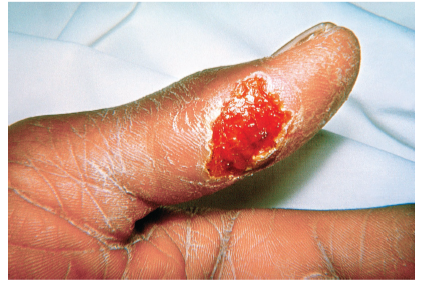
If injury
Ulceration 2-5 days followed by
Lymph node enlarged, fever, chills, ache
Last 1-4 weeks can last months
If inhaled or infected lungs
Pneumonia (PNA) can occur
Dry cough, pain beneath sternum due to enlarged lymph nodes
Rare- but mortality rate 30%
Causative agent
Francisella tularensis
Medium enriched with cysteine( ammonia acids)
Epidemiology
Handlers and eaters of wildlife, northern hemisphere
Pathogenesis
Enters break in skin or mucus membrane
Carried to reginal lymph node- large and tender may become pus filled
Spread via lymphatic and blood vessels
Grows within phagocytic cells
Treatment and prevention
Ciprofloxacin
Streptomycin
Insect repellant- tick removal
Fully cook meat of wild game
Brucellosis – undulant fever, bang disease
Sign/ symptoms
Gradual onset
Fever (mild)- recurrence weeks and months
Sweating, weakness, aches and pains, enlarged lymph nodes, depression and weight loss.
Epidemiology
Domestic animals, category B bioterrorism agent
Pathogenesis
Penetrate mucus membrane or break in the skin
Spread to --- heart, kidney other
Enlarged spleen
Grow in phagocyte cell – avoid antibodies
*** rare complication (osteomyelitis)
Causative agent
Brucella species
B melitensis – goats and sheep
Treatment
Chronic cases 6+ months
Controlled animal environment be cautious of the wild game and animals
Vaccinate animals
Plaque – Black death
Category A bioterrorism
Pneumonic plaque- inhaled respiratory droplets from infected patient/animals
Cough, blood sputum arise within 1-3 day
100% mortality if untreated
Septicemic Plaque – endotoxin- difficult to treat do to being part of cell wall
Shock – decreased b/p, and disseminated intravascular coagulation
Causes bleeding into the skin and organs, red / black patchy rash. 50-80% death without treatment
Sign and symptoms
Develop 2-6 days
Boboes- painful swelling in armpit/groin; size of egg/apple
Vomiting/fever
Bleeding under skin causes blotching on the body
Attacks Nervous system – suffering spasm and terrible pain
Boboes- can burst- this is how the patient survives. However, most patients suffer Terrible death
Virulence factors
There are 7 factors but it is important to know 3
PLA – protease- destroys complement components, promotes clearance of clots
YOPs (proteins) – interferes with phagocytosis, and the immune response.
F1- Forms antiphagocytic capsule 37 degrees C
Pathogenesis
Forms biofilm in the digestive system of infected flea, often blocking the tract. The flea starves- increasing the likelihood that it will feed again- causes bacteria to be regurgitated (vomited) into the bite wound as the flea attempts to fed. Y pestis release bacterial cells in their feces, and these can be introducing into human tissue when a person scratches the flea bite.
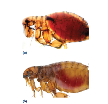 A healthy B. obstruction due to Y pestis infection
Epidemiology
25% population died Europe.
Endemic found in rodents, prairie dogs, squirrels, rats, rabbits, dogs, fleas from rate bite.
Causative agent
Yersinia pestis
Bacteria, safety pin shape
Treatment
Antimicrobial 24 hours after symptoms onset
Prompt diagnosis
Avoid infected rodents and their fleas.
VIRAL DISEASE OF THE LYPHOID AND BLOOD VASCUALR DIEASE
Mononucleosis (mono or kissing disease)
Signs and symptoms
Incubation period 30-60 days
Fever, sore throat – usually goes away in 2 weeks
Swollen lymph nodes and enlarged spleen- 3 weeks gone
Causative agent
Esptein-barr virus (EBV)
Epidemiology
Saliva
Treatment/ Prevention
Analgesic
Cortisone- airway obstructions, fatigue
Avoid saliva- toothbrushes, drinking glasses
No vaccine
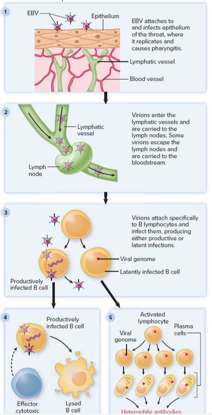
Pathogenesis
Infects via mouth and throat- can cause pharyngitis
Replicates and carried to lymph nodes
B lymphocytes/b cells infected
Productive- virus replicates resulting in death of b- cells
Latent- maintained in plasma or provirus proliferation of b-cells, immunoglobulins produces
Cytotoxic t cells- reproduce and destroy B cells (infected)
Lymph nodes spleen- enlarged
Abnormal size nucleus- Downey cells
Play an important role in some cancers
Burkitt lymphoma, nasopharyngeal carcinoma
Can live in saliva up to 18 months after infection
People with AID can spread virus – continuously shedding
What is a B cell do? Produces antibodies
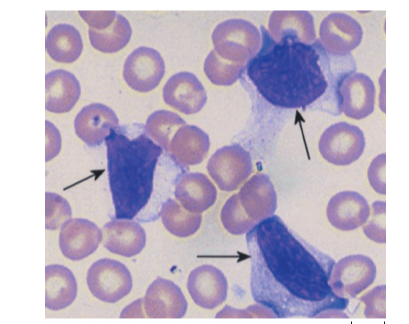 mononucleosis lymphocyte
YELLOW FEVER
Sign and symptoms
Appears after 3-6 days and can range form mild to severe
Accumulated bilirubin (jaundices) found in liver/gallbladder
Mild- fever slight headache
Severe- high fever, nausea, bleeding nose and skin. Black vomit, gastrointestinal bleeding
Causative agent
RNA arbovirus Aedes aegypti
Vector mosquitoes –
Pathogenesis
Bite from infected mosquito, multiplies in host, enters blood stream
Liver damage- jaundice
Decrease clotting proteins
Hemorrhage- destruction small blood vessels
Kidney failure
Disseminated intravascular coagulation DIC
Epidemiology
Infected mosquitoes
No proven treatment
Control vector, attenuated vaccine
PROTOZAN DISEASES
MALARIA- BAD AIR
Signs and symptoms
“flu like”
Fever, headache, joint pain/muscle (2 weeks after bite)
Cold stage: patient feels cold and develops shaking chills for up to an hour
Hot stage: Patient temperature rises sharply for 3-8 hours, 40 degree C
Wet stage: Patients temperature falls; drenching sweating 2 to 4 hours – feels exhausted.
Paroxysm- cycle of intense symptoms of chills, fever and sweat
Epidemiology
Temperature/tropical areas- transmitted from person to person by bite(anopheles)
Causative agent
Plasmodium
P vivax
P fluciparum – most severe
P malariae
P ovale
P knowlesti
Complex life cycle- liver stage ( exoerythrocytic stage) – outside red blood cells
Red blood cell stage- erythrocytic stages
Sporozoites- infectious form of the protozoan
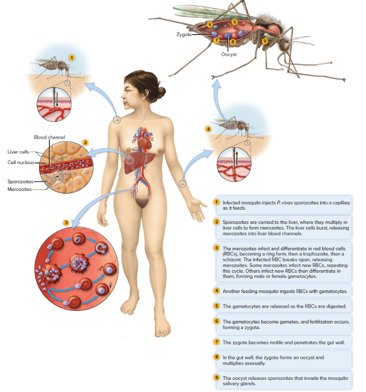 Merozoites- infect and destroy red blood cells- reproduce A sexually
Infected mosquito injects p vivax into capillary as they feed.
Sporozoites carried to liver multiply in the liver cells forming merozoites liver cell burst releasing merozoites into liver blood channels
Merozoites infect and differentiates in red blood cells rings from there trophozoite feed and reproduce
Finally, schizont break open releasing merozoites.
Female/male gametocytes
Another mosquito ingests RBC gametocytes.
Gametocytes released as RBC-to be digested
Gametocytes—gametes-zygotes
Zygotes motile penetrates gut wall, oocyst form multiples asexually invade mosquitos salivary glands.
Treatment/ prevention
Complex depends on the different stages of life cycle
Chloroquine- erythrocytic stage
Primaquine
What is the significance of immune complex formation in infective endocarditis?
They can be deposited in tissue and organs causing damage
Describe disseminated intravascular coagulation.
Extensive, abnormal clotting that occurs throughout the small blood vessels using up all the existing clotting factors- this can cause extensive bleeding and hypoxia
Organ failure due to lack of oxygen and bleeding due to shortage of clotting protein
Why is brucellosis a threat to big game hunters?
Domestic animals are affected, hunters have acquired infections by eating meat from infected elk, moose, bison, caribou, and reindeer. Hunters also encounter disease form skinning them.
Why might the Yersinia pestis from a patient with pneumonic plague be more dangerous than the same organism from fleas?
A person inhales respiratory droplets from an individual or animal. Could also be caused by organisms reaching lungs through the bloodstream. Because of the cough and bloody sputum, it also has built in virulence factors that provide defense against the immune system.
Why might rodent burrows be a source of plague months after they are abandoned?
Y pestis is released bacterial cells into their feces, if there is still feces in their abandoned burrows it could still be contaminated
What type of leukocytes does EBV infect?
B lymphocytes
Travelers to and from which areas of the world should be vaccinated against yellow fever.
Travelers from parts of central and South America, as well as parts of Africa.
Which plasmodium species cause the most dangerous form of malaria?
Plasmodium falciparum
Blood Vascular diseases
Acute bacterial endocarditis
-staphylococcus aureus and streptococcus pneumonia
Subacute endocarditis
Staphylococcus aureus and streptococcus epidermis
Sepsis/septic shock
Gram (-) bacteria containing lipopolysaccharides- endotoxins
If occurs urine output decreases, respiratory and pule increase, arms and legs produce cool dusky color
Bacterial disease of the lymph nodes and spleen
Tularemia -rabbit fever or deer fly fever
Francisella tularensis
Brucellosis- undulant fever bang disease
Brucella species (B Melitensis- goats and sheep)
Category B bioterrorism
** rare complication osteomyelitis
Plaque Black death
Category A bioterrorism
PLA, YOPS, F1 (virulence factors)
Yersinia pestis
Viral disease of the lymphoid and blood vascular disease
Mononucleosis- mono or kissing disease
Esptien-barr virus
Yellow fever
RNA arbovirus
Aedes aegypti
Vector mosquitos
Protozan Diseases
Malraia -Bad air
Cold stage, hot stage, wet stage
Plasmoidum – Vivas, fluciparum(most severe) marliarae, ovale, knowlesti