B1.1 Carbohydrates and lipids Notes
B1.1.1 chemical properties of a carbon atom
Why Is Carbon So Essential?
From the DNA in your cells to the food you eat, carbon forms the foundation of all biological molecules, enabling the complexity of life as we know it.
But why is it the element of life, rather than something more common, like oxygen or nitrogen?
The answer lies in its bonding abilities, versatility, and role in molecular diversity.
Covalent Bonding: The Key to Stability
A covalent bond forms when two atoms share a pair of electrons. Carbon is exceptional because:
Stability: Covalent bonds are the strongest type of bonds in biological systems, perfect for creating durable molecules like DNA and proteins.
Electron Configuration: Carbon has four electrons in its outer shell but needs eightfor stability.
This allows it to form four covalent bonds, giving rise to complex structures.
Tip
Strong covalent bonds make carbon-based molecules resistant to breakdown, ensuring stability in biological systems.
Carbon's Bonding Potential: The Foundation of Molecular Diversity
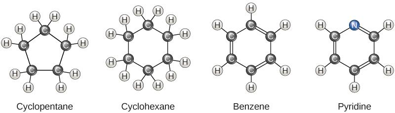
Carbon can form a variety of structures, ranging from simple chains to complex rings, making life's molecular diversity possible.
Single Bonds: Creating Long Chains
Straight chains (e.g., fatty acids in cell membranes).
Branched chains (e.g., isobutane, which affects molecular interactions and function).
Double and Triple Bonds: Adding Rigidity and Reactivity
Double bonds introduce kinks in molecules (e.g., unsaturated fatty acids, which impact membrane fluidity).
Triple bonds (less common but extremely strong) alter molecular properties.
Carbon Rings: The Basis of Life's Chemistry
Single Ring (e.g., glucose): Soluble, making it ideal for energy transport in the body.
Multiple Rings (e.g., cholesterol): Strengthens cell membranes and serves as a precursor for hormones.
Example
Compare ethene (C₂H₄) and ethane (C₂H₅):
Ethene's double bond makes it rigid and reactive.
Ethane's single bonds allow for more flexibility.
This difference affects their physical and chemical behavior in biological systems.
Analogy
Think of carbon chains as scaffolding and rings as specialized rooms.
Together, they form the architectural framework of life's molecules.
Functional Groups: The Source of Chemical Diversity
Definition
Functional groupsFunctional groups are clusters of atoms that dictate a molecule's properties and reactivity.
While the carbon backbone provides structure, functional groups determine a molecule's reactivity and interactions.
The key functional groups in biological molecules are:
Hydroxyl (-OH): Found in sugars and alcohols, making molecules polar and water-soluble.
Carboxyl (-COOH): Found in amino acids and fatty acids, making them acidic and reactive.
Amino (-NH₂): Found in proteins, essential for forming peptide bonds.
Example
Glucose (-OH groups) dissolves in blood for easy transport.
Fatty acids (-COOH groups) help form cell membranes.
Amino acids (-NH₂ and -COOH groups) link together to form proteins.
Warning
Don't confuse functional groups with the carbon backbone as the backbone gives shape, while functional groups control chemical properties and reactivity.
Why Carbon Is the Foundation of Life
Carbon's ability to form four covalent bonds allows for:
Three-dimensional molecular structures (crucial for DNA's double helix).
Complex interactions (essential for enzyme function and signaling pathways).
Versatility in Biological Molecules
Carbohydrates (Sugars): Store and provide energy.
Lipids (Fats): Form membranes and store energy long-term.
Proteins: Enable structural support and enzymatic reactions.
Nucleic Acids (DNA & RNA): Carry genetic information.
Exam_technique
Mnemonic: "C.H.A.R.M." -- Key Features of Carbon Compounds:
C - Covalent Bonds (Carbon forms strong bonds with other atoms)
H - Hydrocarbons (Basic carbon chains, essential for energy storage)
A - Aromatic Rings (Complex ring structures like in glucose and steroids)
R - Rings and Chains (Carbon forms both rings and straight/branched chains)
M - Molecule Diversity (Carbon's versatility leads to endless compound diversity)
Nature of Science (NOS): Scientific Conventions and Units
Scientists use SI units to ensure consistent measurement of carbon-based molecules.
SI Prefixes
kilo- (k) = 10³
centi- (c) = 10⁻²
milli- (m) = 10⁻³
micro- (μ) = 10⁻⁶
nano- (n) = 10⁻⁹
Simplifies expression of very large or very small measurements.
Self Review
Why is carbon uniquely suited to form the backbone of biomolecules?
How do single, double, and triple bonds contribute to molecular diversity?
What role do functional groups play in determining the properties of carbon-based compounds?
Can you name a carbon-based molecule in your body and explain how its structureenables its function?
Tip
Always remember that carbon's four bonds allow it to build the key molecules of life, such as proteins, lipids, carbohydrates, and nucleic acids.
B1.1.2 production of macromolecules by condensation reactions
Condensation Reactions
Definition
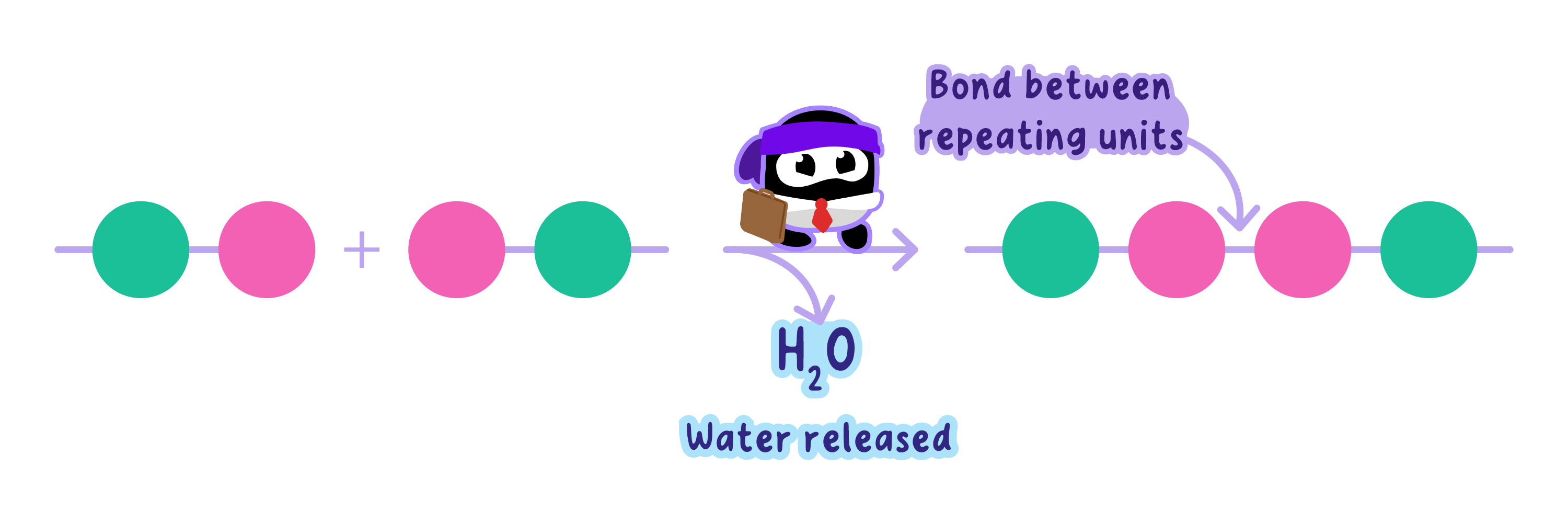
Condensation Condensation is a reaction where two molecules combine, releasing water.
It is enzyme-catalyzed, requires energy input, and typically occurs inside cells.
The removal of H₂O allows the formation of a covalent bond between the reacting functional groups.
These reactions are reversible via hydrolysis, which breaks the polymer back into monomers by adding water.
Analogy
Think of condensation reactions as building a brick wall (polymer) from individual bricks (monomers).
The mortar that holds them together is formed by removing water from the bricks!
Key Macromolecules Formed by Condensation
The main types of biological macromolecules produced through condensation are:
Polysaccharides
Polypeptides (proteins)
Nucleic acids (DNA and RNA)
Each type of macromolecule has distinct monomers, bond types, and biological functions.
1. Polysaccharides (Carbohydrates)
Monomer
Monosaccharides single sugar units with the general formula (CH₂O)n.
Examples: α-glucose, β-glucose, galactose, fructose
Reaction
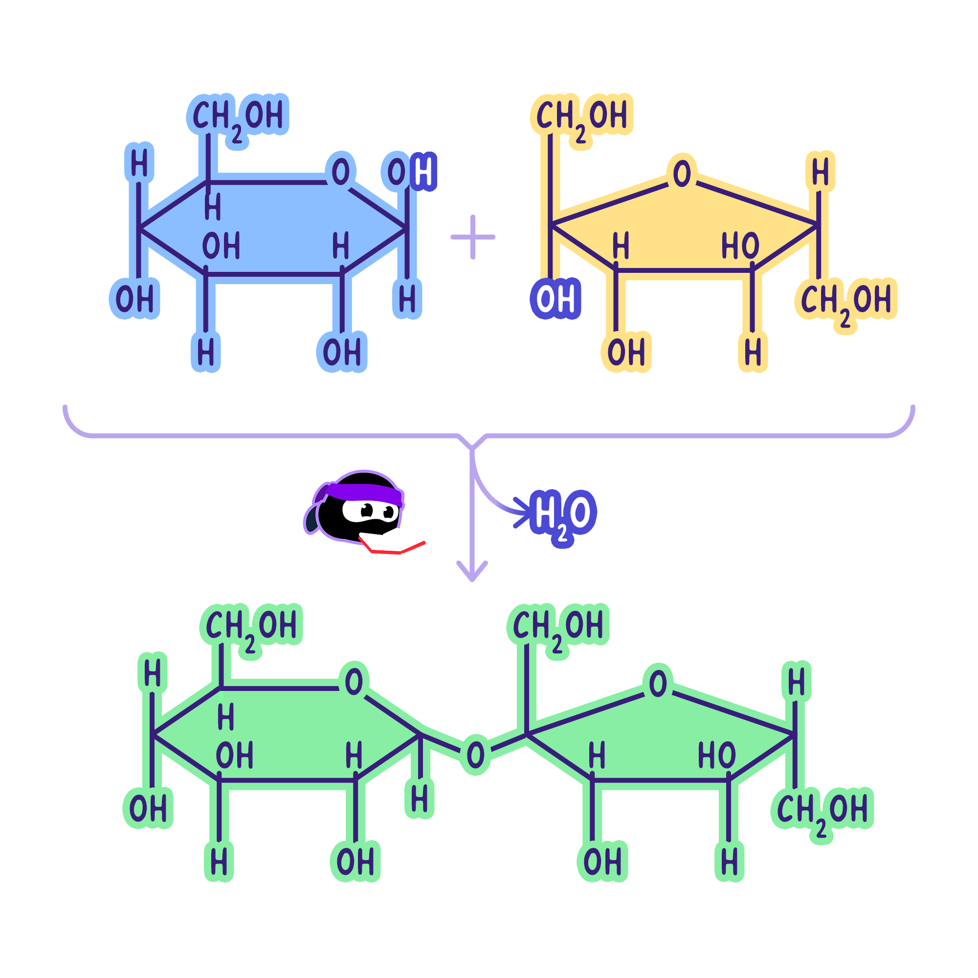
Glycosidic bond formation occurs between hydroxyl (-OH) groups on two monosaccharides.
Enzyme example: maltase catalyzes maltose formation
By-product: one molecule of H₂O per glycosidic bond
Polysaccharide | Monomer | Bondy Type | Function | Structure |
|---|---|---|---|---|
Starch | α-glucose | α(1->4) and α(1->6) | Energy storage in plants | Amylose (unbranched), Amylopectin (branched) |
Glycogen | α-glucose | α(1->4), α(1->6) | Energy storage in animals (liver, muscle) | Highly branched |
Cellulose | β-glucose | β(1->4) | Structural component in plant cell walls | Straight chains; form microfibrils |
2. Polypeptides (Proteins)
Monomer:
Amino acids contain a central carbon (C) bonded to:
An amino group (-NH₂)
A carboxyl group (-COOH)
A hydrogen atom
A variable R group (determines identity)
Reaction:
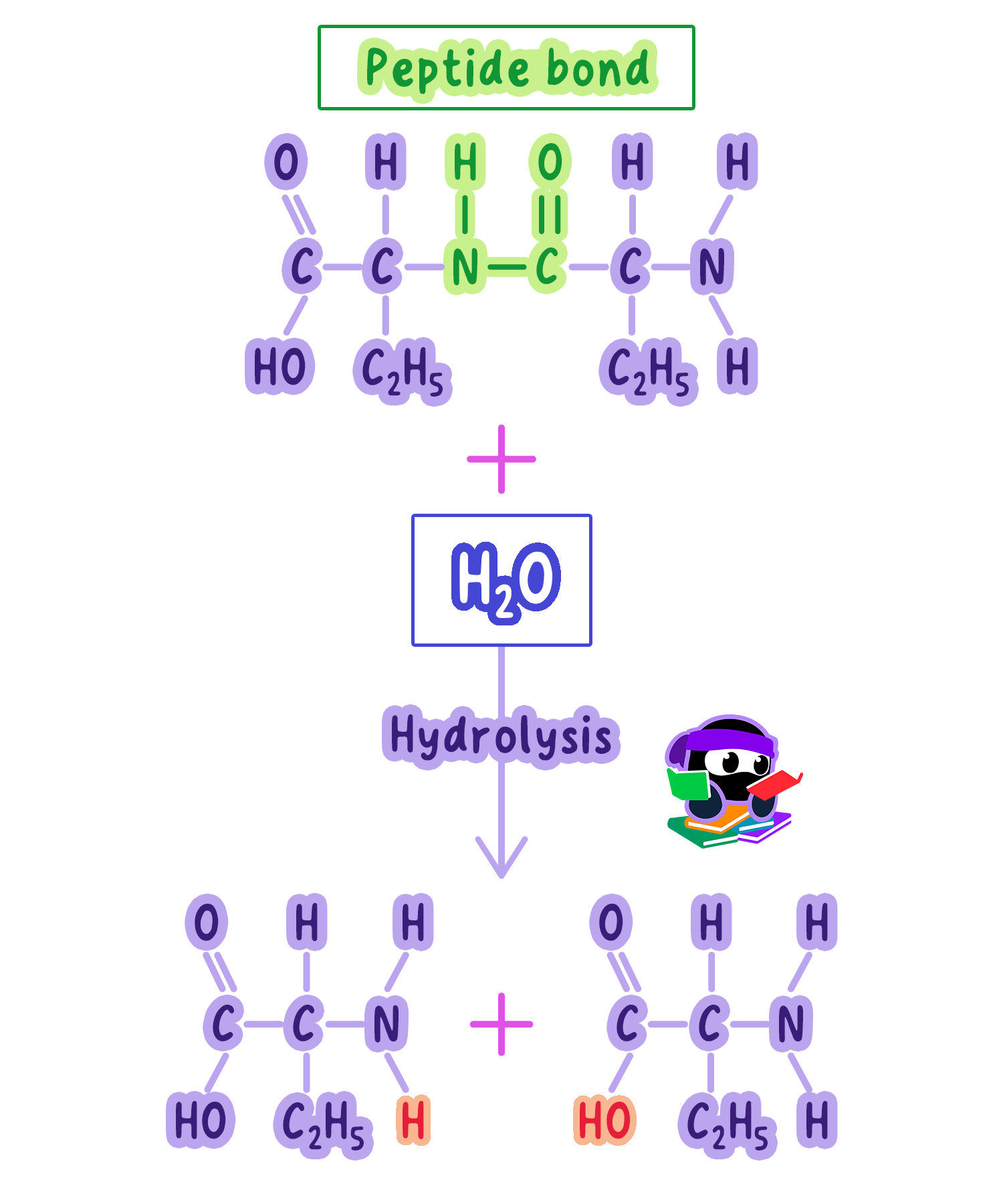
Peptide bond forms between the amino group of one amino acid and the carboxyl group of another.
The reaction removes H₂O (from -OH of carboxyl and H of amino group).
Enzyme-catalyzed during translation (ribosome-mediated)
Definition
Peptide bondA peptide bond is a covalent bond formed between the carboxyl group of one amino acid and the amino group of another.
Example
Dipeptide: two amino acids joined by one peptide bond.
Polypeptide: chain of many amino acids.
Protein: one or more polypeptides folded into a specific 3D structure (primary to quaternary levels).
Protein | Function | Example |
|---|---|---|
Enzymes | Catalyze metabolic reactions | e.g., amylase, pepsin |
Hormones | Regulation of physiology | e.g., insulin |
Structural | Support and integrity | e.g., collagen |
Transport | Movement of molecules | e.g., hemoglobin (O₂ transport) |
Antibodies | Immune defense | Specific binding to antigens |
Tip
The sequence of amino acids determines protein folding, which determines function.
3. Nucleic Acids (DNA and RNA)
Monomer:
Nucleotides, each composed of:
Phosphate group
Pentose sugar (deoxyribose in DNA, ribose in RNA)
Nitrogenous base (A, T/U, G, C)
Reaction:
Phosphodiester bond forms between the phosphate group of one nucleotide and the 3′-OH group of the sugar on another nucleotide.
Water is released during each bond formation.
Forms the sugar-phosphate backbone of DNA/RNA.
Definition
Phosphodiester bondA phosphodiester bond is a covalent bond formed between nucleotides in a nucleic acid chain via condensation.
Polymer:
DNA: double-stranded helix; stores genetic instructions
RNA: single-stranded, involved in protein synthesis (mRNA, tRNA, rRNA)
Tip
Always mention water release and the specific bond type (e.g., glycosidic, peptide, or phosphodiester) in your answers to get full marks!
Note
DNA strands are antiparallel and held together by hydrogen bonds between complementary bases
Self Review
Explain how a glycosidic bond is formed between two glucose molecules.
Describe the formation of a dipeptide from two amino acids, including the functional groups involved.
Outline the role of condensation reactions in the synthesis of DNA.
Explain why water is released during the formation of a phosphodiester bond in a nucleotide chain.
B1.1.3 digestion of polymers into monomers by hydrolysis reactions
What Is Hydrolysis?
Definition
HydrolysisHydrolysis is a chemical reaction where water () breaks down a compound.
A water molecule splits into -H and -OH.
These fragments attach to the exposed ends where the bond was broken, preventing the monomers from reconnecting.
Example
Hydrolysis of a Disaccharide Take sucrose, a common sugar found in many foods.
It's a disaccharide made of two monosaccharides: glucose and fructose.
During hydrolysis, a water molecule is added, breaking the bond between glucose and fructose.
The from water attaches to the glucose, while the attaches to the fructose.
This reaction is facilitated by the enzyme sucrase.
The Role of Hydrolysis in Digestion
Hydrolysis is vital for breaking down large macromolecules (polymers) into smaller monomers that the body can absorb.
Polysaccharides -> Monosaccharides
Starch or glycogen (long chains of glucose) are hydrolyzed by enzymes (e.g., amylase, maltase).
Glycosidic bonds are broken, releasing glucose units.
Polypeptides -> Amino Acids
Proteins (chains of amino acids linked by peptide bonds) are digested by pepsin, trypsin, etc.
Each bond is cleaved by adding water, yielding free amino acids.
Nucleic Acids -> Nucleotides
DNA and RNA (linked by phosphodiester bonds) are broken down by nucleases.
The result is nucleotide monomers (building blocks of genetic material).
Example
For example, starch is first hydrolyzed into maltose (a disaccharide) by amylase.
Maltose is then further hydrolyzed into glucose by maltase, making it available for absorption in the small intestine.
Why Is Water Essential?
Splitting H₂O: The -H and -OH derived from water block the ends of newly formed monomers, stabilizing them.
Preventing Rebonding: These newly attached groups stop the monomers from re-polymerizing.
Universal Solvent: Water's presence in cells makes hydrolysis feasible in aqueousenvironments.
Note
Without water, hydrolysis reactions cannot proceed.
This is why dehydration can disrupt digestion and other critical metabolic processes.
Hydrolysis vs. Condensation
Condensation (Dehydration Synthesis)
Removes water to form bonds, building polymers from monomers.
Hydrolysis
Adds water to break bonds, splitting polymers back into monomers.
Together, hydrolysis and condensation form a biological cycle:
Condensation assembles large molecules for storage or function.
Hydrolysis disassembles them to release nutrients or energy.
Analogy
Think of hydrolysis and condensation as a zipper.
Condensation zips monomers together to form a polymer, while hydrolysis unzips them back into monomers.
Applications of Hydrolysis in the Body
Energy Release: ATP hydrolysis liberates energy for cellular processes.
Detoxification: The liver uses hydrolysis to break down toxic substances.
Biomolecule Recycling: Cells reuse monomers by hydrolyzing older or damaged molecules.
Tip
Enzymes that catalyze hydrolysis reactions are highly specific.
For example, lactase hydrolyzes lactose, while lipase targets triglycerides.
Warning
Students often confuse hydrolysis with hydration.
Hydration involves adding water to a molecule without breaking it, whereas hydrolysis splits the molecule using water.
Self Review
Can you explain how water is used in hydrolysis to break down a polymer into monomers?
Try drawing a diagram to illustrate the process.
B1.1.4—form and function of monosaccharides
Recognizing Monosaccharides: Pentoses vs. Hexoses
Basic Building Blocks
Monosaccharides are the simplest form of carbohydrates.
They provide the foundation for more complex carbohydrates (disaccharides, polysaccharides).
Pentoses (5-Carbon Sugars)
Ribose (C₅ H₁₅O₅ ) and Deoxyribose (C₅ H₁₅O₄).
Crucial components of RNA (ribose) and DNA (deoxyribose).
Form the sugar-phosphate backbone in nucleotides.
Hexoses (6-Carbon Sugars)
Example: Glucose (C₅H₁₂O₅).
Often found in ring forms, which are more stable and biologically active.
Hint
Here is a helpful memory:
"Pentagons Have Hexagons, Both Ring Stable"
Pentagons = Pentoses (5 carbon atoms, e.g., ribose, deoxyribose)
Have = Hexoses (6 carbon atoms, e.g., glucose)
Hexagons = Ring forms (more stable than straight chains)
Both = Both pentoses and hexoses exist in ring form
Ring Stable = Ring form is more biologically functional
The Ring Structure of Glucose (A Hexose)
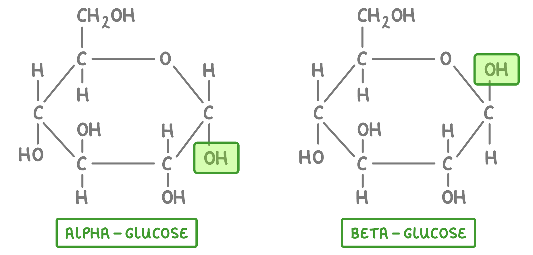
Glucose (C₅H₁₂O₅) is a hexose with 6 atoms in its ring: 5 carbon atoms and 1 oxygenatom.
Glucose has two forms:
α-glucose
β-glucose
They differ in the orientation of the hydroxyl group attached to carbon 1.
Pentoses: Ribose and Deoxyribose
Ribose (C₅ H₁₅O₅ ) and Deoxyribose (C₅ H₁₅O₄) are pentoses.
Ribose is part of the structure of RNA, and Deoxyribose is part of DNA.
These sugars form the backbone of nucleotides in genetic material.
Tip
Pentoses like ribose and deoxyribose are critical for genetic information storage, while hexoses such as glucose are primarily energy sources.
Properties of Glucose and Their Biological Significance
Glucose is highly important in living organisms. Its properties make it perfect for energy transport and storage.
Solubility
Glucose is soluble in water because it has hydroxyl (-OH) groups, forming hydrogen bonds with water.
This allows glucose to dissolve in the blood and be transported to cells.
Analogy
Imagine stirring a sugar cube into water.
Like the sugar cube, glucose dissolves easily, ensuring it can travel through the bloodstream to deliver energy wherever it's needed.
Transportability
Glucose's small size and solubility allow it to be carried through cell membranes using GLUT transporters.
This ensures glucose can be used for cellular respiration to produce ATP.
Tip
The brain relies almost exclusively on glucose for energy, consuming about 20% of the body's total energy despite its relatively small size.
Chemical Stability
Stable under physiological conditions, glucose doesn't react prematurely or degrade during transport.
It reaches its target cells intact.
Energy Yield from Oxidation
Glucose is a high-energy molecule.
In cellular respiration, glucose is oxidized to produce carbon dioxide, water, and ATP.
Up to 36-38 ATP molecules can be generated from one glucose molecule in aerobic respiration.
Note
Although glucose is stable during transport, enzymes like hexokinase activate it for metabolism when energy is required.
Warning
Don't confuse the energy yield of glucose in aerobic respiration (36-38 ATP) with anaerobic respiration, which produces only 2 ATP per glucose molecule.
Linking Structure to Function: Why Glucose Works
Solubility: Glucose's hydroxyl (-OH) groups help it dissolve in blood for efficient transport.
Transportability: Its small size allows glucose to enter cells through GLUT transportersfor energy production.
Chemical Stability: Ensures glucose doesn't break down during transport to cells.
Energy Yield: Through cellular respiration, glucose provides significant ATP (energy) for cellular activities.
Hint
The IB love asking questions relating structure to function, so when you see this make sure you focus on it!
Analogy
Think of glucose as a versatile currency.
It's easy to transport, stable for storage, and can be "spent" as energy or converted into other useful forms as needed.
Applications and Real-World Relevance
Plants: Glucose is produced during photosynthesis and used to make starch and cellulose.
Animals: Stored as glycogen in the liver and muscles for quick energy release.
Medicine: Glucose solutions are used in IV drips to give patients quick energy.
Tok
How does the universal use of glucose across species reflect the shared evolutionary origins of life?
Could the unique properties of glucose have influenced its selection as a primary energy source?
Self Review
What are the structural differences between pentoses and hexoses?
Why is glucose's solubility important for its function?
How does the oxidation of glucose yield energy?
Self Review
Explain how the structure of glucose makes it suitable for transport and energy production in living organisms.
B1.1.5 polysaccharides as energy storage compounds
The Structure of Starch and Glycogen: Compact and Efficient
Starch Is The Plant Energy Reserve
Plants store energy in the form of starch, a polysaccharide composed entirely of α-glucose molecules.
Starch is a mixture of two types of molecules: amylose and amylopectin.
Amylose:
Amylose is an unbranched chain of α-glucose molecules linked by1->4 glycosidic bonds.
Due to the bond angles, amylose forms a helical structure, which makes it compact and efficient for storage.
Its unbranched nature means it has fewer ends where enzymes can act to release glucose, making energy release slower compared to amylopectin.
Amylopectin:
Amylopectin is similar to amylose but includes additional1->6 glycosidic bonds, forming a branched structure.
The branches allow enzymes to access multiple chain ends simultaneously, enabling faster glucose release when energy is needed.
Glycogen Is The Animal Energy Reserve
Animals store energy in the form of glycogen, which has a structure similar to amylopectin but is even more highly branched. In glycogen:
1->4 glycosidic bondslink the α-glucose molecules in the main chain.
1->6 glycosidic bondsform branches approximately every 10 glucose units, compared to every 20 units in amylopectin.
This extensive branching makes glycogen even more compact and allows rapid mobilization of glucose, which is critical for meeting the high and variable energy demands of animals.
Tip
Glycogen is primarily stored in the liver and muscle cells, where it can be quickly broken down to glucose during exercise or fasting.
Why Polysaccharides Are Ideal Energy Storage Molecules
Compactness
The coiling (in amylose) and branching (in amylopectin and glycogen) during polymerization make these polysaccharides highly compact.
This allows a large amount of glucose to be stored in a small space, which is especially important in cells with limited storage capacity.
Analogy
Think of starch and glycogen as tightly packed suitcases filled with glucose "clothes."
Their efficient packing ensures you can store as much as possible without wasting space.
Low Solubility
Unlike glucose, which is highly soluble and would cause osmotic problems if stored in large quantities,
starch and glycogen are relatively insoluble due to their large molecular size. This means they:
Do not dissolve in the cytoplasm.
Avoid drawing water into the cell via osmosis, which could cause the cell to swell and burst.
Warning
Students often think polysaccharides are insoluble because they are hydrophobic.
This is incorrect.
They are insoluble primarily due to their large size, not because they repel water.
Ease of Adding and Removing Glucose
Polysaccharides are dynamic storage molecules. Glucose can be added (viacondensation reactions) or removed (via hydrolysis reactions) as needed:
Condensation: When glucose is abundant, enzymes link α-glucose molecules to the growing polysaccharide chain, releasing water as a byproduct.
Hydrolysis: When energy is needed, enzymes break the glycosidic bonds, releasing glucose monomers for cellular respiration.
Example
For instance, during intense exercise, glycogen in muscle cells undergoes rapid hydrolysis to provide glucose for ATP production, fueling muscle contractions.
Applications of Starch and Glycogen as Energy Stores
Starch in Plants
Starch is stored in chloroplasts and amyloplasts (specialized storage organelles in plants).
It serves as a long-term energy reserve, particularly in seeds, tubers, and roots. For example:
Potatoes store starch in their tubers to provide energy for sprouting new plants.
Seeds like rice and wheat store starch to fuel germination and early growth.
Glycogen in Animals
Glycogen is stored in the liver (to regulate blood glucose levels) and muscles (to provide energy during physical activity). For example:
During fasting, glycogen in the liver is broken down to maintain blood glucose levels.
During a sprint, muscle glycogen is rapidly mobilized to meet the immediate energy demands of muscle cells.
Tok
How do cultural diets that rely on starch-rich foods, such as rice or potatoes, shape human energy metabolism compared to diets rich in fats or proteins?
Consider the biological and societal implications.
Why Branching Matters: Faster Energy Mobilization
The branched structure of amylopectin and glycogen provides numerous chain ends where enzymes can act simultaneously.
This is especially important for animals, which often need to mobilize energy quickly. For instance:
A bear waking from hibernation relies on glycogen to fuel its initial movements.
A sprinter uses glycogen stores to power their muscles during a race.
Self Review
Why is glycogen more suitable than starch for energy storage in animals?
Consider differences in branching and energy demands.
Comparing Polysaccharides to Lipids: Energy Storage
While polysaccharides like glycogen and starch provide short-term energy storage, lipids serve as long-term energy reserves.
The key differences between these two types of molecules in terms of energy storage include:
Energy Density:
Lipids are more energy-dense than polysaccharides, storing approximately 9 kcal/g compared to 4 kcal/g for carbohydrates. This makes lipids a more efficient form of long-term energy storage.
Osmotic Issues:
Glycogen and starch, being hydrophilic, would draw water into the cell if stored in large amounts, leading to potential osmotic problems. In contrast, lipids are hydrophobic, meaning they do not affect cellular water balance.
Mobilization:
Glycogen is more readily mobilized than lipids, which require more complex metabolic processes (e.g., beta-oxidation) for breakdown. This makes glycogen ideal for rapid energy needs (e.g., during exercise or stress), whereas lipids are better suited for slow, sustained energy release.
Self Review
Attempt this commonly examined question:
Explain why glycogen is more suited than lipids for providing rapid energy in animals during short bursts of intense activity. In your answer, include:
The structure of glycogen and how this facilitates its rapid breakdown.
The energy density of glycogen compared to lipids and the implications for rapid energy release.
The metabolic processes involved in glycogen breakdown and the speed at which glucose is made available.
Marks: 6
Key Takeaways
Starch (in plants) and glycogen (in animals) are polysaccharides used for energy storage.
Both are composed of α-glucose, making them easily broken down for energy.
Their compact, branched structures allow efficient storage and rapid mobilization of glucose.
Their large size makes them relatively insoluble, avoiding osmotic problems in cells.
Glucose can be added or removed through condensation and hydrolysis reactions, making these molecules dynamic energy reserves.
Self Review
How might the structure of glycogen differ in animals with high energy demands, such as migratory birds, compared to sedentary animals?
What are the limitations of using starch or glycogen as energy stores compared to lipids, which provide more energy per gram?
B1.1.6 structure of cellulose
The Molecular Structure of Cellulose
Polysaccharide Framework
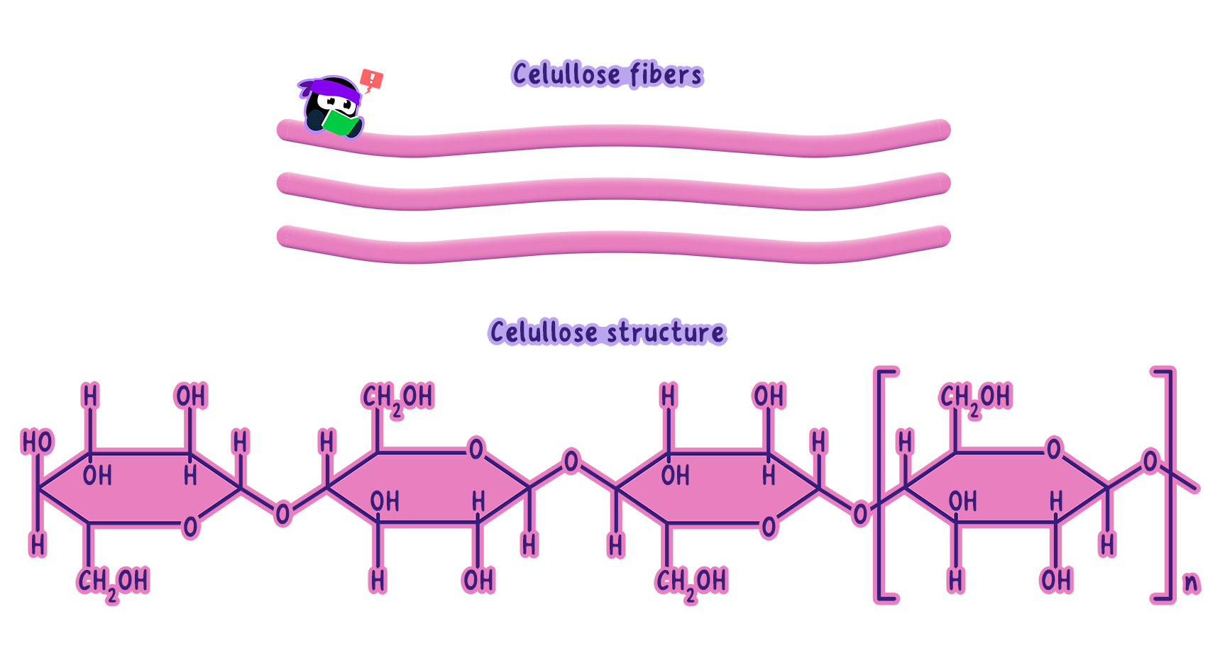
Cellulose is a polymer of β-glucose monomers linked by β-1,4-glycosidic bonds.
This alternating orientation (rotation of each subsequent glucose by 180°) yields straight, unbranched chains.
β-1,4-Glycosidic Bonds
The hydroxyl (-OH) on carbon 1 of one glucose is above the ring plane, and on carbon 4 of the next glucose it is below.
This arrangement forms long, linear cellulose chains rather than the coiled or branched structures seen in starch or glycogen.
Hydrogen Bonding
Each cellulose chain exposes -OH groups at regular intervals.
Adjacent chains form hydrogen bonds, creating microfibrils.
Microfibrils bundle into larger fibers, lending mechanical strength and stability to the plant cell wall.
Function of Cellulose in Plant Cell Walls
High Tensile Strength
Cellulose microfibrils confer resistance to stretching.
This strength prevents cells from bursting when they absorb water (turgor pressure) and helps maintain rigidplant structures.
Structural Support for Growth
The rigidity of cellulose permits plants to stand upright, develop stems, branches, and compete for light.
Enables complex plant architectures (e.g., tall trees, sprawling vines).
Cross-Linking in Cell Walls
Cellulose microfibrils are embedded in a matrix of hemicellulose and pectin.
These cross-links add both strength and flexibility, helping cell walls adapt to environmental stresses.
Example
Picture a young tree sapling swaying in the wind.
The cellulose in its cell walls enables it to withstand mechanical stress, keeping it upright and allowing it to grow.
Why Cellulose Isn't an Energy Source
Enzymatic Resistance
The β-1->4 glycosidic bonds in cellulose are difficult to hydrolyze without cellulase.
Most organisms (including humans) lack this enzyme, so they cannot break down cellulose for energy.
Durability: This resistance ensures cellulose's function as a structural molecule, rather than an energy reserve (like starch or glycogen).
Note
Hydrogen bonding is essential for cellulose's strength.
Although weak individually, the collective network of bonds creates a durablestructure.
Self Review
What structural features of cellulose allow it to form strong microfibrils?
How does this relate to its function in plant cell walls?
B1.1.7 role of glycoproteins in cell–cell recognition
Glycoproteins Are The "ID Badges" of Cells
Definition
GlycoproteinA protein with carbohydrate chains attached, which are involved in cell recognition, signalling, and adhesion.
Structure: The carbohydrate chains typically protrude from the cell surface, acting as identifiers for other cells.
Function
Cell-Cell Recognition: Like ID badges, glycoproteins enable cells to recognize and distinguish each other as "self" or "non-self."
Tissue Organization: During embryonic development, glycoproteins ensure cells attach in the correct location to form tissues.
Immune Defense: Immune cells use glycoprotein "barcodes" to detect and eliminatepathogens or infected cells.
Blood Type Compatibility: Glycoproteins on RBCs determine blood groups (e.g., ABO system).
How Does Cell-Cell Recognition Work?
Carbohydrate "Barcodes": The carbohydrate portion of glycoproteins is diverse, providing a unique marker for each cell type.
Receptor Interactions
When one cell encounters another, receptor proteins on the first cell's membrane bind to specific carbohydrate chains on the glycoprotein of the second.
This lock-and-key interaction ensures high specificity in recognizing cell identity.
Example
Consider how immune cells use glycoproteins to distinguish between the body's own cells (self) and foreign invaders (non-self).
If the glycoprotein's carbohydrate chain is unfamiliar, the immune system launches an attack to neutralize the potential threat.
Tip
Glycoproteins are dynamic.
Their carbohydrate chains can be modified in response to changing cellular conditions, allowing cells to adapt their recognition signals as needed.
ABO Blood Group System: A Case Study in Glycoprotein Recognition
Antigens on RBCs
Type O: Carries a base glycoprotein (antigen O).
Type A: Has an N-acetylgalactosamine added to antigen O.
Type B: Has a galactose added to antigen O.
Type AB: Possesses both modifications (A and B).
Why It Matters
Type O is called the "universal donor" since it lacks A or B modifications that might trigger an immune response.
Type AB is the "universal recipient," having both antigens and therefore no antibodies against A or B.
Immune Response
People naturally produce antibodies against non-self antigens.
Mismatched blood transfusions can lead to agglutination (RBC clumping) and severe complications.
How Blood Type Determines Compatibility
Type O Blood: Contains only antigen O and is often called the "universal donor" because it lacks the additional sugars (A or B) that could trigger an immune response.
Type A Blood :Contains antigen A. If transfused into someone without antigen A (e.g., type B or O individuals), the immune system will recognize it as foreign and reject the blood.
Type B Blood: Contains antigen B. Similarly, it will be rejected by individuals without antigen B (e.g., type A or O individuals).
Type AB Blood: Contains both antigens A and B and is often called the "universal recipient," as individuals with this blood type can accept blood from any group without triggering an immune response.
Warning
It's a common misconception that type O blood has "no antigens."
In fact, it has the O antigen, but this structure is less likely to provoke an immune response because it lacks the additional sugars found in A and B antigens.
Why Does the Immune System Reject Foreign Blood?
The immune system produces antibodies that specifically target foreign antigens.
For example:
A person with type A blood produces antibodies against antigen B.
A person with type B blood produces antibodies against antigen A.
A person with type O blood produces antibodies against both A and B antigens.
A person with type AB blood does not produce antibodies against either A or B antigens.
When incompatible blood is transfused, these antibodies bind to the foreign antigens, causing red blood cells to clump together (a process called agglutination).
This can block blood vessels and lead to life-threatening complications.
Self Review
Which blood type is considered the universal recipient, and why?
Broader Implications of Glycoproteins in Recognition
Immune System Regulation
Glycoproteins help immune cells detect aberrant cells (e.g., cancer, virus-infected).
Errors in recognition can prompt autoimmune disorders.
Viral Infections: Certain viruses (e.g., HIV) attach to host glycoproteins to gain entry into cells.
Autoimmune Diseases: Misinterpretation of "self" glycoproteins can lead the immune system to attack normal tissues.
Tok
How does the concept of cell-cell recognition challenge our understanding of "self" and "non-self"?
Consider its implications for organ transplantation or autoimmune diseases.
Conclusion: Glycoproteins as Gatekeepers of Cellular Communication
Glycoproteins serve a critical role in cell-cell interactions, enabling organisms to differentiate friends from foes, maintain tissue integrity, and control immune responses.
The ABO blood group system highlights how slight differences (e.g., one sugar attachment) can have major consequences (compatible vs. incompatible transfusions).
Self Review
How do glycoproteins contribute to the immune system's ability to distinguish between self and non-self?
Provide an example.
B1.1.8 hydrophobic properties of lipids
What Makes Lipids Hydrophobic?
Dominance of Non-Polar Bonds
Lipids predominantly contain long hydrocarbon chains or rings, consisting of carbon (C) and hydrogen (H) atoms.
These non-polar covalent bonds lack partial charges, meaning they cannot form hydrogen bonds with polarwater molecules.
Exclusion from Water Network
Water molecules form a cohesive hydrogen-bonded network.
Lipids are pushed out of this network, causing them to cluster and separate in aqueous environments (e.g., oil droplets in water).
Analogy
Think of water molecules as a group of friends holding hands at a party.
Lipids, being strangers who don't know the handshake, are left to cluster together on the sidelines.
Classes of Lipids and Their Hydrophobicity
Fats and Oils (Triglycerides)
Composed of three fatty acid chains bonded to glycerol.
Fats are typically solid at room temperature (more saturated chains).
Oils are liquid at room temperature (richer in unsaturated chains).
Both readily form droplets in water due to hydrophobic tails.
Waxes
Longer hydrocarbon chains than fats, highly water-repellent.
Found as protective coatings (e.g., plant cuticles, bird feathers).
Steroids
Have four fused rings of hydrocarbons (e.g., cholesterol, testosterone).
Mostly non-polar, though small polar functional groups (like -OH) can slightly reducehydrophobicity.
Note
Cholesterol, for example, has a small hydrophilic hydroxyl group, but its large hydrophobic structure dominates, making it insoluble in water.
Example
Think of butter (a fat) and olive oil (an oil). Their inability to mix with water is a direct result of their hydrophobic triglyceride structure.
Biological Significance of Hydrophobic Lipids
Energy Storage
Lipids' non-polar nature lets them form compact, water-excluding droplets in adipose tissue.
High energy density: Lipids release twice as much energy per gram as carbohydrates.
Tip
Because lipids are hydrophobic, they do not interfere with the cell's water balance, unlike hydrophilic carbohydrates.
Membrane Formation
Phospholipids are amphipathic (hydrophobic tails + hydrophilic heads).
They self-assemble into a bilayer, with tails facing inward (forming a hydrophobic core) and heads facing aqueous environments.
This bilayer forms the cell membrane, selectively controlling substance movement.
Waterproofing and Insulation
Waxes on leaves/fur repel water.
Triglycerides in adipose tissue provide thermal insulation.
Hormone Transport
Steroid hormones (hydrophobic) easily diffuse through the hydrophobic membrane core, regulating growth, metabolism, and reproduction.
Why Are Lipids Hydrophobic While Carbohydrates Are Not?
Carbohydrates
Contain many hydroxyl (-OH) groups, making them polar and water-soluble(hydrophilic).
Lipids
Primarily hydrocarbon chains/rings with few or no polar groups.
Cannot form hydrogen bonds with water -> remains insoluble (hydrophobic).
Warning
It's a common misconception that lipids are "repelled" by water.
In reality, water excludes lipids because water molecules prefer to interact with each other rather than with non-polar lipids.
Reflection
Lipids' hydrophobic nature shapes their roles in energy storage, membrane structure, waterproofing, and hormone transport.
This non-polar chemistry is central to lipid function in organisms, illustrating how molecular structure underpins biological roles.
Self Review
Why are lipids hydrophobic, and how does this property affect their solubility in water?
How does the hydrophobic nature of lipids contribute to their role as energy storage molecules?
Compare the hydrophobic properties of triglycerides, waxes, and steroids. How do these differences relate to their biological functions?
Tok
To what extent does the hydrophobic nature of lipids influence their role in the evolution of complex life forms?
For example, how might the formation of cell membranes have depended on this property?
B1.1.9 formation of triglycerides and phospholipids by condensation reactions
Triglycerides: Energy Storage Molecules
Building Blocks
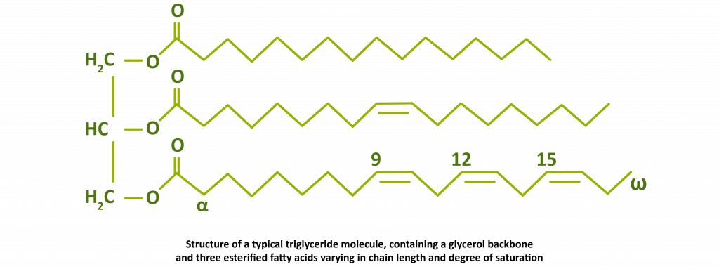
Glycerol: A three-carbon molecule with three hydroxyl (-OH) groups.
Fatty Acids: Long hydrocarbon chains ending in a carboxyl (-COOH) group.
Saturated (no C=C double bonds), monounsaturated (one double bond), or polyunsaturated(multiple double bonds).
Formation via Condensation
Each fatty acid reacts with a hydroxyl on glycerol, releasing water (H₂O) and forming an ester bond.
Glycerol can bond with three fatty acids -> triglyceride (3 ester bonds, 3 H₂O molecules released).
The carboxyl and hydroxyl groups are consumed, making the resulting triglyceride hydrophobic.
Properties and Functions
Hydrophobic: Insoluble in water, forming compact droplets for energy storage.
Energy Density: Higher energy content per gram than carbohydrates.
Insulation and Protection: In animals, stored in adipose tissue to cushion organs and retain heat.
Condensation Reactions in Lipid Formation
Water Release: Each ester bond formed expels 1 H₂O molecule.
Energy Requirement: Formation typically needs energy input (e.g., from ATP) to build covalent bonds.
Covalent Bond Stability: Ester bonds provide the robust link between glycerol and fatty acids (or phosphate group).
Importance of Triglycerides and Phospholipids
Triglycerides
Long-Term Energy Storage: Higher energy density vs. carbohydrates.
Protection/Insulation: Fat deposits help organisms maintain thermal balance and protect organs.
Phospholipids
Membrane Formation: Essential for cell boundary and compartmentalization.
Membrane Fluidity: Adjusted by fatty acid saturation, key to proper cellular function.
Tip
Triglycerides store approximately twice as much energy per gram as carbohydrates, making them an efficient energy reserve for organisms.
Phospholipids Are The Building Blocks of Membranes
Phospholipids, another class of lipids, are critical for forming cell membranes.
They share similarities with triglycerides but have a distinct feature: one fatty acid chain is replaced by a phosphate group.
The Structure of Phospholipids
Glycerol Backbone: Like triglycerides, phospholipids have a glycerol molecule.
Two Fatty Acids: Two fatty acids are attached to the glycerol via ester bonds.
Phosphate Group: The third hydroxyl group of glycerol is linked to a phosphate group, which is often bonded to additional polar molecules.
Amphipathic Nature
Phospholipids are amphipathic, meaning they have both hydrophobic and hydrophilic regions:
Hydrophobic Tails: The two fatty acid chains are nonpolar and repel water.
Hydrophilic Head: The phosphate group, often carrying a charge, interacts with water.
This dual nature allows phospholipids to self-assemble into bilayers, the structural basis of cell membranes.
In these bilayers, the hydrophobic tails face inward, shielded from water, while the hydrophilic heads face outward toward the aqueous environment.
Example
Think of a soap bubble.
Soap molecules have hydrophilic heads that interact with water and hydrophobic tails that avoid it.
Similarly, phospholipids organize into bilayers to create a stable barrier between the inside and outside of a cell.
The Role of Condensation Reactions in Lipid Formation
The synthesis of both triglycerides and phospholipids depends on condensation reactions.
These reactions are a cornerstone of biochemistry, used to build macromolecules such as carbohydrates, proteins, and nucleic acids.
Key Features of Condensation Reactions
Water Release: Each bond formation produces a molecule of water.
Energy Requirement: The process requires energy, often supplied by ATP.
Covalent Bonds: Strong covalent bonds, such as ester bonds, stabilize the resulting molecules.
Note
Condensation reactions are the reverse of hydrolysis reactions, which break down macromolecules by adding water.
Why Are Triglycerides and Phospholipids Important?
Triglycerides
Energy Storage: Triglycerides are a dense energy source, storing more energy per gram than carbohydrates.
Insulation and Protection: In animals, triglycerides stored in adipose tissue provide thermal insulation and cushion vital organs.
Phospholipids
Membrane Structure: Phospholipid bilayers form the foundation of cell membranes, regulating the movement of substances in and out of cells.
Membrane Fluidity: The degree of saturation in the fatty acid tails influences membrane fluidity, which is crucial for cellular function.
Self Review
Can you identify how the amphipathic nature of phospholipids contributes to their role in forming cell membranes?
Reflection and Connections
The contrast between triglycerides (energy reservoirs) and phospholipids (membrane builders) highlights the versatility of lipid chemistry.
Condensation reactions underpin not just lipid synthesis, but also the formation of carbohydrates, proteins, and nucleic acids--showcasing a unifying theme in biochemistry.
By appreciating how molecular structure drives function, we see potential insights for materials science, biotech, and medicine--fields that may harness lipid properties for innovative applications.
Tok
How does the hydrophobic nature of lipids influence their role in biological systems?
Consider its impact on energy storage, membrane formation, and even the evolution of life in aqueous environments.
B1.1.10 difference between saturated, monounsaturated and polyunsaturated fatty acids
The Structure of Fatty Acids: A Quick Overview
All fatty acids share a common structure: a long hydrocarbon chain (a chain of carbon atoms bonded to hydrogen) with a carboxyl group (-COOH) at one end. However, these chains can vary in two key ways:
Length: The number of carbon atoms in the chain (typically between 14 and 20 in biological systems).
Saturation: The number and position of double bonds between carbon atoms.
Hint
The term "saturation" refers to how many hydrogen atoms are bonded to the carbon chain:
Saturated fatty acids: No double bonds. Every carbon atom is fully "saturated" with hydrogen atoms.
Unsaturated fatty acids: One or more double bonds between carbon atoms. These are further divided into:
Monounsaturated fatty acids (MUFA): Contain one double bond.
Polyunsaturated fatty acids (PUFA): Contain two or more double bonds.
Saturated Fatty Acids: The Straight Chains
Characteristics
No double bonds: A rigid, linear structure.
High melting point: Tightly packed molecules require more energy (heat) to separate, making saturated fats solid at room temperature.
Sources: Found in animal fats (e.g., butter, lard) and some plant oils like coconut oil.
Example
Stearic acid: A saturated fatty acid with 18 carbon atoms and no double bonds.
Biological Role
Saturated fats are ideal for long-term energy storage and thermal insulation in animals.
For example, marine mammals like whales rely on blubber, rich in saturated fatty acids, to retain heat in cold waters.
Warning
Don't assume "saturated" automatically means "unhealthy."
While excessive intake is linked to health risks, saturated fats play essential roles in energy storage and insulation.
Monounsaturated Fatty Acids: The First Bend
Analogy
Now, considere a rope with a single knot.
This is how monounsaturated fatty acids (MUFA) appear, a single double bond introduces a "kink" in the chain, preventing tight packing.
Characteristics
One double bond: The kink reduces the molecule's ability to stack.
Moderate melting point: MUFAs are typically liquid at room temperature but may solidify when cooled.
Sources: Found in plant-based oils like olive oil, avocado oil, and nuts.
Example
Oleic acid: A monounsaturated fatty acid with 18 carbon atoms and one double bond.
Biological Role
MUFAs are common in plant oils, which are liquid at room temperature.
These oils provide energy storage in seeds and are easier to mobilize during germination.
Analogy
Think of olive oil: its liquid state at room temperature is due to the "kink" introduced by monounsaturated fatty acids.
Self Review
Why does a single double bond in monounsaturated fatty acids prevent them from being solid at room temperature?
Polyunsaturated Fatty Acids: The Curvy Chains
Analogy
Finally, think of a rope with multiple knots.
This is the structure of polyunsaturated fatty acids (PUFA), with two or more double bonds creating multiple "kinks" in the chain.
Characteristics
Two or more double bonds: The chain is highly curved and cannot pack tightly.
Low melting point: These fats remain liquid even at low temperatures.
Sources: Found in fish oils (e.g., salmon, mackerel) and plant oils like sunflower and flaxseed oil.
Example
Linoleic acid: A PUFA with 18 carbon atoms and two double bonds.
Omega-3 and Omega-6 fatty acids: Essential PUFAs required for brain function and cell membrane integrity.
Biological Role
Polyunsaturated fats are common in plants and cold-water fish.
Plants store them in seeds for energy, while fish benefit from their fluidity, which keeps cell membranes functional in cold environments.
Tip
Omega-3 fatty acids are abundant in cold-water fish like salmon.
Including them in your diet supports brain health and reduces inflammation.
Melting Points and Energy Storage: Why It Matters
The melting point of a fatty acid depends on how tightly the molecules can pack:
Saturated fats: Straight chains pack tightly, forming solid fats (e.g., butter).
Monounsaturated fats: A single kink reduces packing, making oils semi-liquid (e.g., olive oil).
Polyunsaturated fats: Multiple kinks prevent packing, keeping oils liquid (e.g., sunflower oil).
How Double Bonds Affect Melting Point
Double bonds in fatty acids introduce kinks in the hydrocarbon chain, disrupting their ability to pack tightly together.
This reduces the strength of van der Waals interactions between molecules.
As a result, unsaturated fatty acids with one or more double bonds require less energy to separate, leading to lower melting points.
The more double bonds present, the greater the disruption, making polyunsaturated fatty acids liquid at lower temperatures compared to monounsaturated and saturated fatty acids.
Case_study
Interesting Science Behind Double Bonds
Double bonds in fatty acids are chemically reactive, making unsaturated fats more prone to oxidation.
This is why polyunsaturated oils, like flaxseed oil, tend to go rancid faster than saturated fats like butter.
Adding antioxidants such as vitamin E can delay this process, which is why many oils include them as preservatives.
Example
Sunflower oil is liquid at room temperature because it is rich in polyunsaturated fatty acids.
In contrast, butter is solid due to its high saturated fat content.
The Bigger Picture: Health and Function
The type of fatty acid in your diet affects health:
Saturated fats: Excessive intake raises LDL cholesterol, increasing the risk of heart disease.
Monounsaturated fats: Improve heart health by lowering LDL cholesterol while maintaining HDL cholesterol.
Polyunsaturated fats: Essential for brain function but prone to oxidation, leading to rancidity.
Tok
How do cultural and economic factors influence the types of fats consumed globally, and
How should dietary guidelines adapt to these differences?
Self Review
What structural feature distinguishes saturated, monounsaturated, and polyunsaturated fatty acids?
How does the number of double bonds affect the melting point of a fatty acid?
Why are unsaturated fats more common in plants, while saturated fats dominate in animals?
B1.1.11 triglycerides in adipose tissues for energy storage and thermal insulation
he Structure of Fatty Acids: A Quick Overview
All fatty acids share a common structure: a long hydrocarbon chain (a chain of carbon atoms bonded to hydrogen) with a carboxyl group (-COOH) at one end. However, these chains can vary in two key ways:
Length: The number of carbon atoms in the chain (typically between 14 and 20 in biological systems).
Saturation: The number and position of double bonds between carbon atoms.
Hint
The term "saturation" refers to how many hydrogen atoms are bonded to the carbon chain:
Saturated fatty acids: No double bonds. Every carbon atom is fully "saturated" with hydrogen atoms.
Unsaturated fatty acids: One or more double bonds between carbon atoms. These are further divided into:
Monounsaturated fatty acids (MUFA): Contain one double bond.
Polyunsaturated fatty acids (PUFA): Contain two or more double bonds.
Saturated Fatty Acids: The Straight Chains
Characteristics
No double bonds: A rigid, linear structure.
High melting point: Tightly packed molecules require more energy (heat) to separate, making saturated fats solid at room temperature.
Sources: Found in animal fats (e.g., butter, lard) and some plant oils like coconut oil.
Example
Stearic acid: A saturated fatty acid with 18 carbon atoms and no double bonds.
Biological Role
Saturated fats are ideal for long-term energy storage and thermal insulation in animals.
For example, marine mammals like whales rely on blubber, rich in saturated fatty acids, to retain heat in cold waters.
Warning
Don't assume "saturated" automatically means "unhealthy."
While excessive intake is linked to health risks, saturated fats play essential roles in energy storage and insulation.
Monounsaturated Fatty Acids: The First Bend
Analogy
Now, considere a rope with a single knot.
This is how monounsaturated fatty acids (MUFA) appear, a single double bond introduces a "kink" in the chain, preventing tight packing.
Characteristics
One double bond: The kink reduces the molecule's ability to stack.
Moderate melting point: MUFAs are typically liquid at room temperature but may solidify when cooled.
Sources: Found in plant-based oils like olive oil, avocado oil, and nuts.
Example
Oleic acid: A monounsaturated fatty acid with 18 carbon atoms and one double bond.
Biological Role
MUFAs are common in plant oils, which are liquid at room temperature.
These oils provide energy storage in seeds and are easier to mobilize during germination.
Analogy
Think of olive oil: its liquid state at room temperature is due to the "kink" introduced by monounsaturated fatty acids.
Self Review
Why does a single double bond in monounsaturated fatty acids prevent them from being solid at room temperature?
Polyunsaturated Fatty Acids: The Curvy Chains
Analogy
Finally, think of a rope with multiple knots.
This is the structure of polyunsaturated fatty acids (PUFA), with two or more double bonds creating multiple "kinks" in the chain.
Characteristics
Two or more double bonds: The chain is highly curved and cannot pack tightly.
Low melting point: These fats remain liquid even at low temperatures.
Sources: Found in fish oils (e.g., salmon, mackerel) and plant oils like sunflower and flaxseed oil.
Example
Linoleic acid: A PUFA with 18 carbon atoms and two double bonds.
Omega-3 and Omega-6 fatty acids: Essential PUFAs required for brain function and cell membrane integrity.
Biological Role
Polyunsaturated fats are common in plants and cold-water fish.
Plants store them in seeds for energy, while fish benefit from their fluidity, which keeps cell membranes functional in cold environments.
Tip
Omega-3 fatty acids are abundant in cold-water fish like salmon.
Including them in your diet supports brain health and reduces inflammation.
Melting Points and Energy Storage: Why It Matters
The melting point of a fatty acid depends on how tightly the molecules can pack:
Saturated fats: Straight chains pack tightly, forming solid fats (e.g., butter).
Monounsaturated fats: A single kink reduces packing, making oils semi-liquid (e.g., olive oil).
Polyunsaturated fats: Multiple kinks prevent packing, keeping oils liquid (e.g., sunflower oil).
How Double Bonds Affect Melting Point
Double bonds in fatty acids introduce kinks in the hydrocarbon chain, disrupting their ability to pack tightly together.
This reduces the strength of van der Waals interactions between molecules.
As a result, unsaturated fatty acids with one or more double bonds require less energy to separate, leading to lower melting points.
The more double bonds present, the greater the disruption, making polyunsaturated fatty acids liquid at lower temperatures compared to monounsaturated and saturated fatty acids.
Case_study
Interesting Science Behind Double Bonds
Double bonds in fatty acids are chemically reactive, making unsaturated fats more prone to oxidation.
This is why polyunsaturated oils, like flaxseed oil, tend to go rancid faster than saturated fats like butter.
Adding antioxidants such as vitamin E can delay this process, which is why many oils include them as preservatives.
Example
Sunflower oil is liquid at room temperature because it is rich in polyunsaturated fatty acids.
In contrast, butter is solid due to its high saturated fat content.
The Bigger Picture: Health and Function
The type of fatty acid in your diet affects health:
Saturated fats: Excessive intake raises LDL cholesterol, increasing the risk of heart disease.
Monounsaturated fats: Improve heart health by lowering LDL cholesterol while maintaining HDL cholesterol.
Polyunsaturated fats: Essential for brain function but prone to oxidation, leading to rancidity.
Tok
How do cultural and economic factors influence the types of fats consumed globally, and
How should dietary guidelines adapt to these differences?
Self Review
What structural feature distinguishes saturated, monounsaturated, and polyunsaturated fatty acids?
How does the number of double bonds affect the melting point of a fatty acid?
Why are unsaturated fats more common in plants, while saturated fats dominate in animals?
B1.1.12 formation of phospholipid bilayers
Amphipathic Nature of Phospholipids
Phospholipids are remarkable molecules with a dual personality: they are amphipathic, this unique structure consists of:
Hydrophilic Head: The phosphate group in the "head" region is polar and interacts readily with water.
Hydrophobic Tails: The two fatty acid chains in the "tail" region are non-polar and avoid water.
This combination allows phospholipids to interact with both water and other lipids, making them perfect for forming cell membranes.
Definition
Amphipathic Amphipathic: A molecule that has both hydrophilic (water-attracting) and hydrophobic (water-repelling) regions.
Analogy
Imagine a phospholipid as a person standing on the beach, with their head in the water (hydrophilic) and their feet buried in the sand (hydrophobic).
This dual preference explains why phospholipids behave the way they do in water.
Why is Amphipathic Nature Important?
When phospholipids encounter water, their amphipathic nature drives them to self-assemble into specific structures.
The hydrophilic heads face the water, while the hydrophobic tails hide from it. This results in the formation of a phospholipid bilayer, the key structural component of cell membranes.
Tip
Think of the bilayer as a sandwich: the hydrophilic heads are like the bread, facing outward, while the hydrophobic tails are the filling, tucked safely inside.
Formation of the Phospholipid Bilayer
Spontaneous Assembly in Water
When phospholipids are exposed to water, they spontaneously organize into a bilayer. Here's how it works:
Hydrophilic Heads Face Outward: The phosphate heads interact with water molecules on both sides.
Hydrophobic Tails Face Inward: The fatty acid tails cluster together, avoiding contact with water.
This arrangement minimizes energy, creating a stable structure.
Biological Significance of the Phospholipid Bilayer
Role in Cell Membranes
The phospholipid bilayer forms the backbone of all cell membranes. It provides:
Compartmentalization: Separates the cell's internal environment from the external environment, allowing specialized processes to occur.
Selective Permeability: Allows small, non-polar molecules like oxygen and carbon dioxide to pass through, while blocking larger or charged molecules.
Fluidity: Enables the membrane to remain flexible, accommodating changes in cell shape and facilitating the movement of proteins.
Example
oxygen molecules diffuse freely across the bilayer, enabling cellular respiration, while ions like sodium require specialized protein channels to cross.
Practical Example: Formation of Liposomes
Liposomes are spherical vesicles formed when phospholipids are mixed with water.
They mimic natural cell membranes, with a bilayer enclosing an aqueous core.
Liposomes are widely used in medicine for drug delivery because:
The hydrophilic core can carry water-soluble drugs.
The hydrophobic bilayer can carry lipid-soluble drugs.
Example
For instance, liposomes are used to deliver chemotherapy drugs directly to cancer cells, reducing side effects on healthy tissues.
Note
Liposomes demonstrate how the self-assembly of phospholipids can be harnessed for practical applications, bridging biology and technology.
Common Misunderstandings About Phospholipid Bilayers
Note
Hydrophobic tails are not actively "repelled" by water, as they simply prefer interactions with other non-polar molecules over interactions with water.
Warning
The bilayer is not a static structure. Phospholipids and proteins within it are constantly moving, contributing to the membrane's dynamic nature.
Reflection and Broader Implications
Tok
How does the amphipathic nature of phospholipids illustrate the relationship between structure and function in biology?
Can you think of other examples in biology where molecular structure determines function?
Self Review
Why do phospholipids spontaneously form bilayers in water?
How does the bilayer's structure contribute to its function as a selective barrier?
What factors influence the fluidity of the phospholipid bilayer?
B1.1.13 ability of non-polar steroids to pass through the phospholipid bilayer
Introduction to the Phospholipid Bilayer
The phospholipid bilayer is a fundamental component of all cell membranes, acting as a selective barrier that regulates the passage of substances into and out of cells.
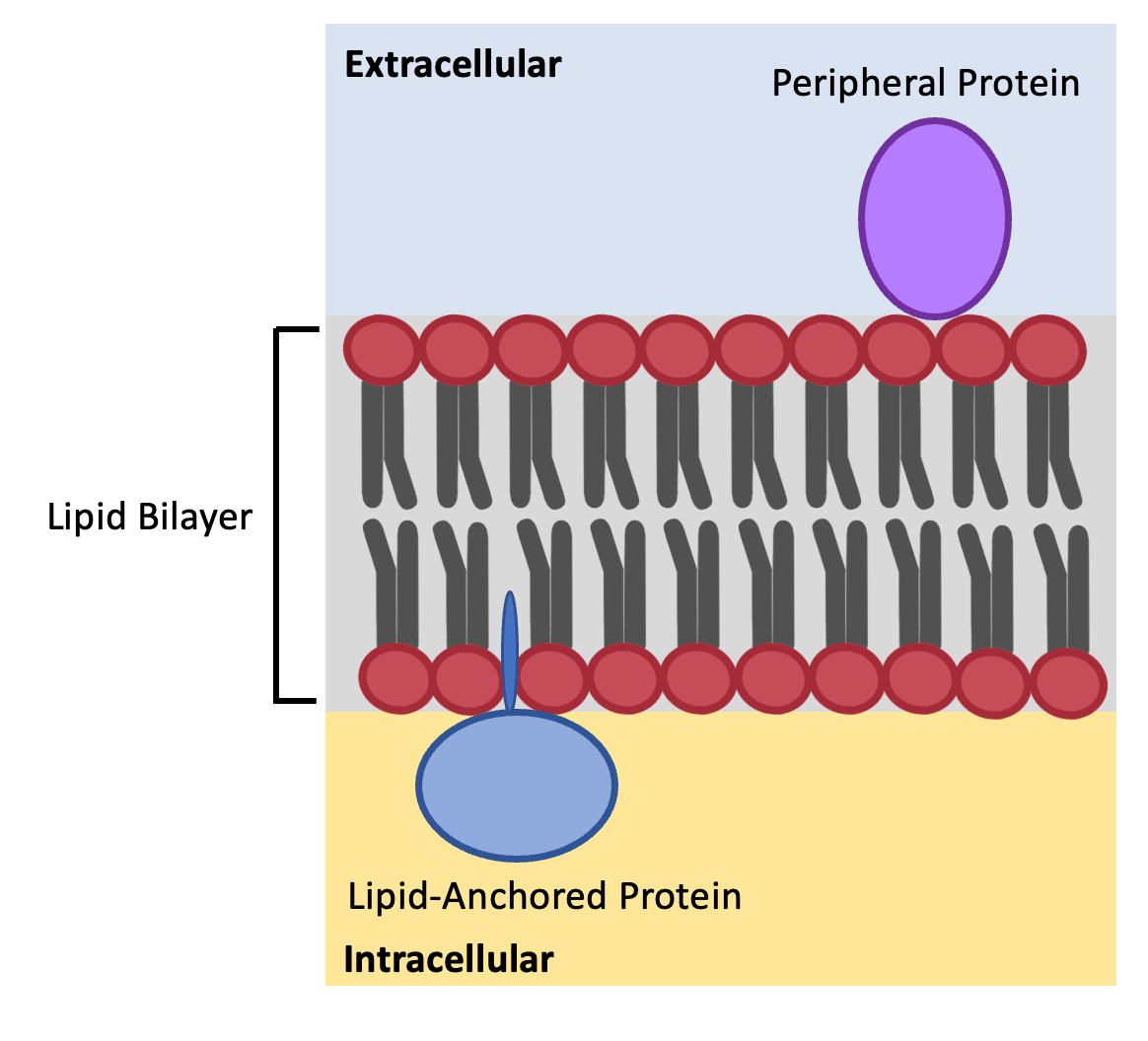
It is composed of phospholipid molecules arranged in two layers, with their hydrophilic (water-attracting) heads facing outward and hydrophobic (water-repelling) tails facing inward.
This unique structure creates a semi-permeable membrane that allows certain molecules to pass through while blocking others.
Think of the phospholipid bilayer like a security fence with water-loving spikes on the outside and water-fearing bars on the inside. Only certain things can slip through the gaps.