Anatomy & Physiology of Hearing Study Notes
Anatomy & Physiology of Hearing
Pinna
Pinna (auricle) landmarks include:
- Helix and antihelix
- Concha (entrance to External Auditory Meatus, EAM)
- Scaphoid fossa
- Tragus
- Antitragus
- LobuleFunction: Channels sound waves into the ear canal.
Tissue: Covered with epithelial tissue invested with fine hair.
External Auditory Meatus (EAM)
Length: Approximately 2.5 cm long from the concha.
Lining: Lined with epithelial tissue (same tissue as pinna).
Composition:
- Outer 1/3 is the Cartilaginous Meatus (cartilage, courses upwards).
- Inner 2/3 is the Osseous Meatus (temporal bone, courses downwards) which protects the deeper parts of the ear.Function: Channels sound waves towards the tympanic membrane (ear drum).
Additional Function: Hair and cerumen (earwax) trap particles to protect the ear.
Tympanic Membrane (TM)
Role: Acts as a boundary between the outer and middle ear.
Size: Approximately 55 mm² in area.
Layers of the TM:
- Outer Layer: Continuation of the EAM and pinna lining (epithelial tissue).
- Intermediate Layer: Composed of fibrous (connective) tissue that enhances strength and flexibility.
- Inner Layer: Continuous with middle ear mucosa (epithelial tissue).Key Landmarks:
- Umbo: Attachment point of the TM to the malleus.
- Manubrium: Visible behind TM, aids in orientation.Function: The TM vibrates with sound waves and transfers these vibrations to the ossicles in the middle ear.
Middle Ear Ossicles
Ossicular Chain: Composed of three small bones:
- Malleus (Hammer)
- Largest ossicle.
- Attaches to the tympanic membrane.
- Transmits sound vibrations from the TM to the incus.
- Incus (Anvil)
- The middle bone acts as a bridge between malleus and stapes.
- Transmits sound vibrations along the chain.
- Stapes (Stirrup)
- Smallest bone in the body.
- Connects to the oval window of the inner ear through its footplate.
- Final point of sound transmission in the middle ear.
Middle Ear Muscles
Tensor Tympani
- Function: Dampens loud sounds by pulling the malleus inward, thus tightening the tympanic membrane.
- More active in response to self-generated sounds (e.g., chewing, speaking).
- Innervation: Mandibular branch of the Trigeminal Nerve (CN V).Stapedius
- Function: Pulls the stapes away from the oval window, stiffening the ossicular chain and reducing movement of the stapes to protect the inner ear from loud sounds.
- This protective mechanism is termed the "acoustic reflex", minimizing potential damage from loud noises.
- Innervation: Facial Nerve (CN VII).Both the tensor tympani and stapedius work together to manage sound vibrations entering the inner ear.
Inner Ear
Contains structures for hearing and balance.
Main parts include:
- Cochlea (hearing)
- Semicircular Canals (balance)Function of Cochlea: The hearing organ, where sound vibrations are converted into electrical signals for the brain.
Sound vibrations enter through the oval window connected to the stapes.
Vibrations create waves in the cochlear fluid; different frequencies activate specific regions in the cochlea.
Presbycusis: Age-related hearing loss, commonly characterized by a gradual decrease in the ability to hear high-frequency sounds, affecting many individuals as they grow older.
trouble with fricatives
Cochlear Anatomy
Inside the cochlea lies the Organ of Corti which contains sensory hair cells (stereocilia).
Movement of cochlear fluid bends these hair cells, which leads to the release of neurotransmitters.
These neurotransmitters activate the auditory nerve (part of CN VIII).
Auditory Pathway
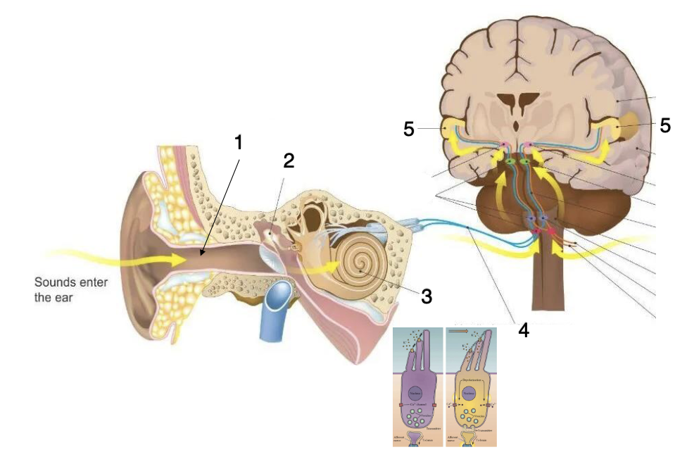
The auditory nerve carries signals through the brainstem, which helps process basic sound information.
Signals reach the primary auditory cortex for interpretation of sound as speech, music, or environmental noise.
Auditory Transduction
Outer Ear: Collects and directs sound waves to the eardrum.
Middle Ear: The tympanic membrane vibrates with sound waves, and tiny bones (ossicles) enhance strength through impedance matching, transmitting the vibrations to the oval window.
Inner Ear: Sound vibrations reach the cochlea, creating waves in cochlear fluid that cause hair cells to bend, triggering neurotransmitter release and activating the auditory nerve fibers.
Initial Signal Processing: Signals travel along the auditory nerve to the brainstem for initial sound analysis, aiding in sound localization and pitch recognition.
Advanced Processing: Signals are processed in the primary auditory cortex, where they are interpreted as recognizable sounds such as speech or music.