1.6 Plasma Membrane
Why Cells Need a Membrane?
Separate interior from exterior - protection, support
Very thin boundary
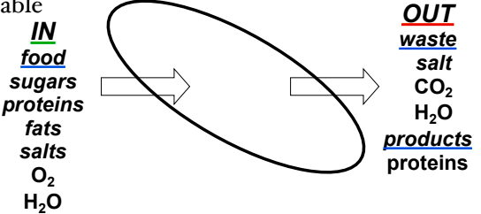
Allows some materials to cross more easily - selectively permeable
 What is it Made of?
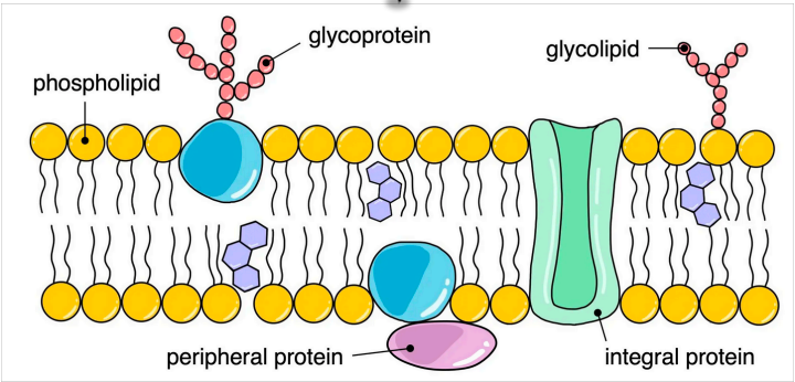
Phospholipids
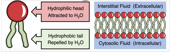
Spontaneously arrange into bilayer (two parallel layers)
Amphipathic due to lipophilic and hydrophilic regions
Bilayer held together by weal hydrophobic interactions between tails
 Proteins
Proteins may be permanently or temporarily attached
Diverse in structure, position, and function
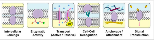
Functions include: junctions, enzymes, transport, recognition, anchorage, and transduction
 Integral Proteins – permanently attached to membrane and are typically transmembrane (span across bilayer)
Peripheral Proteins – temporally attached to noncovalent interactions and associate with one surface
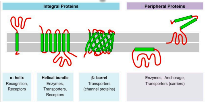
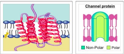
Non-polar amino acids are located within membrane
Anchors protein into membrane
Polar amino acids located on outer surface of membrane
Extend into extracellular fluid & cytosol
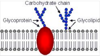
Carbohydrates
Glycolipid
lipid with a carbohydrate chain attached
help cell to recognize other cells of body
Glycoprotein
protein with carbohydrate chain attached
help in cell to cell communication and transport
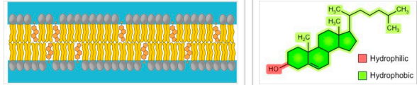
Cholesterol
Amphipathic molecule found in animal membranes
Immobilizes outer surface - reducing fluidity
Makes less permeable to small water-soluble molecules
Separates phospholipid tails - prevents crystallization
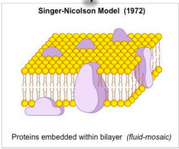
 Fluid Mosaic Model
Singer-Nicolson Model
Membranes are fluid - not fixed in position (can adopt
amorphous shapes)
Fluid - phospholipids can move
Mosaic - embedded with proteins
 Fluidity
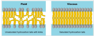
Fluidity increases with temperature and shorter fatty acid tails - changes to kinetic energy and decreased viscosity
Fluidity increases with double bonds - harder to pack
At high temps cholesterol stabilizes membrane, at low temps it prevents clustering of phospholipids
 Davson-Danielli Model
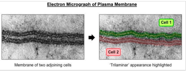
Lipo-protein sandwich’, lipid layer sandwiched between two protein layers - trilaminar appearance
Falsification of this model led to Singer-Nicolson model
Proteins are embedded within lipid bilayer rather than existing as separate layer