Central and Peripheral Nervous Systems Flashcards
6.15 Central Nervous System: Brain
Terminology:
Axon: extension of a neuron that transmits signals away from the cell body.
Nerve: bundle of axons located in the peripheral nervous system (PNS), facilitating communication between the CNS and the rest of the body.
Pathway/Tract/Commissure(left/right brain link): These are bundles of axons located within the central nervous system (CNS), serving as routes for neural communication.
CNS Pathways:
Long Neural Pathways: directly transmit information between the brain and the spinal cord, enabling rapid communication for essential functions.
Multisynaptic Pathways: involve complex neural processing through multiple synapses, allowing for intricate integration of information.
due to more synapses, multisynaptic pathways perform more complex neural processing
Information Flow: The flow of information within the nervous system is essential for maintaining homeostasis by coordinating various physiological processes.
Ganglia vs. Nuclei:
Ganglia: clusters of neuron cell bodies located in the peripheral nervous system (PNS), often serving as relay points for nerve signals.
Nuclei: clusters of neuron cell bodies located within the central nervous system (CNS), playing key roles in neural processing and integration.
Brain Development: The brain develops from a neural tube, with the anterior portion differentiating into the forebrain, midbrain, and hindbrain.
Brain Regions & Subdivisions:
Forebrain: The forebrain consists of the cerebrum, responsible for higher-level cognitive functions, and the diencephalon, which includes the thalamus and hypothalamus.
Midbrain: The midbrain is a single division involved in motor control, sensory processing, and relaying information between the forebrain and hindbrain.
Hindbrain: The hindbrain includes the pons, medulla, and cerebellum, which are responsible for vital functions such as respiration, movement coordination, and balance.
Brainstem: The brainstem comprises the pons, medulla, and midbrain, serving as a critical relay center for information between the brain, spinal cord, and peripheral nervous system.
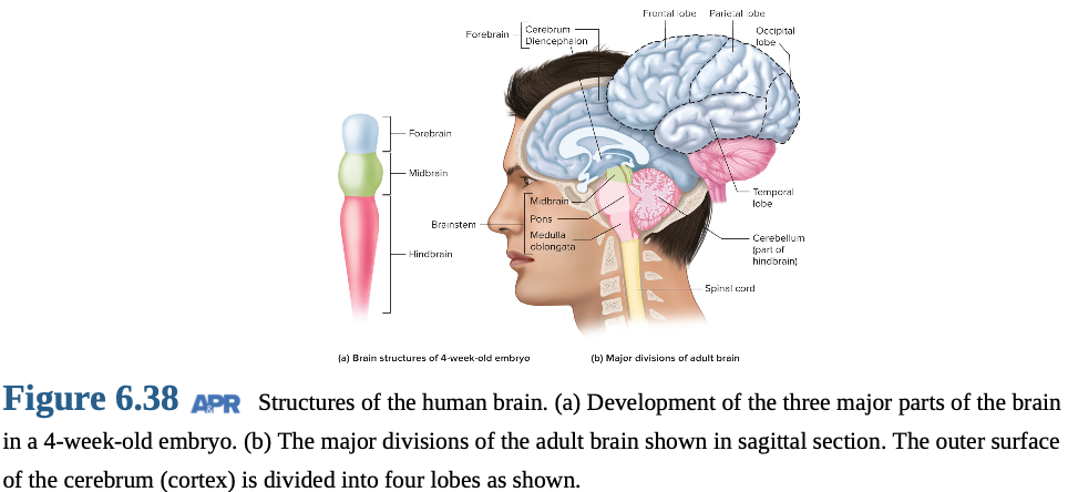
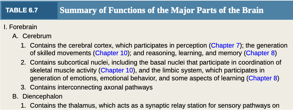
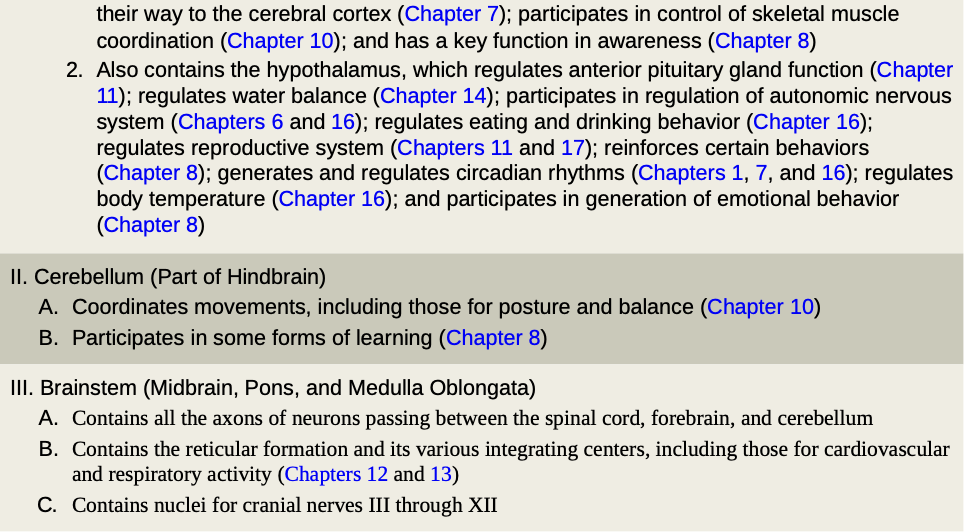
Table 6.7: Brain Functions Summary
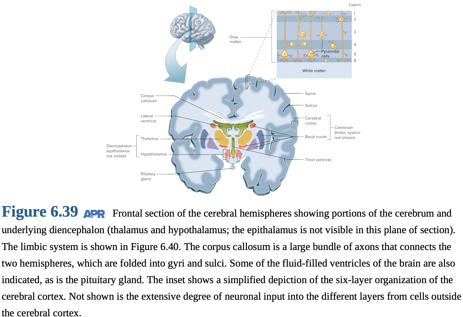
Cerebral Ventricles: four interconnected cavities filled with fluid and provide support for brain
Cerebrum: largest part of the forebrain; includes the cerebral hemispheres and other structures.
Cerebral Hemispheres:
Cerebral Cortex: outer layer of the cerebrum, composed of gray matter, and is responsible for higher cognitive functions.
also contains white matter, which contains myelinated axons that facilitate communication between different brain regions.
Subcortical Nuclei: Clusters of gray matter located beneath the cerebral cortex.
hypothalamus, thalamus, basal ganglia
*those 3 above r tracts (bundles of neurons)
Corpus Callosum: large bundle of axons (commisure) that connects the right and left cerebral hemispheres, enabling communication between them.
Cerebral Cortex
Lobes: four lobes: frontal, parietal, occipital, and temporal, each associated with specific functions.
Folding: folding of the cerebral cortex increases its surface area, allowing for more complex neural processing.
Gyri: Ridges on the surface of the cerebral cortex.
Sulci: Grooves on the surface of the cerebral cortex.
Cell Layers: T6 distinct layers, each with unique cellular composition and functions.
Cell Types:
Pyramidal Cells: primary output cells of the cerebral cortex, responsible for transmitting signals to other brain regions.
Nonpyramidal Cells: primarily function as inputs to, and for processing of information within, the cortex.
Specialization: The specialization of different cortical areas allows for increased neuron integration and efficient processing of information.
Evolution: The complexity of the cerebral cortex parallels the evolutionary development of cognitive abilities.
more evolved → more layers
Basal Nuclei
Subcortical Nuclei: Heterogeneous gray matter groups located beneath the cerebral hemispheres.
Basal Nuclei: play a critical role in movement and posture control, as well as influencing behavior.
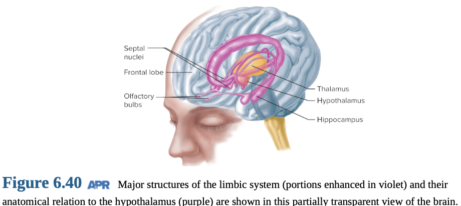
Limbic System
Functional System: includes parts of the frontal lobe, temporal lobe, thalamus, and hypothalamus; works together to regulate emotion and memory.
fiber pathways connect parts
Connections: limbic system is interconnected with various parts of the central nervous system (CNS), enabling its diverse functions.
Functions: The limbic system is involved in learning, emotion, behavior, and visceral/endocrine functions.
Forebrain: The Diencephalon
Diencephalon: This region of the brain includes the thalamus, hypothalamus, and epithalamus, each with distinct functions.
Thalamus:
relay and integrating center for inputs to the cerebral cortex.
It plays a role in arousal and attention, helping to focus cognitive resources.
Filters sensory information, prioritizing relevant stimuli.
Hypothalamus:
serves as a command center for coordinating homeostatic regulation.
It regulates preservation behaviors, ensuring survival.
Pituitary regulation: The hypothalamus controls the release of hormones from the pituitary gland, influencing various bodily functions.
Epithalamus: The epithalamus contains the pineal gland, which regulates circadian rhythms by secreting melatonin.
Hindbrain: The Cerebellum
Cerebellum:
It has a cerebellar cortex (outer layer) + deeper cell clusters.
Coordinates movements, and maintains posture/balance.
Receives information from muscles, joints, skin, eyes, viscera, and brain.
Involved in learning motor skills.
Brainstem: The Midbrain, Pons, and Medulla Oblongata
Brainstem:
Axons relay signals between the brain and spinal cord.
Contains the reticular formation.
Reticular Formation:
Integrates input from ALL regions of CNS.
Involved in motor control, cardio/resp control, sleep/wake cycles, and attention.
Uses biogenic amine neurotransmitters to modulate brain activity.
Regulates arousal and wakefulness.
Directs attention to relevant stimuli.
Receives spinal cord influence.
Interacts with the forebrain, spinal cord, and cerebellum.
Brainstem nuclei: Contains centers for cardio, resp, swallowing, and vomiting reflexes.
Eye-movement control, and helps maintain body orientation.
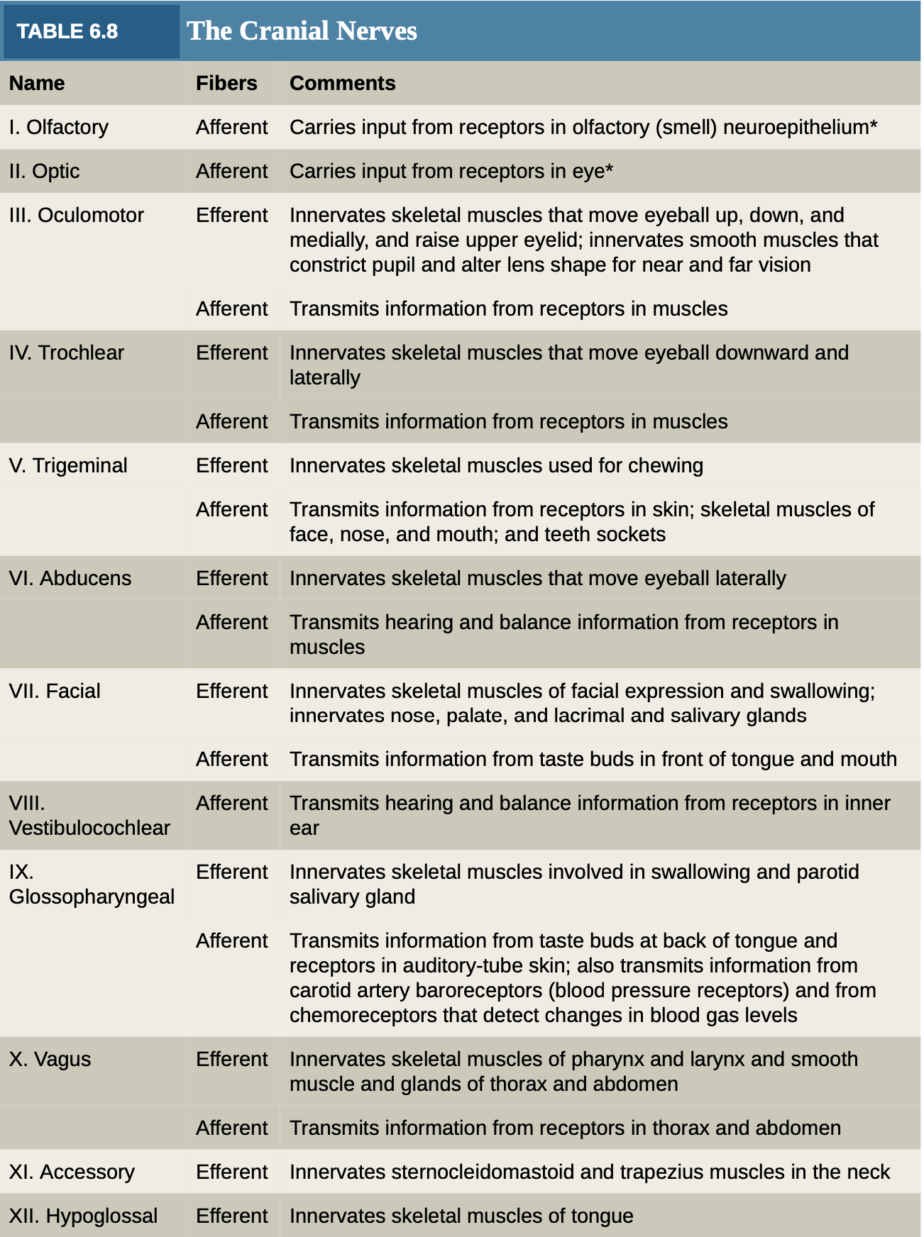
Cranial nerve nuclei (10/12) are located in the brainstem.
6.16 Central Nervous System: Spinal Cord
Location: The spinal cord is located within the vertebral column, providing protection and support.
Structure: It is a cylinder of soft tissue that extends from the brainstem to the lumbar region of the vertebral column.
Gray Matter: butterfly-shaped and contains interneurons, efferent neuron bodies, afferent axons, and glial cells.
Dorsal Horns: located at the back of the spinal cord and primarily process sensory information.
Ventral Horns: located at the front of the spinal cord and contain motor neurons that control muscle movement.
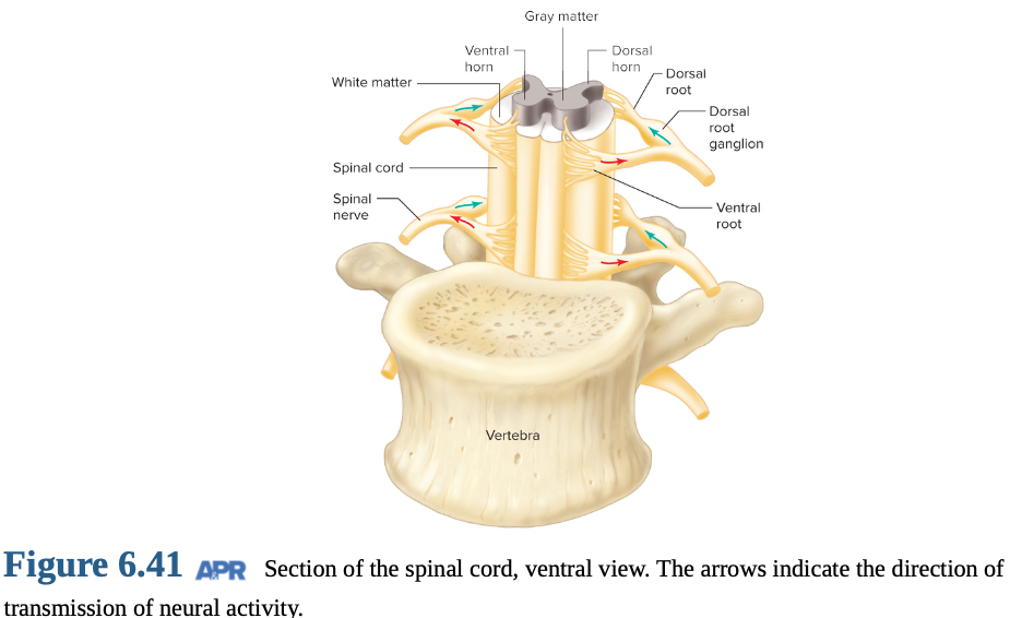
Dorsal Roots: Afferent (sensory) axons enter the spinal cord via the dorsal roots, with cell bodies located in the spinal ganglia.
Ventral Roots: Efferent (motor) axons leave the spinal cord via the ventral roots, carrying motor commands to muscles and glands.
Spinal Nerve: The dorsal and ventral roots combine to form a spinal nerve, which carries both sensory and motor information.
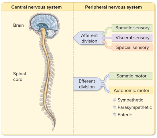
6.17 Peripheral Nervous System
Function: The peripheral nervous system (PNS) functions to transmit signals between the central nervous system (CNS) and the rest of the body.
Nerves: Nerves are bundles of axons that transmit signals throughout the body.
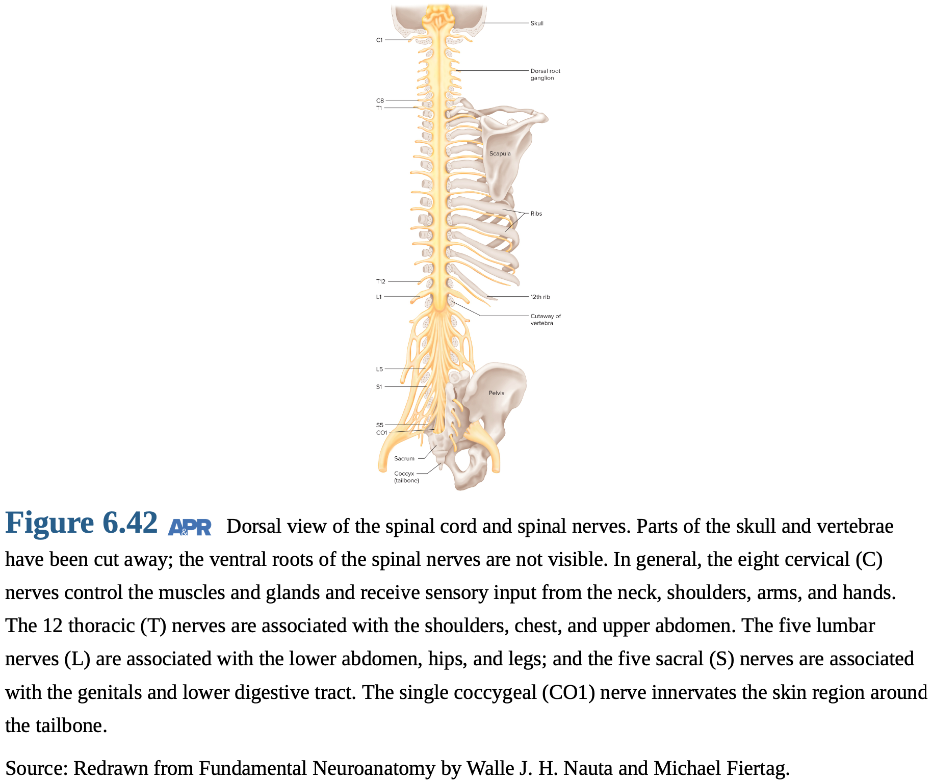
Composition: The PNS consists of 43 nerve pairs: 12 cranial nerves and 31 spinal nerves, which connect with spinal cord.
Spinal Nerves: These are categorized into cervical, thoracic, lumbar, sacral, and coccygeal nerves, based on their location along the spinal column.
Cervical Nerves: innervate the neck, shoulders, arms, and hands, controlling movement and sensation in these areas.
Thoracic Nerves: innervate the chest and upper abdomen, controlling muscles involved in breathing and providing sensory information.
Lumbar Nerves: innervate the lower abdomen, hips, and legs, controlling movement and sensation in these regions.
Sacral Nerves: nnervate the genitals and lower digestive tract, controlling sexual function and bowel movements.
Coccygeal Nerves: Coccygeal nerves primarily innervate the skin around the tailbone.
All Spinal Nerves: They contain both afferent and efferent fibers, allowing them to transmit sensory information to the CNS and motor commands from the CNS.
Afferent Neurons: These neurons transmit sensory information from sensory receptors to the CNS.
Primary/first-order neurons are the initial sensory neurons in the pathway.
Efferent Neurons: These neurons transmit signals from the CNS to effector organs.
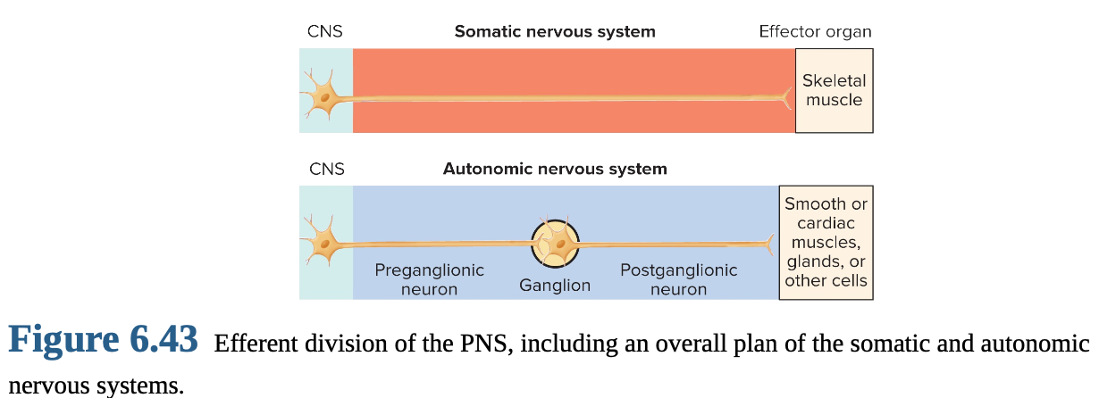
Efferent Division: The efferent division of the PNS is divided into the somatic and autonomic nervous systems.
Somatic Nervous System: The somatic nervous system controls skeletal muscles, enabling voluntary movement.
Autonomic Nervous System: The autonomic nervous system regulates smooth muscle, cardiac muscle, glands, and GI neurons, controlling involuntary functions.
Somatic
Single neuron: extends from the CNS to skeletal muscle, facilitating rapid and direct control.
Innervates skeletal muscle, enabling voluntary movement.
Muscle excitation only: Somatic motor neurons trigger muscle contraction.
Autonomic
Two-neuron chain: extends from the CNS to effector organs, with a synapse in an autonomic ganglion.
Innervates smooth/cardiac muscle, glands, and GI neurons, controlling involuntary functions.
Excitatory or inhibitory: Autonomic neurons can either stimulate or inhibit their target organs.
Somatic Portion: \ consists of motor neurons that extend from the CNS to skeletal muscle cells.
Cell bodies: The cell bodies of somatic motor neurons are located in the brainstem and ventral horn of the spinal cord.
Large, myelinated axons: These axons transmit signals rapidly, enabling quick muscle contractions.
Acetylcholine neurotransmitter is released at the neuromuscular junction to trigger muscle contraction.
Motor neurons: Motor neurons stimulate muscle contraction.
Muscle relaxation: Relaxation occurs when motor neuron activity is inhibited, reducing muscle tension.
6.18 Autonomic Nervous System
Function: provides efferent innervation to organs other than skeletal muscle, regulating involuntary functions.
Enteric Nervous System: network of neurons within the GI tract that regulates digestive functions independently of the CNS.
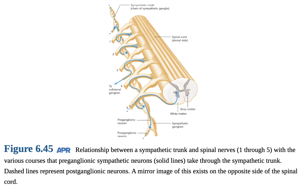
Structure: two neurons in series: a preganglionic neuron and a postganglionic neuron.
Autonomic Ganglion: This is where the synapse occurs between the preganglionic and postganglionic neurons outside the CNS.
Preganglionic Neurons: These neurons extend from the CNS to the autonomic ganglia.
Postganglionic Neurons: These neurons extend from the ganglia to the effector cells in target organs.
ANS is divided into the sympathetic and parasympathetic divisions, which often have opposing effects on target organs.
Sympathetic(thoracolumbar) Division: Preganglionic neurons originate from the thoracic and lumbar regions of the spinal cord.
Parasympathetic(cranial) Division: parasympathetic neurons arise from the brainstem and sacral regions of the spinal cord.

Ganglia Location:
Sympathetic Ganglia: located near the spinal cord, forming the sympathetic chain.
form sympathetic trunks
Collateral Ganglia: located in the abdominal cavity, innervating digestive organs.
celiac superior mesenteric inferior mesenteric ganglia
Parasympathetic Ganglia: located near or within the target organs.
Activation Pattern:
Sympathetic: Activation tends to be body-wide, preparing the body for "fight or flight."
Parasympathetic: Activation is more specific to individual organs, promoting "rest and digest" functions.
Neurotransmitters:
Acetylcholine: It is the neurotransmitter used by preganglionic neurons in both the sympathetic and parasympathetic divisions.
Nicotinic Acetylcholine Receptors: These receptors are found on postganglionic cells in autonomic ganglia.
Parasympathetic: Acetylcholine is also used by postganglionic neurons to signal to effector organs.
Sympathetic: Norepinephrine is used by most postganglionic neurons to signal to effector organs.
Nonadrenergic/Noncholinergic Neurons: Some autonomic neurons use nitric oxide or other neurotransmitters.
Receptors:
Nicotinic Receptors: These are located in autonomic ganglia, where preganglionic neurons synapse onto postganglionic neurons.
Muscarinic Receptors: These are found on postganglionic targets and mediate the effects of acetylcholine.
Adrenal Medulla:
It is a modified sympathetic ganglion.
It releases epinephrine and norepinephrine into the bloodstream.
Catecholamines act as hormones, prolonging and amplifying the sympathetic response.
Dual Innervation: Many organs, such as the heart, glands, and smooth muscles, receive input from both the sympathetic and parasympathetic divisions, allowing for fine-tuned control.
6.19 Protective Elements Associated with the Brain
Meninges: protective membranes surrounding the brain and spinal cord, consisting of the dura mater, arachnoid mater, and pia mater.
dura mater is thicc
Subarachnoid Space: filled with cerebrospinal fluid (CSF) and lies between the arachnoid mater and pia mater.
Functions: These elements protect the brain and spinal cord, provide support, and circulate cerebrospinal fluid (CSF).
Meningitis: It is an infection of the meninges, leading to inflammation and increased pressure within the skull.
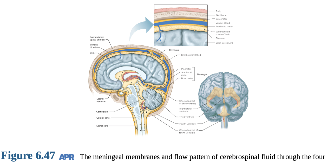
Choroid Plexus: The choroid plexus produces cerebrospinal fluid (CSF) within the ventricles of the brain.
CSF Flow: CSF flows through the ventricular system of the brain and into the subarachnoid space, where it cushions and nourishes neural tissue.
CSF Sampling: CSF can be sampled from the spinal canal below the L2 vertebra for diagnostic purposes.
Function: The CNS floats in CSF, which cushions and protects the brain from mechanical injury.
Hydrocephalus: condition which occurs when CSF accumulates in the ventricles, leading to increased intracranial pressure.
Brain Metabolism:
Glucose is the primary substrate metabolized by the brain to meet its energy demands, with most energy from glucose oxidation transferred to ATP.
Negligible glycogen stores mean the brain requires a continuous supply of glucose from the blood.
Stroke: It is the death of neurons due to vascular disease, leading to impaired blood flow and oxygen deprivation.
Blood Supply:
The brain accounts for approximately 2% of body weight but receives 12-15% of the body's blood supply, highlighting its high metabolic demands.
Reduced blood flow leads to membrane pump failure, disrupting ion gradients and neuronal function.
Blood–Brain Barrier:
The blood-brain barrier selectively controls the substances that enter the brain, protecting it from harmful compounds.
Astrocytes and specialized blood vessel cells form tight junctions that limit permeability.
Lipid solubility: Substances with high lipid solubility can rapidly enter the brain.
Morphine vs Heroin:
Heroin is more lipid-soluble than morphine, allowing it to cross the blood-brain barrier more easily and produce a faster, more intense effect.
Transport Proteins: These facilitate the entry of essential substances into the brain.
CSF Barrier: The CSF barrier selectively secretes substances into the cerebrospinal fluid, regulating its composition.