IB Biology HL Unit 8&9
Cell Cycle and Mitosis
Cell and Nuclear Division
DNA packaging
In eukaryotic cells, DNA is contained in the nucleus
Each cell contains 2 meters of DNA within the nucleus
DNA is “packaged” to fit all of the DNA within the nucleus of each cell
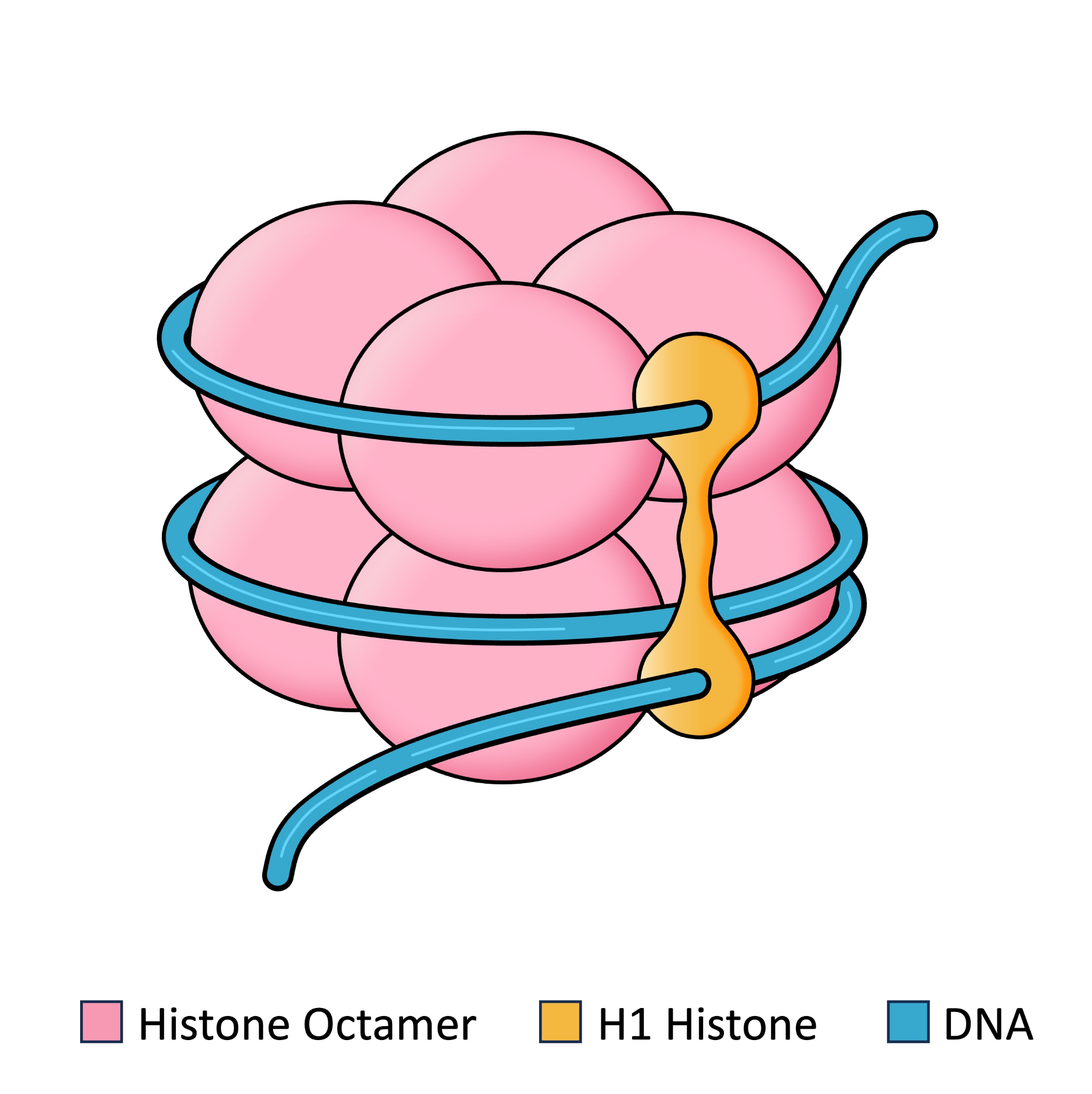
Nucleosomes
A nucleosome is a stretch of DNA that is wrapped around a core of 8 histone proteins
Two copies each histone protein: H2A, H2B, H3, and H4
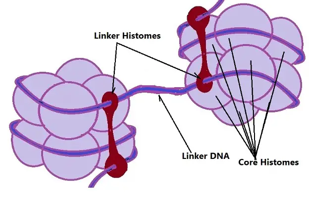
A 9th histone protein (H1) holds the DNA around the nucleosome
Nucleosomes are connected with Linker DNA - a stretch of DNA wrapped around histones that connect neighboring nucleosomes
This is often referred to as “beads on a string”
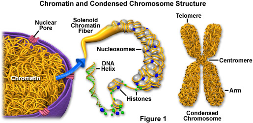
Chromatin and Chromosome
The DNA is further packaged into chromatin - loosely packed DNA found in non-dividing cells
When a cell is actively dividing, the chromatin condenses into chromosome - tightly wound DNA found in actively dividing cells
Chromosome Structure
After DNA replication occurs, the 2 DNA molecules (chromatids) are tethered to each other at the centromere
When the DNA packs into chromosomes, we see the x-shaped structure of a duplicated chromosome (replicated chromosome)
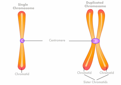
Chromatids are sister chromatids if they are connected at the centromere
DNA packaging and Gene Expression
Tightly packaged DNA (chromosomes) makes the genes inaccessible
No transcription
No gene expression
One way to control gene expression is to manipulate the level of DNA packing; looser packaging = more gene expression
Unicellular Organisms
Unicellular organisms:
Cell division = reproduction because a new organism is produced
It is a type of asexual reproduction because new cells are genetically identical to each other and the original cell
Cell proliferation in multicellular organisms
Growth
Increase the size and complexity of multicellular organisms by producing more cells
Cell replacement
Tissues such as skin cells require routine replacement
Helps to maintain healthy tissues and replace dead cells that are lost
Tissue repair
Wound healing requires cells to proliferate to repair the damaged tissues

Cell proliferation ensures continuity of genetic information across all cells within an organism
The cell cycle
The sequence of events that a cell undergoes, including growth, DNA replication, and nuclear and cytoplasmic division.
Two parts:
Interphase
Miotic phase (M phase)
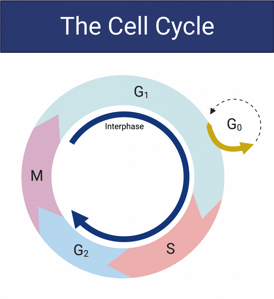
Interphase
Interphase is composed of 3 subphases:
G1 - gap 1 - Involves cell growth and normal metabolic functions
S - Synthesis - DNA replication occurs
G2 - gap 2 - involves cell growth and preparation for nuclear division
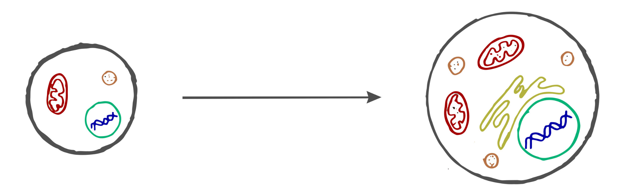
Interphase: G1
the cell approximately doubles in size
Metabolic processes occur such as protein synthesis
Mitochondria (and chloroplasts in plants) divide using binary fission
Interphase: S
DNA is replicated within the nucleus
Remember: DNA replication is semi-conservative
Interphase: G2
Continued growth
Synthesizing microtubules (proteins0 and other proteins necessary for cell division
Mitotic Phase (M Phase)
The mitotic phase (M phase) is composed of 2 subphases:
Mitosis - the division of the nucleus
Cytokinesis - the division of the cytoplasm
Cytokinesis happens simultaneously with the end of mitosis
Mitosis
The division of the nucleus
At the end of mitosis and cytokinesis, 2 genetically identical daughter cells have been produced
Genetically identical to each other
Genetically identical to the original parent cell
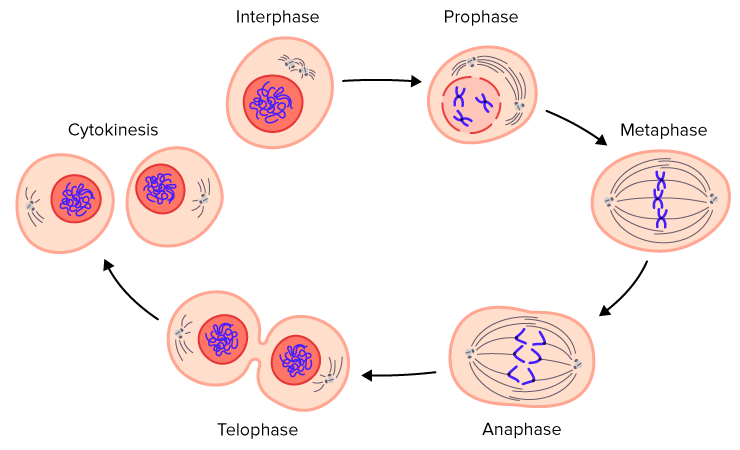
Stages of mitosis
Mitosis is divided into 4 stages:
Prophase
Metaphase
Anaphase
Telophase
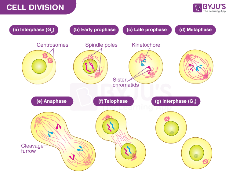
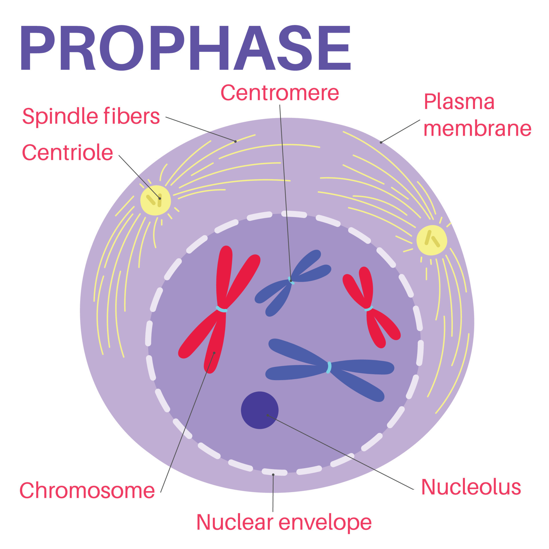
Prophase
Chromatin condenses into chromosomes - remember, they are duplicated chromosomes because they have just been replicated during the S phase
The nuclear membrane breaks down
Spindle fibers form
Spindle fibers are made of microtubules and are responsible for the movement of chromosomes during mitosis
Sometimes they are referred to as “the mitotic spindle”
Plant cells use microtubule organizing centers (MTOCs) to organize the spindle
Animal cells use centrosomes (a type of MTOC that contains centrioles)
During prophase, the MTOCs migrate to the poles of the cell
Metaphase
Duplicated chromosomes line up along the metaphase plate at the equator of the cell
spindle fibers attach to the chromosomes at the kinetochore - motor proteins that are located in the centromere region
Anaphase
Centromere splits and sister chromatids separate and move away from each other towards the poles of the cell
Spindle fibers are responsible for chromosome movement
Once the sister chromatids have separated, they are considered 2 unduplicated chromosomes
Telophase
Chromosomes decondense into chromatin
The nuclear membrane reforms around the two new nuclei
Spindle fibers
Cytokinesis
The division of the cytoplasm
Separates the parent cell into 2 daughter cells
Sometimes simultaneous with telophase
Proceeds differently in plant cells v. animal cells
Animal cell cytokinesis
Actin and myosin proteins form a contractile ring at the center of the cell
The contractile ring pinches the cell membrane in and forms a cleavage furrow
The furrow deepens so it eventually splits the cell into 2
Plant cell cytokinesis
Vesicles carrying cell wall material assemble into the cell plate
The cell plate grows outwards and will eventually form the cell wall between te 2 daughter cells
Binary Fission
Type of cell division performed by prokaryotes (and mitochondria & chloroplasts)
Steps:
1. DNA replication
2. Cell elongation
3. Cytoplasm divides
4. Daughter cells produced
Mitosis and Binary fission produce genetically identical cells - in general, they do NOT increase genetic diversity… except for mutations during DNA replication
Cell cycle control, cancer, and the mitotic index
The cell cycle must be tightly regulated to maintain healthy tissues
Cell cycle control
The cell cycle contains a series of checkpoints that help to regulate the cell cycle
G1 checkpoint
G2 checkpoint
M checkpoint
Internal and external controls are molecules that act as stop-and-go signals at the different checkpoints
G1 Checkpoint
The G1 checkpoint determines if the cell will eventually go on through the rest of the cell cycle
A “go” signal at the G1 checkpoint will allow the cell to continue through the rest of the cell cycle
A “stop” signal at the G1 checkpoint will cause the cell to exit the cell cycle and enter G0 - a phase where the cell is not preparing to divide
Some cells can re-enter the cell cycle from G0
Other cells are in “terminal G0” and cannot re-enter the cell cycle
G2 Checkpoint
The G2 checkpoint checks for:
Has the cell grown enough?
Has the DNA replicated fully?
Has the cell produced enough energy, proteins, organelles, etc. in preparation for cell division?
A “go” signal at the G2 checkpoint allows the cell to enter the mitotic phase
M checkpoint
The M checkpoint occurs during the metaphase of mitosis
Check to make sure all of the chromosomes have:
Attached to spindle fibers
Lined up at the metaphase plate
A “go” signal at the M checkpoint allows the cell to enter anaphase
External regulators
response to events outside the cell, often directing cells to speed up or slow down the cell cycle
Examples;
Growth factors
Anchorage Dependence
Density-dependent inhibition
Growth factors
an important group of external regulatory proteins
Stimulate the growth and division of cells
Important during embryo development and wound healing
Example:
PDGF (platelet-derived growth factor) stimulates the division of human fibroblast cells in culture
Anchorage dependence and Density-dependent inhibition
most animal cells exhibit anchorage dependence, in which they must be attached to a substratum to divide
Density-dependent inhibition:
Crowded cells stop dividing
Prevents excess cell division
Internal regulators
Response to events inside a cell and allow the cell cycle to proceed only when certain events have occurred
Example:
Cyclins and Cyclin-dependent kinases (CDKs)
Cyclins and CDKs
Cyclins are a family of proteins that regulate the cell cycle by activating cyclin-dependent kinases
Cyclin-dependent kinases (CDKs) are enzymes that, when activated phosphorylate proteins to progress the cell cycle
CDKs are always present, but not always active
Cyclins activate CDKs by binding to them creating a cyclin-CDK complex
Different cyclins accumulate during different stages of the cell cycle, thus activating different CDKs at different times
Once the cyclin-CDK complex has completed its task, the cyclin is degraded and the CDK is deactivated
The cyclin must reach a critical concentration for the cell to progress to the next stage
Fluctuations of cyclin concentrations control the cell cycle
Cancer
Cell cycle control genes
the proteins that act as regulators for the cell cycle are coded for by genes
When those genes are mutated, that can cause a breakdown in the cell cycle control system
Mutations are any change to the DNA sequence
Proto-Oncogenes
Proto-oncogenes are genes that code for proteins that help promote cell growth and division
Mutations to proto-oncogenes can lead to the proteins being overexpressed, leading to uncontrolled cell division
When a proto-oncogene becomes mutated, it is called an oncogene
Tumor suppression genes
Tumor suppression genes are genes that code for proteins that normally slow down or prevent cell division
They can also trigger apoptosis - programmed cell death
Mutations in tumor suppressor genes will lead to malfunctioning proteins - the absence of the protective function of these proteins leads to uncontrolled cell division
Benign Tumors
Uncontrolled cell division can lead to the accumulation of an abnormal mass of cells - tumor
Benign tumors - abnormal growth of cells that are not cancerous
Grow slowly, well-defined margins, don’t metastasize (spread to other parts of the body)
Can still cause issues depending on location and size
Malignant tumors
Malignant tumors - cancerous, growing and dividing more rapidly than benign tumors
Lack well defined borders
Undergo metastasis
Primary tumor - original tumor
Secondary tumor - a tumor that forms in a new location
Treatments for tumors
Benign tumors are often treated with surgery
Malignant tumors are often treated with a combination of:
Surgery
Radiation
Chemotherapy
Mitotic index
Mitotic index
Measure of the proportion of actively dividing cells in a population
Value of 0 to 1, or it can be written as a percentage (0% - 100%)
The more cell division, the larger the mitotic index
A high mitotic index is NOT always an indicator of a cancerous tumor
Some cells/tissues/developmental stages will have high rates of cell division
Embryonic development
Epithelial cells (skin & lining of the digestive tract)
Meristem cells in plants (areas of growth in roots/shoots)
Mitotic index can be used as a diagnostic tool for cancerous tumors - must be compared to the MI of healthy tissues
Mitotic Index
Meiosis
Purpose of reproduction
Reproduction is one of the processes of life
The way by which organisms pass on their genes to future generations
Ensure the continuity of their species
Also necessary for natural selection to occur
Asexual reproduction
The production of genetically identical offspring from a single source of genetic information
It occurs in prokaryotes, fungi, many plants, and some animals
Binary fission - a type of cell division performed by prokaryotes
Mitosis & cytokinesis - asexual reproduction performed by unicellular eukaryotes
Budding - a new organism develops as an outgrowth (bud) from the parent organism, eventually detaches and becomes an independent organism
Fragmentation - The parent organism breaks into fragments and each fragment develops into a new organism
Asexual reproduction - Advantages
A large number of offspring is produced in a short amount of time
An advantage in a stable environment if the parent organism is well adapted to the environment
Less costly in; time, energy, and resources
Less complex process
Asexual reproduction - disadvantages
Does not increase genetic variation/diversity
No genetic variation means that if the environment changes, the species could be completely wiped out
Harmful mutations have a widespread effect
Sexual reproduction
The production of genetically different offspring from two sources of genetic information
The offspring inherit some genetic information from each source (parent)
Occurs in multicellular plants and animals
Sexual reproduction - advantages
Increases genetic variation/diversity - offspring are similar but not identical to each other and the parents
Advantages in changing environments - more genetic variation increases the chance of survival of the species
Sexual reproduction - disadvantages
In general, fewer organisms produced in a longer period of time
More costly in; time, energy, and resources
More complex process
Harder to accomplish because gametes must fuse
Process of sexual reproduction
Production of gametes - using meiosis
Fertilization - fusion of haploid gametes to create a diploid zygote
Development of offspring-cell proliferation
Sources of genetic variation
In sexual reproduction there are 4 sources of genetic variation:
Crossing over during meiosis 1
Independent assortment of homologous chromosomes during meiosis 1
Random orientation of homologous chromosomes during meiosis 1 and sister chromatids during meiosis 2
Random fertilization during the fusion of gametes
Meiosis
A process of cell division that produces 4 haploid gametes that are genetically unique
Meiosis is considered a reduction division because it takes 1 diploid cell and creates 4 haploid cells by separating the homologous chromosomes
Gametes are cells used in sexual reproduction
Review: Chromosome number
Diploid cells contain 2 copies of each chromosome -2n
Ex. Humans 2n = 46
Body cells are somatic cells and they are diploid
Haploid cells contain 1 copy of each chromosome - n
Ex. Humans n = 23
Gametes (sex cells - sperm and eggs) are haploid
A duplicated chromosome is still only 1 chromosome
This means that a diploid cell is still diploid before and after S phase (synthesis)
DNA replication does not increase the number of chromosomes, it increases the number of chromatids
Review: Homologous chromosomes
Diploid cells have pairs of chromosomes
The chromosomes within the pair are homologous chromosomes
Homologous chromosomes:
are the same length
have the same gene loci in the same order and location
have the centromere region in the same location
In humans, the pairs of autosomes (chromosomes 1-22) are homologous
Before Meiosis
DNA replication must occur before meiosis can proceed
At the beginning of meiosis 1, the parent cell is a diploid cell with duplicated chromosomes (2 sister chromatids in each)
Meiotic Divisions
Meiosis consists of 2 rounds of division: Meiosis1 and Meiosis 2
Each round of division progresses through prophase, metaphase, anaphase, telopase, and cytokinesis
Meiosis 1
Meiosis 1: Prophase 1
The nuclear membrane breaks down
Spindle fibers start to form
Microtubule organizing centers (MTOCs) move away from each other toward opposite poles of the cell
Chromatin condenses into (duplicated) chromosomes
Homologous chromosomes pair up during synapsis creating tetrads/bivalents
Tetrads remain paired up until anaphase 1
Crossing over occurs when non-sister chromatids exchange equivalent segments of DNA
The exchange of DNA happens at the chiasmata - the x-shaped regions where crossing over has occurred
Crossing over creates recombinant chromatids
Meiosis 1: Metaphase 1
Tetrads line up along the metaphase plate
Independent assortment of tetrads - each tetrad pair lines up independently of the other pairs - there is a random orientation towards the poles
Spindle fibers attach to the kinetochores on either side of the homologous pairs
Meiosis 1: Anaphase 1
Homologous chromosomes separate from each other
Sister chromatids remain connected at the centromere during Anaphase 1
Spindle fibers are responsible for moving the homologous chromosomes away from each other
Meiosis 1: Telophase 1/cytokinesis 1
Homologous chromosomes have reached the poles of the cell
Chromosomes decondense into chromatin
Nuclear membrane reforms around the 2 new (haploid) nuclei
Spindle fibers break down
Cytokinesis occurs by producing 2 daughter cells that are:
Haploid with duplicated chromosomes
Non-identical (genetically unique)
After Meiosis 1 is complete interkinesis occurs - a period of rest, no DNA replication occurs
Meiosis 2
Meiosis 2: Prophase 2
Meiosis 2 is very similar to mitosis, each daughter cell produced after Meiosis 1 will proceed through Meiosis 2
Chromatin recondenses into chromosomes
The nuclear membrane breaks down
MTOCs migrate to opposite poles of the cell
Spindle fibers begin to form
Meiosis 2: Metaphase 2
Spindle fibers attach to the kinetochores at the centromeres of the duplicated chromosomes
Duplicated chromosomes (sister chromatids) line up along the metaphase plate
Sister chromatids show random orientation toward the poles - remember they are no longer identical
Meiosis 2: Anaphase 2
Centromeres split
Sister chromatids separate from each other and move towards the opposite poles of the cell
Spindle fibers are responsible for the movement of chromosomes
Remember. once the sister chromatids are separated, they are called unduplicated chromosomes
Meiosis 2: Telophase 2/Cytokinesis 2
Chromosomes reach the opposite poles of the cell
Chromosomes decondense into chromatin
Nuclear membranes form around the 2 (haploid) nuclei
Spindle fibers break down
Cytokinesis occurs by producing a total of 4 daughter cells that are:
Haploid gametes with unduplicated chromosomes
Non-identical (genetically unique)
These daughter cells will mature into the gametes
Review
Review: Karyograms
A karyogram shows an image of an organism’s chromosomes
In humans:
Chromosomes 1-22 are autosomes and each pair is homologous to each other
Chromosome 23 are the sex chromosomes and are not homologous
Aneuploidy
The presence of an abnormal number of chromosomes in a cell
Trisomy - 3 copies of a chromosome
Monosomy - 1 copy of a chromosome
The most common type of aneuploidy compatible with life is trisomy 21 which causes Down syndrome
Nondisjunction
An aneuploidy is created if nondisjunction occurs
Nondisjunction is a failure of homologous chromosomes to separate during meiosis 1 or a failure of sister chromatids to separate during meiosis 2
Creates gametes with a missing chromosome or an extra chromosome
If that gamete is involved in fertilization, then the zygote (and therefore the offspring) will have a monosomy or trisomy
Nondisjunction during mitosis does not cause aneuploidy in offspring
Karyotyping
A karyotype is created when analyzing a karyogram - it can determine if there is an aneuploidy
When a woman is pregnant, a karyotype can be done on the fetus by collecting fetal cells using:
Non-invasive prenatal testing (NPT)
Blood draw
Aminocentesis - the collections of amniotic fluid
Chorionic Villus Sampling - the collection of placental tissue
Mendelian Genetics
Gregor Mendel
Heredity is the passing on of characteristics/traits from parents to offspring
Genetics is the field of biology that deals with heredity
He investigated 7 pairs of traits, performed crosses, and observed the offspring
Model Organisms: Pea Plants
Mendel had easy access to pea plants because of his monastery
They were a good model organism because:
They can reproduce sexually
They can self pollinate and Mendel could control the cross-pollination between plants
They have a relatively short reproductive cycle and produce many offspring
Mendelian Traits of Pea Plants
Each trait had 2 different alleles - different versions of the same gene
Flower color (purple or white)
Seed color (yellow or green)
Sed shape (round or wrinkled)
Pod color (green or yellow)
Pod shape (smooth or bumpy)
Flower position (mid-stem or end of stem)
Plant height (tall or short)
Alleles
Alleles are different versions of the same gene
Alleles are created by mutations to a gene sequence
Many alleles are due to single nucleotide polymorphisms (SNPs)
Mendel’s Peas
P generation
Mendel created true-breeding plants - a line of plants that only produced plants with a specific trait when allowed to self-pollinate
Mendel’s P generation consisted of 2 true-breeding plants of opposite traits (ex. a true-breeding purple flower plant and a true-breeding white flower plant)
F1 Generation
Mendel performed the first cross with the P generation where he cross-pollinated the true-breeding plants
The resulting offspring were called the F1 generation
He called these the hybrids (monohybrids)
100% of the hybrids exhibited only 1 of the two versions of each trait (ex. purple flowers crossed with white flowers produced 100% purple flowers)
F2 Generation
Mendel allowed the hybrids (F1 generation) to self-pollinate to produce the F2 generation
The offspring in the F2 generation exhibited both versions of the traits in predictable ratios (ex. ¾ purple flowers and ¼ white flowers - 3:1 ratio)
1st concept of Mendelian genetics
2 alleles
Traits that exhibit the patterns of Mendelian genetics only have 2 alleles
Within each organism, there are 2 copies of each gene, one on each chromosome in a homologous pair (at the same loci)
Those copies could both be the same, or they could be different
2nd concept of Mendelian genetics
Biparental Inheritance
For each trait, an organism inherits 2 alleles, 1 from each parent/gamete
Remember - these alleles could both be the same or they could be different
Autosomal traits
Traits that exhibit Mendelian genetics have genes that are found on the autosomes (chromosomes 1-22)
this allows for them to be inherited in a biparental pattern
Traits determined by genes that are found on the sex chromosomes (X or Y) are not considered to be “Mendelian”
3rd concept of Mendelian genetics
Dominance vs. Recessiveness
of the 2 different alleles for any trait, one is dominant and one is recessive
When an organism has 2 different alleles, the dominant allele will be the one that is expressed
The recessive allele is only expressed in the absence of the dominant allele - aka when an organism has two copies of the recessive allele
4th concept of Mendelian genetics
Law of segregation
In the parent, the alleles of each trait segregate (or separate) into each gamete so each gamete only receives one copy of each gene
This is because the homologous chromosomes separate during Meiosis 1
Segregation of Alleles and Biparental Inheritance
These 2 concepts are tied directly to the process of sexual reproduction: Gamete formation and fertilization
During gamete formation, the homologous chromosomes separate during Meiosis 1- (segregation of alleles) - because gametes are haploid, each gamete to only contribute 1 copy of each gene to their offspring (biparental inheritance)
During fertilization, the fusion of sperm and egg creates a diploid zygote
The fertilization process provides the offspring with 1 copy of each gene from each parent/gamete (biparental inheritance)
Tied directly to the chromosome numbers:
Before Meiosis: Diploid cell
After Meiosis: Haploid gametes
After fertilization: Diploid zygote
Writing Alleles - Autosomal traits
The alleles for an autosomal trait are given letters
Capital for the dominant allele
Lowercase for the recessive allele
Genotype - Autosomal Traits
The combination of alleles is the genotype of an individual
Genotypes for 1 trait are written with 2 letters to represent the 2 alleles on the homologous chromosomes
Possible combinations: FF, Ff, and ff
Organisms are homozygous if they have 2 copies of the same allele
FF = homozygous dominant
ff = homozygous recessive i
Organisms are heterozygous if they have 1 copy of each allele"
Ff = heterozygous
Genotype and Phenotype
the genotype determines the phenotype (with a few exceptions)
The phenotype of an individual is the expression of the genes (ex. physical appearance or metabolic characteristics, etc.)
Homozygous dominant and heterozygous genotypes will result in the dominant phenotype
Homozygous recessive genotypes will result in the recessive phenotype
Recessive Disorders
Many genetic disorders are inherited in an autosomal recessive pattern - the gene’s locus is on an autosome, and the allele is recessive, so an individual must be homozygous recessive to express the recessive disorder
If someone is heterozygous for a recessive disorder, they are said to be a “carrier”
Carriers have the normal (dominant) phenotype but have 1 copy of the recessive allele that they can pass on to their offspring
Many partners will get genetically tested to see if they are carriers of the same trait
Example: Phenylketonuria or PKU
Phenylketonuria (PKU)
PKU is caused by a mutation in a gene on chromosome 12 (autosomal)
This gene codes for an enzyme called phenylalanine hydroxylase (PAH)
PAH converts the amino acid phenylalanine (Phe) into tyrosine (Tyr)
The mutation creates a malfunctioning enzyme
Individuals that are homozygous recessive are unable to break down phenylalanine (Phe)
Phenylalanine builds up to toxic levels and can cause: a musty odor from the skin and urine, fair skin, eczema, seizures, tremors, and hyperactivity
If the condition is left untreated, brain damage can occur
PKU can be managed with a tightly controlled, low-protein diet
5th concept of Mendelian genetics
Law of independent assortment
Each pair of alleles segregates independently of each other pair of alleles during gamete formation
AKA the way each tetrad lines up during metaphase 1 has zero impact on the other tetrads
Assumption: the gene loci are on different chromosomes
Law of independent assortment
If an organism is heterozygous for 2 traits, their genotype would be AaBb
Remember: Each gamete only gets 1 copy of each gene (aka 1 of each type of letter)
When we perform a 2 trait cross, we assume that the traits are independent (and therefore Mendelian)!
Pedigrees
Pedigree charts
Illustrate the inheritance of a trait through a family’s history
Useful for illustrating genetic disorders and to help predict the probability of future generations having the disorder
Pedigree Symbols
Circles are females
Squares are males
Filled in shape = individual has the trait
Unfilled in shape = individual without the trait
Horizontal line between a circle and square = mating lines (children branch off of the mating lines)
Identifying individuals
Generations are labeled with Roman numerals starting at the top row
Individuals are labeled with Arabic numerals moving left to right
Types of Inheritance
Autosomal dominant
AA, Aa = has trait
aa = doesn’t have the trait
Autosomal recessive
AA, Aa = doesn’t have the trait
aa = has the trait
Sex-linked recessive
XAXA = female without trait
XAXa = female without trait (carrier)
XaXa = female with trait
XAY = male without trait
XaY = male with trait
Determining Inheritance
Autosomal Dominant
Means filled in shapes are either AA or Aa
If a child has the trait, at least one parent must also have the trait
If both parents have the trait, and at least one of the children doesn’t have the trait, it must be autosomal dominant
Means filled in shapes are either AA or Aa
If neither of the parents has the trait, the offspring will not exhibit it either
If both parents do have the trait, but the offspring does not, then the child will be homozygous recessive and the parents will be heterozygous or carriers
Autosomal recessive
This means filled-in shapes are aa
If both parents are affected (filled in), the offspring should be also
If the parents are both unaffected, but the offspring has the trait, then the parents must be heterozygous
X-linked recessive
Means filled-in shapes are XaXa (female) and XaY (male)
Mostly males with the trait suggest X-linked, but sex linkage cannot be confirmed
A female with the trait must have a father with the trait and 100% of her sons would also have the trait
An unaffected mother can have affected sons if she is a carrier (heterozygous