BIOL_250_EText_Ch_10_-_MUSCLE_TISSUE
Introduction to Muscle Tissue
Muscle tissue is essential for active movement of the body and the transportation of materials within the body. It is composed of specialized cells capable of contraction. The three primary types of muscle tissue are skeletal muscle, cardiac muscle, and smooth muscle, each with distinct structures, functions, and locations within the body.
Types of Muscle Tissue
Skeletal Muscle: This type of muscle is responsible for moving the skeleton and is under voluntary control, meaning it can be consciously controlled by an individual. Skeletal muscle fibers are long, cylindrical, and striated due to the organized arrangement of contractile proteins actin and myosin.
Cardiac Muscle: Found exclusively in the heart, cardiac muscle is responsible for pumping blood through the circulatory system. It is involuntary and striated like skeletal muscle but consists of shorter, branched fibers with one or two central nuclei. Intercalated discs connect adjacent cardiac muscle cells, allowing for rapid electrical signal transmission and synchronized contractions.
Smooth Muscle: Located in the walls of hollow organs and passageways, such as the intestines, blood vessels, and urinary bladder, smooth muscle controls involuntary movements, including the contraction of blood vessels and the movement of food through the gastrointestinal tract. It is spindle-shaped, contains a single nucleus, and lacks striations, making it distinct from the other two types.
Chapter Objectives
Explain the organization and classification of muscle tissue.
Describe the structures and functions of skeletal, cardiac, and smooth muscle.
Explain how muscles work with tendons to facilitate body movement.
Define muscle metabolism and the processes involved in muscle relaxation.
Explore the connections between exercise and muscle performance, including adaptation to training.
Outline the development and regeneration processes of muscle tissue throughout life.
Overview of Muscle Tissues
All muscle tissues share the property of excitability, which is the ability to respond to stimuli and undergo contractions as a result. Muscle membranes can change their electrical states and conduct action potentials, allowing them to contract. Unlike skeletal muscle, which is directly influenced by the nervous system, both cardiac and smooth muscles can also respond to hormonal signals and local stimuli.
Muscle Contraction Mechanism
The primary proteins responsible for muscle contraction are actin and myosin. The presence of calcium ions (Ca++) is crucial for the activation of the contraction mechanism in all muscle types. In skeletal muscle, contractions are initiated by neural signals transmitted from motor neurons, whereas the cardiac muscle can contract autonomously due to pacemaker cells embedded within the heart.
Elasticity in muscle allows it to recoil
Extensibility in muscle allows for it to stretch
Contractility allows muscle tissue to pull on its attachment point and shorten with force
Skeletal Muscle Structure
Control: Skeletal muscles are subject to voluntary control, allowing conscious movement.
Composition: Skeletal muscle fibers are encapsulated by three connective tissue layers: epimysium (outer layer), perimysium (surrounding bundles of fibers), and endomysium (covering individual muscle fibers).
Fascicle are bundles of muscle fibers bundled up by the perimysium
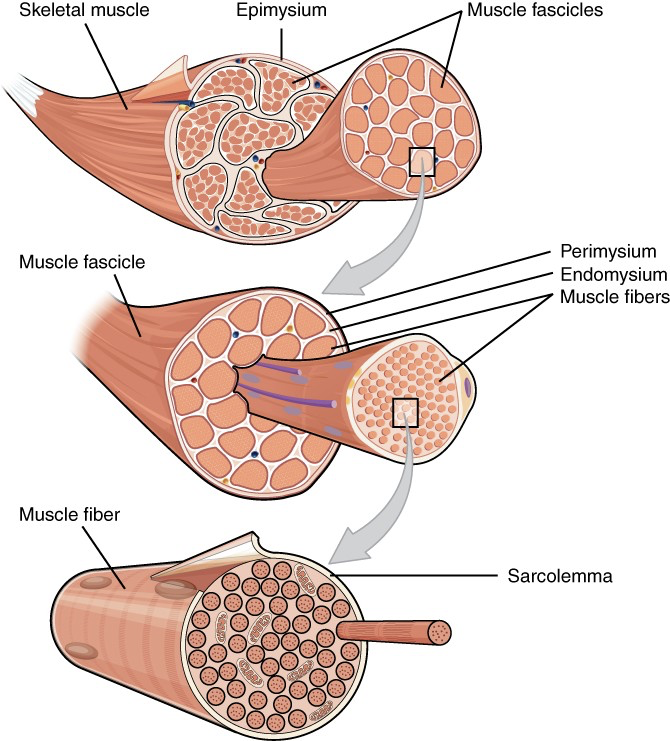
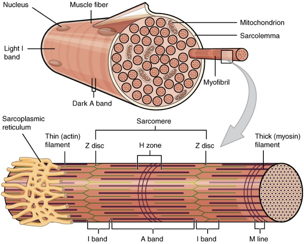
Striations: Myofibrils, located inside muscle fibers, are responsible for the striated appearance of skeletal muscles, resulting from the organized arrangement of actin and myosin filaments.
Functions of Skeletal Muscle
Movement: Facilitate voluntary movements and maintain posture.
Protection: Provide structural support and shield internal organs from injury.
Heat Production: Generate heat during contraction, which is critical for maintaining body temperature.
Structural Details
Skeletal muscle fibers are multinucleated and contain specialized structures:
Sarcolemma: The plasma membrane of muscle fibers that participates in action potential propagation.
Sarcoplasm: The cytoplasm of muscle fibers, containing organelles such as the sarcoplasmic reticulum (SR) for calcium storage.
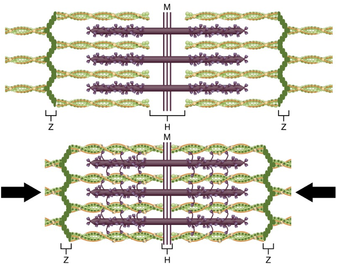
Sarcomeres: The functional unit of muscle contraction, organized into repeating units within the myofibrils.
Neural Control of Muscle Contraction
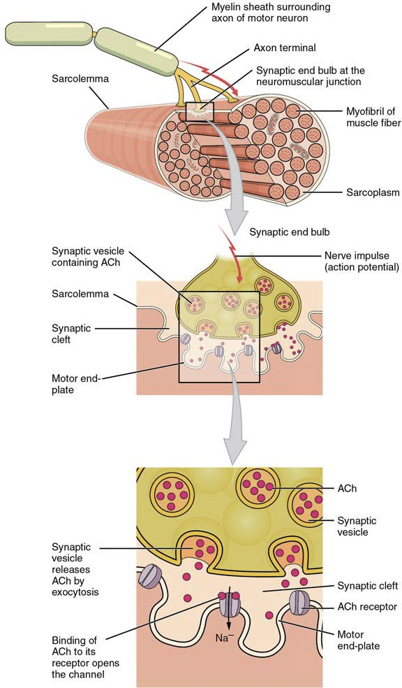
At the neuromuscular junction (NMJ), motor neurons communicate with muscle fibers. The action potentials generated by motor neurons lead to calcium release from the sarcoplasmic reticulum, initiating muscle contraction.
Contraction Cycle
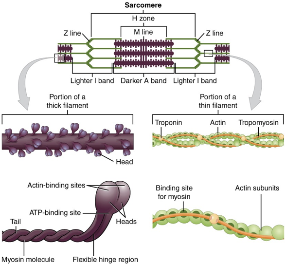
Upon stimulation, calcium ions expose binding sites on actin filaments—this allows myosin heads to bind to actin and form cross-bridges, facilitating the sliding filament model of contraction. ATP is vital for both the contraction and relaxation phases of the muscle cycle.
Cell membrane potential is the -60 to -90 mV, which is used to generate electrical signals
action potential travels from axon to motor neuron, then terminates at the NMJ, leading to the release of acetylcholine, which binds to receptors on the sarcolemma, causing depolarization and the initiation of the action potential in the muscle fiber.
This depolarization wave travels along the sarcolemma and down the T-tubules, triggering the release of calcium ions from the sarcoplasmic reticulum into the cytoplasm, further promoting the contraction process.
Muscle contraction stops when signaling from the motor neuron ends which repolarizes the sarcolemma and T-tubules
Muscle needs ATP to continue contracting
In contraction, the thin filaments slide past the thick filaments, a process known as the sliding filament theory, which shortens the sarcomeres and ultimately leads to muscle shortening.
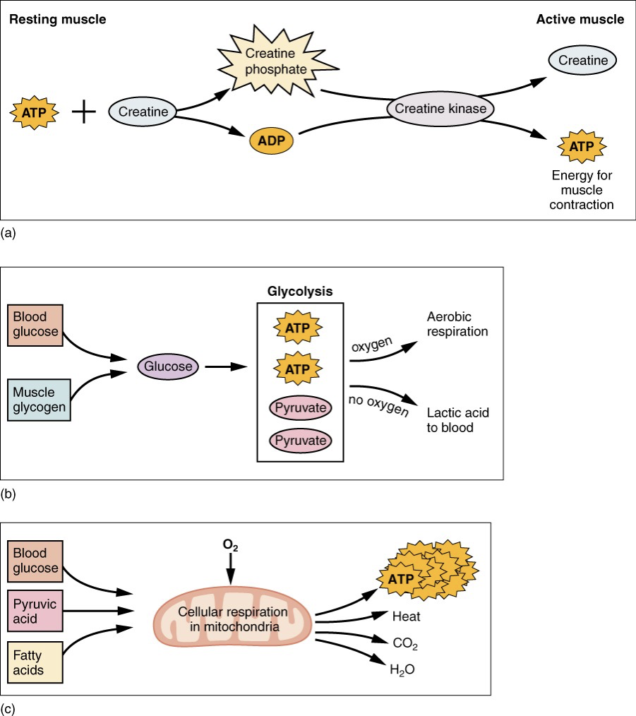
ATP is generated through creatine phosphate metabolism, anaerobic glycosis, fermentation and aerobic respiration
oxygen debt is the amount of oxygen required to restore the body to its normal resting state after exercise, including replenishing ATP and creatine phosphate stores, clearing lactic acid, and restoring oxygen levels in the blood and muscle tissues.
Muscle Tension
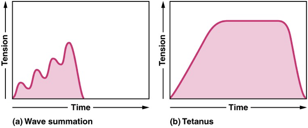
Muscle tension refers to the force generated by muscle fibers during contraction, which can vary in response to different stimuli. This tension is influenced by factors such as the number of motor units activated, the frequency of stimulation, and the length-tension relationship of the muscle fibers.
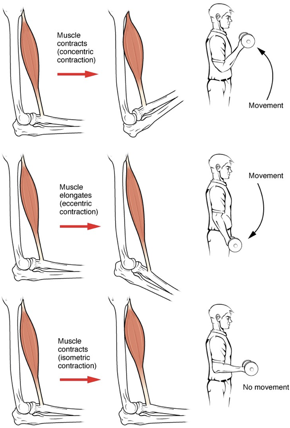
isotonic contractions: involve the muscle tension staying constant while the muscle changes length, allowing movement to occur, such as lifting a weight or walking.
concentric contraction: involves muscle shortening to move a load
eccentric contraction: involves muscle tension diminishes as the muscle lengthens
isometric contraction: produces muscle tension without changing the angle of skeletal joint, which is essential for maintaining posture or stabilizing joints during various activities.
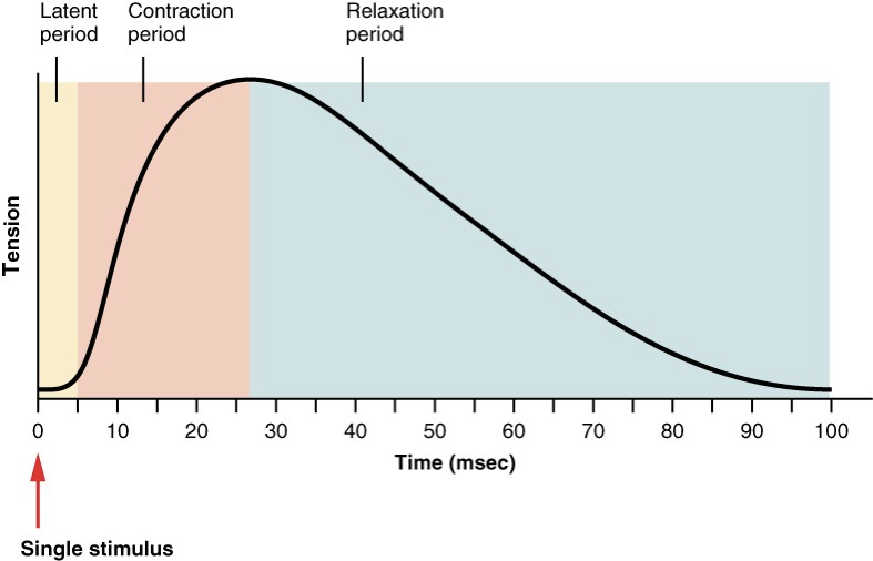
A twitch or single action potential goes through the phases of the latent period, contraction phase, and relaxation phase
latent period: the initial phase where the muscle fiber does not respond to stimulation, as it is preparing for contraction by releasing calcium ions from the sarcoplasmic reticulum.
contraction phase: calcium ions have bound to troponin, causing a conformational change that moves tropomyosin away from the actin binding sites, allowing myosin heads to attach to actin and initiate the power stroke, which leads to muscle shortening.
relaxation phase: the period following contraction where calcium ions are reabsorbed into the sarcoplasmic reticulum, troponin returns to its original shape, and tropomyosin re-covers the actin binding sites, leading to the detachment of myosin heads from actin and allowing the muscle fiber to return to its resting length.
muscle tone
hypotonia: a condition characterized by decreased muscle tone, resulting in reduced resistance to passive movement and often leading to weakness and floppiness in the affected muscles.
hypertonia: a condition characterized by increased muscle tone, resulting in heightened resistance to passive movement and often leading to stiffness and rigidity in the affected muscles.
Muscle Fibers
skeletal muscle fibers
slow oxidative fibers: contract slow and use aerobic respiration to produce ATP, making them well-suited for endurance activities such as long-distance running and maintaining posture.
fast oxidative fibers: contract quickly and utilize both aerobic and anaerobic respiration, allowing for powerful bursts of activity, making them ideal for activities like sprinting and high-intensity exercises.
fast glycolytic fibers: contract rapidly and primarily rely on anaerobic respiration, which enables them to generate quick and explosive bursts of energy, making them suitable for short-duration, high-intensity activities such as weightlifting and sprinting.
Exercise and Muscle Performance
hypertrophy: the increase in muscle size and strength by added structural proteins that occurs in response to resistance training and high-intensity exercise, resulting from the enlargement of muscle fibers as they adapt to the increased load.
atrophy: the decrease in muscle size and strength from loss of structural protein that occurs when muscles are not used regularly, often due to immobilization, aging, or a sedentary lifestyle, leading to a reduction in muscle fiber size and overall muscle mass.
sarcopenia: atrophy that occurs with age
angiogenesis: the process by which new blood vessels form from existing ones, which is crucial for supplying nutrients and oxygen to growing muscle tissue during hypertrophy and recovery from exercise.
Cardiac Muscle Structure and Function
Cardiac muscle, responsible for heart contractions, displays the following characteristics:
Location: Exclusively found within the heart.
Structure: Striated with shorter, branched fibers and one or two central nuclei.
Intercalated Discs: These specialized connections allow rapid electrical signal transmission and maintain structural integrity during contractions.
Rhythmic Contractions: Controlled by pacemaker cells that generate electrical impulses autonomously.
Smooth Muscle Characteristics
Smooth muscle is present in various organs, displaying the following:
Shape: Spindle-shaped with a single nucleus, lacking striations.
Contraction Initiation: Not initiated by neural signals; can respond to various stimuli such as stretching and hormonal signals.
Calcium Interaction: Contraction is regulated by calcium that interacts with calmodulin, a distinct mechanism compared to striated muscles.
Latch-bridges: These structures allow smooth muscle to maintain prolonged contractions with minimal energy expenditure, contributing to the efficiency of muscle function. They keep thin and thick filaments in close proximity, allowing for sustained contraction without the need of ATP.
varicosity: A site on the autonomic nerve fibers where neurotransmitters are released, facilitating communication between the nerve and smooth muscle cells, thereby influencing contraction.
pacesetter cell: Specialized smooth muscle cells that generate rhythmic contractions, establishing the pace for the entire muscle tissue, particularly in the gastrointestinal tract.
visceral muscle : A type of smooth muscle found in the walls of internal organs, such as the stomach and intestines, that is involuntary and responsible for the movement of substances through the body.
stress-relaxation response: A phenomenon in smooth muscle where prolonged tension leads to a gradual decrease in muscle tension, allowing the muscle to maintain a constant volume and adapt to changes in its environment.
Types of Smooth Muscle
Single-Unit Smooth Muscle: Cells connected by gap junctions, allowing synchronized contractions. Commonly found in internal organs.
Multi-Unit Smooth Muscle: Cells act independently, lacking gap junctions. Found in locations such as arterioles and respiratory passages.
Muscle Metabolism
The sources of ATP for muscle contraction include:
Creatine Phosphate Metabolism: Provides an immediate but limited supply of ATP.
Anaerobic Glycolysis: Produces ATP rapidly but is less efficient in the long term.
Aerobic Respiration: Fuels prolonged activities and generates a greater yield of ATP over time.
Muscle Fatigue Overview
Muscle fatigue occurs due to:
Depletion of ATP
Accumulation of lactic acid
Disturbance in ion balance within muscle cells
Development and Regeneration of Muscle Tissue
Muscle tissues originate from embryonic mesoderm. Satellite cells assist in the repair and regeneration of skeletal muscle, although it has limited regenerative capacity. Smooth muscle exhibits a better regenerative ability due to pericytes. Aging leads to muscle atrophy as muscle fibers decrease and are replaced by connective tissues.
myoblasts cells: precursor cells that fuse to form muscle fibers during development and play a crucial role in muscle growth and repair.
myotube: a multinucleated cell formed by the fusion of myoblasts, which subsequently matures into muscle fibers, contributing to the overall structure and function of muscle tissue.
Key Terms
Acetylcholine (ACh): The primary neurotransmitter at the neuromuscular junction, facilitating nerve impulses to muscle fibers.
Sarcoplasmic Reticulum (SR): Organelle that stores calcium ions, essential for muscle contraction signaling.
Myofibril: Thread-like organelles within muscle fibers that contain actin and myosin.
Tetanus: A sustained contraction resulting from a rapid series of stimuli without relaxation.
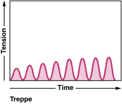
Treppe: The staircase effect observed in muscle contractions where increased strength is noted with successive stimuli.