B2.1 - Membranes and membrane transport
Standard level
B2.1.1 Lipid bilayers as the basis of cell membranes
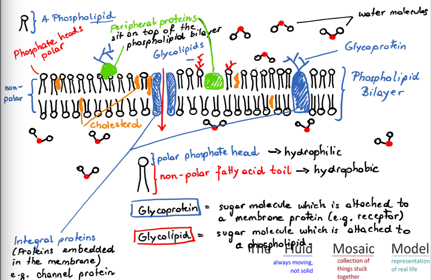
Membranes are important components of cells. They form the border between a cell and its environment, protecting its contents and proving a surface for exchange. Membranes are made up of phospholipids (phospholipid bilayer).
Membranes are also found inside of eukaryotic cells, where they compartmentalize organelles such as mitochondria or rER.
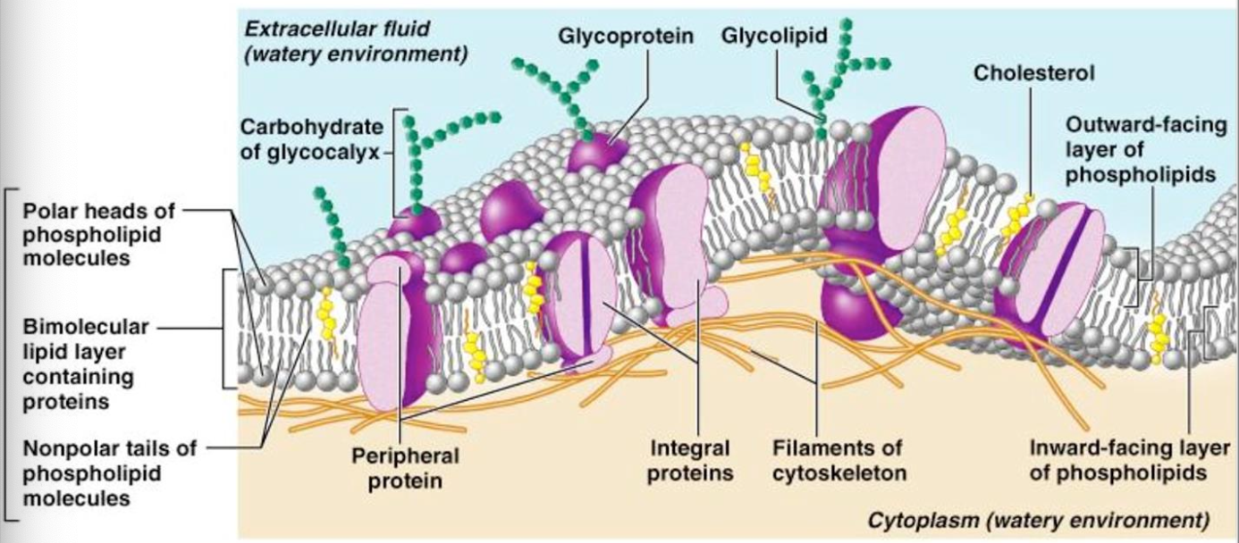
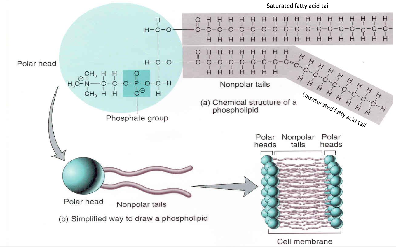
Phospholipids have hydrophobic hydrocarbon tails that form the core of a membrane and hydrophilic phosphate heads which are in contact with the water on both sides of the membrane.
The hydrophilic phosphate heads are attracted to water.
The hydrophobic hydrocarbon tails repelled by water and point away from it.

Hydrophilic - loves water
Substances which are hydrophilic either carry a positive or negative charge (e.g. Na+ or Cl-) or are polar due to a partial charge (caused by a difference in electronegativity). Water is a polar substance and therefore attracts other hydrophilic molecules.
Hydrophobic - repells water
Substances which are hydrophobic are usually non-polar – they don’t carry a charge and are therefore not attracted to the polar water molecules.
B2.1.2 Lipid bilayers as barriers
Because any phospholipids molecule show both characteristics, hydrophobic and hydrophilic, they are said to be amphipathic.
Two phospholipid layers arrange to position the hydrophilic heads towards the water, while the tails form a core of low permeability to large molecules and hydrophilic particles. This arrangement of molecules forms an effective barrier between aquaeous solutions.
B2.1.3 Simple diffusion across membranes
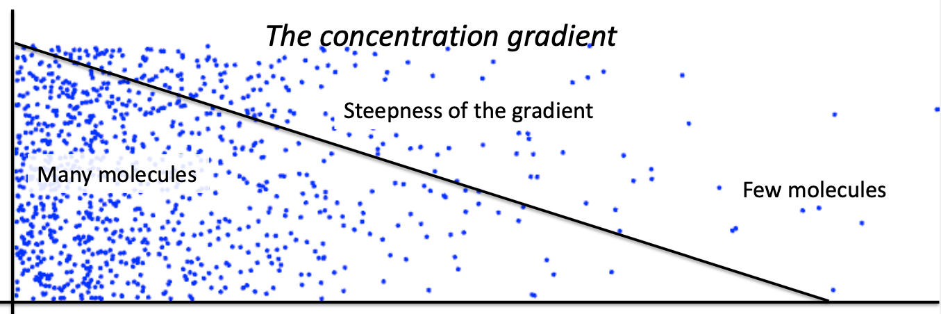
Diffusion is the passive transport (= passive net movement, no ATP) of molecules from a region of high concentration to a region of lower concentration across a semi-permeable membrane until a dynamic equilibrium is reached.
The net movement refers to the overall movement, i.e. all particles are moving all the time – out of the cell and into the cell.
The passive transport of molecules takes place along a concentration gradient, which shows the difference in concentration between two areas. The steepness of the concentration gradient (=the slope) determines the rate at which particles diffuse.
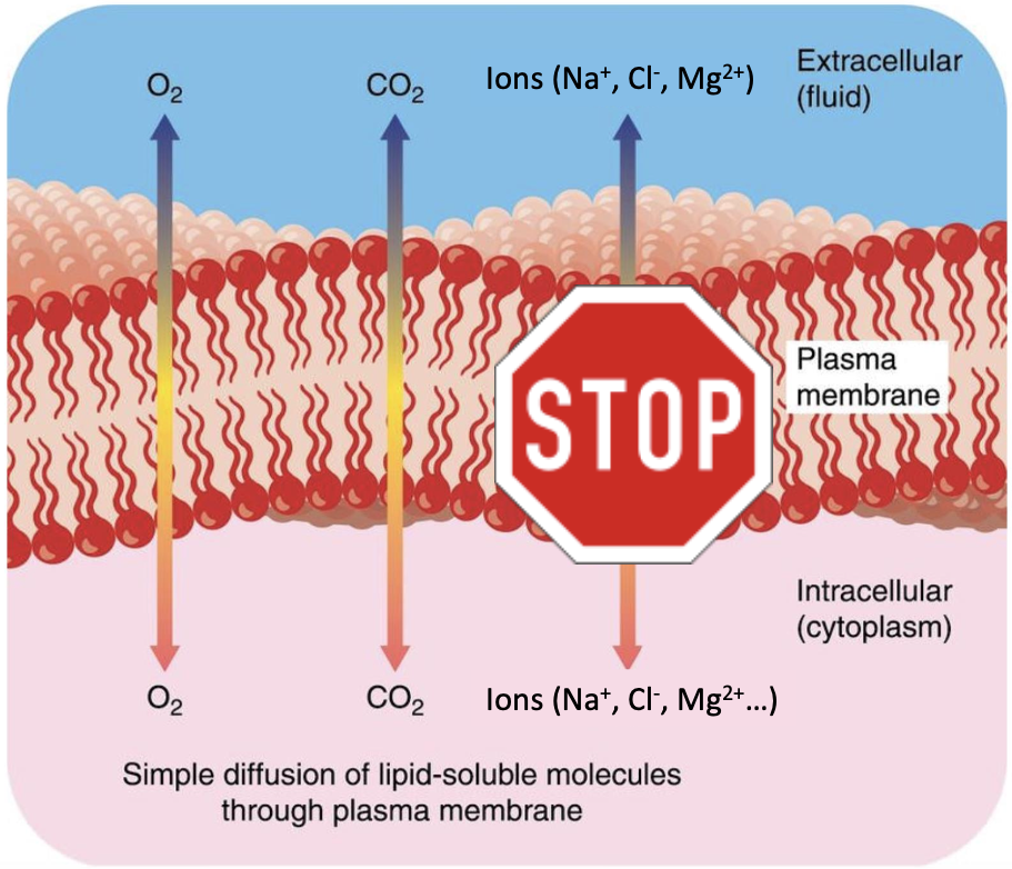
Simple diffusion through membranes happens for molecules for which the phospholipid bilayer is permeable. This is the case for molecules which are non-polar and small, such as O2 and CO2, or polar and but still small (ethanol).
Charged molecules (ions) or large molecules which are polar (glucose, fructose) or nonpolar do not diffuse through the membrane.
Factors affecting the rate of diffusion:
Concentration
The higher the concentration, the steeper the concentration gradient and the faster diffusion takes places
Size and type of molecules
Small and polar/non-polar molecules move fast, larger and non-polar move slower, very large and charged ones not at all
Diffusion pathway
The longer the pathway (e.g. the thicker the membrane) the slower diffusion
Surface area
The bigger the surface area (of a membrane) the more particles can pass though (the more space is available) and the faster diffusion becomes
Moisture
A wet film on the surface of a membrane helps molecules to dissolve and move faster
B2.1.4 Integral and peripheral proteins in membranes
The cell membrane also has membrane proteins which can be grouped into two categories depending on their location in the membrane:
Integral proteins
Integral proteins are embedded in the phospholipid bilayer
The portions of an integral membrane protein found inside the membrane are hydrophobic
Those that are exposed to the cytoplasm or extracellular fluid tend to be hydrophilic.
Proteins that extend all the way across the membrane are called transmembrane proteins.
Peripheral proteins
Peripheral proteins are on the surface of the phospholipid bilayer
These proteins are hydrophilic on their surface and so become attached to the surface of integral proteins or the hydrophilic phosphate heads of the bilayer.
The protein content of membranes is variable and dependent on the function of cells. The more active a membrane, the higher is the protein content. Most plasma membranes have 50% proteins with functions embedded. Chloroplasts and mitochondria have a protein content 75%.
Channel or carrier proteins:
These proteins span the membrane and, transport large or impermeable substances across the membrane.
Protein pumps:
Used for active transport, to shuttle materials from one side to the other. Occurs against the concentration gradient. Needs energy (ATP)
Enzymatic proteins:
Either on the interior or exterior of the membrane surface. They act as enzymes which catalyze metabolic reactions.
Hormone binding membrane proteins:
Membrane proteins may have a binding site for signaling molecules such as hormones or neurotransmitters causing a message to be transmitted to the inside
Cell to cell communication:
Glycoproteins can serve as identification tags that are recognized by membrane proteins of other cells.
Cell adhesion proteins:
Membrane proteins of adjacent cells may hook together to form tissues.
B2.1.5 Movement of water molecules across membranes by osmosis and the role of aquaporins
It is the passive movement of water (H2O) from an area of low solute concentration to an area of high solute concentration through a semi-permeable membrane.
Osmosis is due to differences in the concentration of substances dissolved in water (solutes).
When soluble substances (solutes) dissolve they become surrounded by water molecules forming hydrogen bonds and restricting the movement of water molecules.
This means that areas with higher solute concentration have fewer water molecules available to move than regions with a lower solute concentration.
Net movement of water from regions of lower solute concentration to higher concentration occurs.
Water can move in and out of cells freely because despite being hydrophilic and polar, is very small and easily passes through the membrane.
Some cells have aquaporins – channel proteins which specifically allow water to pass through. The permeability of water is therefore greatly increased. Aquaporins are found in the kidneys or root hair cells.
Aquaporins facilitate a much faster rate of water transport in response to solute concentrations and their levels can be regulated to help control the osmotic conditions of a cell
B2.1.6 Channel proteins for facilitated diffusion
Ions and charged and/or polar particles diffuse using facilitated diffusion by passing through membrane (channel) proteins.
Movement of particles is down a concentration gradient
Passive (no energy required)
Most allow only one type of molecule or ion to diffuse through them. The most common ones are K+ channels.
Most ion channels are gated (voltage, pressure,…) entry/exit is controlled.
All substances which are fat soluble (hydrophobic), will pass through the semi-permeable membrane unhindered, while charged, polar, hydrophilic and large substances require transmembrane proteins for the transport. This makes the cell membrane selectively permeable.
Simple diffusion and facilitated diffusion:
Similarities
Both involve the movement of molecules
Both processes involve passive transport (no use of energy)
Both involve the the movement across a cell membrane
Both move substances along he concentration gradient
Movement can occur in both directions
Differences
Simple diffusion
Fat soluble molecule (hydrophobic or non-polar)
Molecules pass directly though the membrane
No “action” other than concentration gradient
Facilitated diffusion
Water-soluble or hydrophilic/polar molecules
Molecules need a membrane protein to pass through
Each molecule needs a specific protein
Control of types of molecules
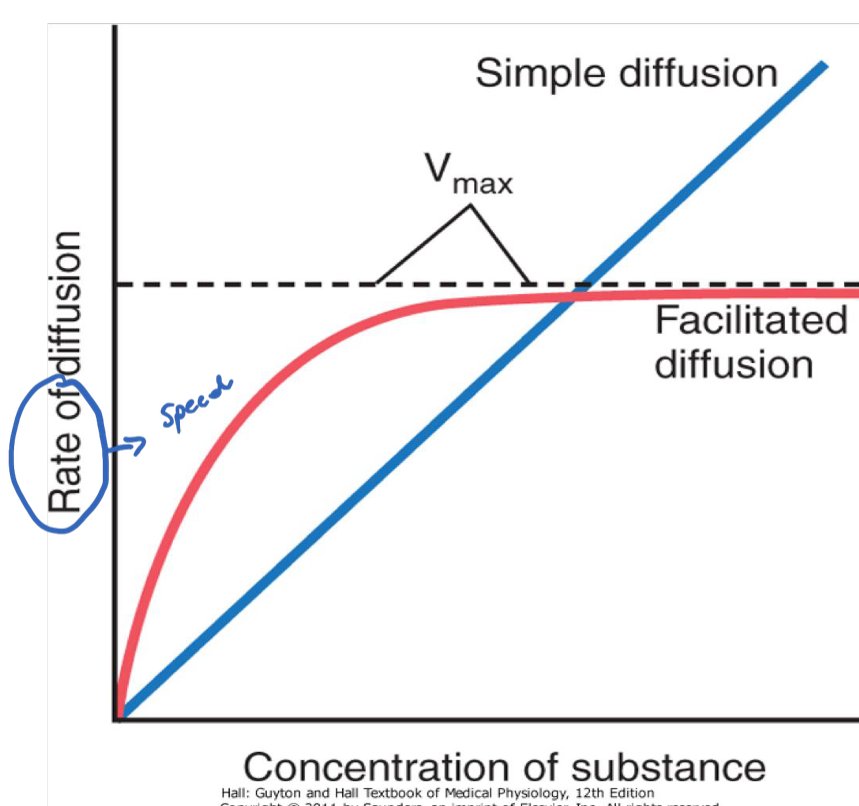
Can you explain why with an increase in substance concentration the rate in facilitated diffusion reaches a plateau, while in simple diffusion rate increases?
Speed of diffusion increases indefinitely with simple diffusion, while with facilitated diffusion it only increases in the beginning and then slows down. This is because facilitated diffusion is limited by membrane proteins which only allow a certain number of molecules to pass through.
B2.1.7 Pump proteins for active transport
Not all particles diffuse easily across the membrane. In particular, hydrophilic and charged molecules have difficulties with passive movement and are not permeable to the membrane. Sometimes molecules also need to be delivered against a concentration gradient.
Can you explain how this type of transport works?
The release of energy from ATP is used to induce a shape (conformational) change in the channel protein. The substance can pass through
Protein pumps within the phospholipid bilayer transport substances only in one direction
This mechanism requires energy in the form of ATP (from cell respiration)
Molecules are usually transported against a concentration gradient
How is active transport using protein pumps different from facilitated diffusion using protein channels?
Similarities:
Both require integral membrane proteins
Both transport substances from one side to the other
Both transport substances which do not simply diffuse (e.g. large or polar molecules)
Differences:
Active transport using protein pumps
Against the concentration gradient
Requires energy (active)
Facilitated diffusion using protein channels
Following the concentration gradient
Passive - no energy required
Protein pumps can come in great variety. A uniporter carries one specific ion or molecule. A symporter carries two different ions or molecules, both in the same direction. An antiporter also carries two different ions or molecules, but in different directions.
B2.1.8 Selectivity in membrane permeability
Every cell needs substances from the outside and it needs to get rid of waste materials. The membrane is a selectively permeable which means that only some molecules pass easily through it, while others need some help.
What substances does a cell need to let in or out?
Glucose, needed for cell respiration to produce energy
Water (for various functions)
Oxygen (aerobic respiration)
Proteins
Fats
Vitamins, minerals (Fe, K, Ca, Na…)
CO2 (gets out → waste)
B2.1.9 Structure and function of glycoproteins and glycolipids
Glycoproteins and glycolipids are important components of the cell membrane. They are involved in cell-to-cell recognition, formation of tissues by forming a glycocalyx and play a role in the immune system. Cell recognition is an important feature of the immune system, as it helps cells to distinguish between self- and non-self cells.
Glycoproteins and glycolipids form the glycocalyx – a carbohydrate rich layer on the outer surface of the plasma membrane of animal cells, with an aqueous solution in the gaps between the sugars.
Glycoproteins:
Proteins with a carbohydrate attached to it
Part of the plasma membrane of cells
The protein part is embedded in the membrane, and the carbohydrate part project into the exterior environment
Cell communication and recognition
Protection
Adhesion and interaction
Transport
Glycolipids:
Carbohydrates (sugar) linked to lipids (of the cell membrane).
They are part of the plasma membrane of cells
The carbohydrate part projects into the exterior environment.
Structural component of the cell membrane
Cell recognition
Protection and lubrication
B2.1.10 Fluid mosaic model of membrane structure
Higher level
B2.1.11 Relationships between fatty acid composition of lipid bilayers and their fluidity
The phospholipids of cell membranes have saturated and unsaturated fatty acids. The composition of them affects the fluidity of membranes.
The fatty acids of the the phospholipid bilayer make the membrane very flexible. Movement around the horizontal plane takes place on many occasions – this contributes to membrane fluidity. Movement along the vertical plane (flip-flopping) is rare.
The fluidity of membrane is affected by the types of fatty acids. Two main factors affect this:
The more unsaturated fatty acids the phospholipid bilayer have, the more fluid the membrane is – they are more spaced out. The less double bonds, the more viscous the membrane.
The shorter the hydrocarbon tails, the more fluid the membrane. The more fluid a membrane, the lower its melting/freezing point – the membrane will freeze less likely.
The ideal composition depends on the temperature of the surrounding of the organism – a membrane should have the right ratio to not become too visous.
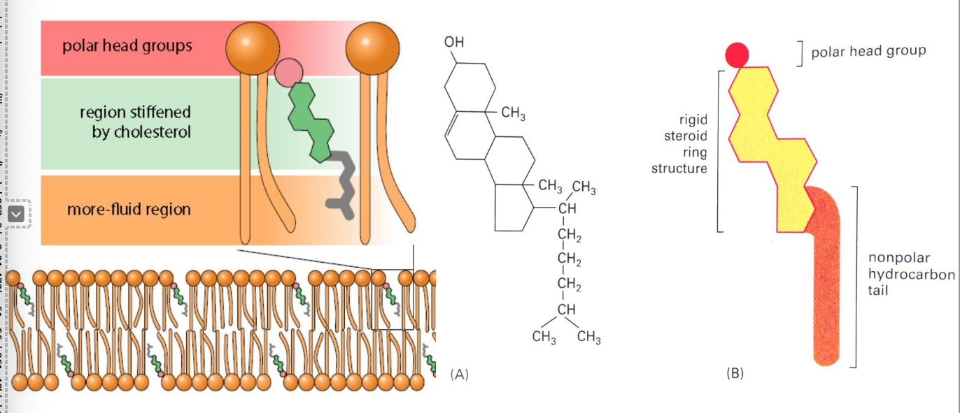
B2.1.12 Cholesterol and membrane fluidity in animal cells
Cholesterol is an important component in cell membranes of animals, and it contributes to the stability and fluidity of membranes at temperature outside the optimum (so at low and high temperatures). This is important, as otherwise the permeability of membranes would be compromised.
Cholesterol is a steroid
The phosphate head of the phospholipid is polar and hydrophilic.
The head group of cholesterol is a hydroxyl group (-OH), making this part slightly polar & hydrophilic.
The fatty acid tails of the phospholipid are non-polar and hydrophobic.
Cholesterol’s steroid ring and its hydrocarbon tail are largely nonpolar & hydrophobic.
The hydrophilic phosphate group is attracted to the hydroxyl group of the cholesterol molecule, and the hydrophobic hydrocarbon tail of cholesterol is attracted to the fatty acid tails of the phospholipid. This causes its rigid, steroid rings to interact with the regular packing of the hydrocarbon tails.
The similar chemical properties of cholesterol and phospholipids hold the molecules together like glue. This ensures the right kind of membrane fluidity. Without cholesterol, cell membranes would be too fluid, not firm enough, and too permeable to some molecules. In other words, it keeps the membrane from turning to mush.
In high temperatures, phospholipids normally become more mobile and fluid compromising the permeability of the membrane, but cholesterol constrains the motion of fatty acid tails and therefore decreases fluidity.
In low temperature the bilayer would normally become less fluid & rigid, but with cholesterol the close packing of the nonpolar fatty acid chains is disrupted and therefore fluidity is increased.
B2.1.13 Membrane fluidity and the fusion and formation of vesicles
Vesicular transport - type of active transport
Vesicles are fluid filled sacs composed of a single phospholipid bilayer which surrounds the fluid/solutes inside. They have multiple functions:
Movement of materials into (endocytosis) and out of a cell (exocytosis).
Transport of substances internally.
Transport of proteins from the rER to the Golgi apparatus and finally to the membrane.
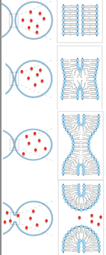
The fluidity of the membrane allows fusion of phospholipids
The vesicle approaches the plasma membrane. All membranes are made of the phospholipid bilayer, so share the same properties.
The membranes begin to fuse. This step requires ATP. Remember the fluidity of the plasma membrane – the phospholipids can flow around each other.
For a moment, there is a single phospholipid bilayer at the point of contact.
The membrane pores opens, allowing the contents to pass through. Notice that through the whole process, there is never an unbroken section of the bilayer.
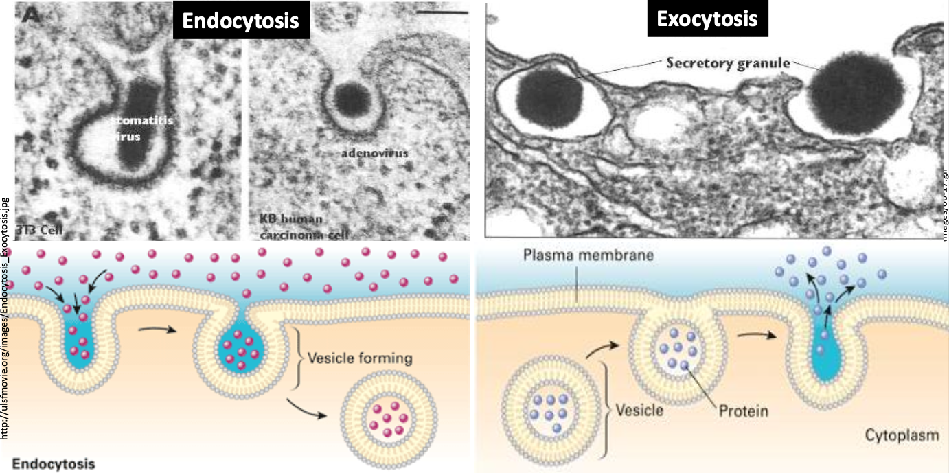
Endocytosis and Exocytosis:
Proteins in the membrane carry out both processes, using energy from ATP.
Endocytosis:
Endocytosis occurs by invagination (infolding) of the plasma membrane, which then forms vesicles that become detached and enter the cytoplasm. Endocytosis can involve the uptake of solid substances (phagocytosis) or liquid substances (pinocytosis).
Examples:
Transport of large hydrophilic particles e.g. food molecules in single-celled organism (amoeba, paramecium, bacteria)
Fetus take up antibodies in mothers womb
Phagocytosis of bacteria by phagocytes
Uptake of viruses by white blood cells
Exocytosis:
Exocytosis occurs by fusion of the vesicle membrane and the plasma membrane, followed by release of the vesicle contents to the outside of the cell.
Examples:
Protein synthesis in rER with vesicles transporting polypeptides to Golgi apparatus
Secretion of enzymes from the pancreas into the pancreatic duct and from these into the intestine
Release of neurotransmitters from synapse (acetylcholin) in neurons
Secretion of sweat from sweat glands
Secretion of breast milk
Similarities:
Both involve active transport across the membrane
Both require vesicle transport inside the cell
Both use ATP
Differences:
Endocytosis
Transport of material from the outside to the inside of the cell (outside → inside)
Exocytosis
Inside → outside
No receptor mediated
B2.1.14 Gated ion channels in neurons
An axon is a long, slender projection of a nerve cell (neuron) that conducts electrical impulses away from the neuron's cell body. The axon transmit information to different neurons, muscles and glands. This involves rapid movements of sodium and potassium ions across the axon membrane, which establish and re-establish a potential difference (voltage) across the axon membrane.
Voltages across cell membranes are due to an imbalance of positive and negative charges inside (cytoplasm) and outside the membrane.
The voltage across membranes is measurable. If the voltage is below a certain value (-50 mV in neurons) the ion gates channels stay closed. If it rises, it triggers Na+ ion gated channels to open, and if it rises even more (+ 40mV) K+ ion gated channels open.
As an example for facilitated diffusion, gated ion channels allow specific ions to pass across a membrane in either direction. They open and close reversibly, being switched on and off. In the axon fibers of neurons these channels are selective for sodium (Na+) and potassium (K+) ion.
When there is a change in voltage, the K+ channel opens, allowing K+ ions to diffuse through.
The channel closes again quickly–due to an extra globular protein subunit (“ball”) which is attached to a flexible chain of amino acids.
The ball fits inside the open pore, closing the channel again.
Some gated channels respond to the binding of a ligand rather than a change in voltage. These channels are equipped with Nicotinic acetylcholine receptors. The receptors bind to neurotransmitters (Acetylcholine).
The gated channel allows positively charged ions (Na+) to pass upon binding of a neurotransmitter (Acetylcholine).
When the ligand (the neurotransmitter) dissociates the conformation of the ion channel closes and does not allow transport.
B2.1.15 Sodium–potassium pumps as an example of exchange transporters
For a neuron to convey a nerve impulse and for ion-gated channels to function, there must be a concentration gradient of Na+ and K+ ions across the membrane. This is achieved by the sodium potassium pump, which actively transports ions against their concentration gradient.
The concentration of Sodium (Na+) outside the cell is higher than inside. The concentration of Potassium (K+ ) is higher inside than outside.
The interior of the pump is open to the inside of the axon – 3 Na+ ions enter the pump and attach to binding sites
The release of energy from the phosphorylation of ATP causes a conformational change in the protein – the pump closes.
The protein opens to the outside, and the Na+ ions are released.
2 K+ ions can now attach. With the help of ATP the channel protein changes in shape and releases the K+ ions to the inside.

B2.1.16 Sodium-dependent glucose cotransporters as an example of indirect active transport
Sodium dependent glucose cotransporters in the small intestine:
Glucose cannot pass through the plasma membrane by simple diffusion because it is polar and hydrophilic. Therefore, a cotransport system together with Na+ (Sodium ions) is put in place.
Maltose is hydrolysed into glucose.
Coupled transport proteins transfer Na+ and Glucose together into the cytoplasm of the villus via facilitated diffusion. This transport depends on the concentration gradient of Na+.
Na+/K+ pumps transfer Na+ ions from the cytoplasm of the villus cell out (active transport, Na/K+ pump) creating a low concentration of Na+ inside the villus → conc.gradient.
Glucose then moves via facilitated diffusion (protein channels) from the villus into the capillary.
Sodium dependent glucose cotransporters in the kidney:
In the kidney, the same system of inactive or secondary transport is in place. The Na+ outside the tubules is increased through active transport, allowing a concentration gradient to build up.
At the beginning, the fluid in the proximal convoluted tubule contains glucose, amino acids, vitamins, hormones, urea, salt ions and water. Re-absorption of glucose and salts (Na+) back into the blood take place at least partly by active transport. Water and some ions (Cl-) are following a concentration gradient.
The kidney’s initial filtrate contains nearly 5.5kg of glucose at the point where it is first formed. Urine does not contain anywhere near as much glucose – it is reabsorbed back in the blood so that it can be utilized where needed.
Na+/K+ pumps transfer Na+ ions from the cytoplasm of the kidney cell out (active transport, Na/K+ pump) creating a low concentration of Na+ inside the cell. This establishes a concentration gradient.
Coupled transport proteins transfer Na+ and Glucose together into the cytoplasm of the kidney cell via facilitated diffusion. This transport depends on the concentration gradient of Na+
B2.1.17 Adhesion of cells to form tissues
Cells in tissues are linked by cell-to-cell junctions which depend on molecules (cell-adhesion-molecules, CAM) in the plasma membranes of adjacent cells. Different types of CAM molecules exist depending on the function of the molecule.
CAMs are proteins with one part embedded in the phospholipid bilayer and other parts projecting outwards into the extracellular environment. These extracellular components bind with the ones of adjacent cells to form a complex.