UNIT FOUR: CELL COMMUNICATION
Cell Communication
Cells communicate using chemical signals. These signals are secreted from the cell and a neighboring cell must have the right receptor for that cell.
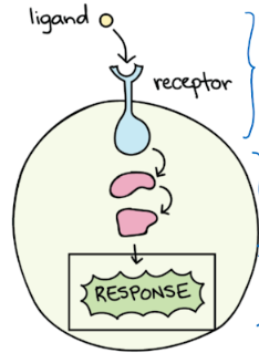
Cell communication has three steps:
Reception: a cell detects (with a receptor) a signalling molecule called a ligand. Receptor: proteins. Ligand: Most hormones (P/L), carbs, or ions
Transduction: this is the cell’s transmission of the molcule that signals from the cells exterior to it’s interirior
Response: the signal from transduction triggering a specific reaction from the cell.
Forms of Signaling
Cell to cell signaling involves the transmission of a signal from one sending cell to a recieving cell. There are different types of signaling however.
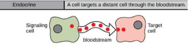
Endocrine: A cell targets a different cell through the bloodstream
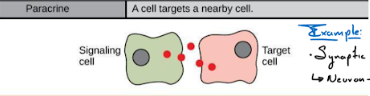
Paracrine: a cell targets a nearby cell (no contact however) (Ex: Synaptic Signaling)
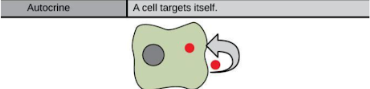
Autocrine: a cell targets itself
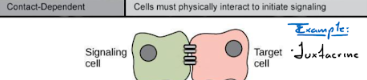
Contact-Dependent: Two cells must physically interact to start signaling (Another word for this would be justacrine)
Types of Receptors
Receptors can be divided into two categories:
Intracellular Receptors: These receptors are found inside of the cell, usually in the cytoplasm or nucleus. The ligands are small and hydrophobic, because they need to cross the cell membrane. An example of this would be lipid based hormones (steroids).
Cell Surface Receptors: These receptors are membrane-bound (outside of the cell). Since the receptor is outside of the cell, the ligands are large and hydrophillic because they do not need to cross the cell membrane. The three most common receptors are the 1. Ligand-gated Ion Channels, w. Receptor Tyrosine Kinases, and 3. G protein-coupled receptors.
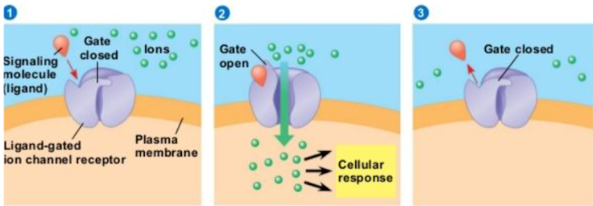
Ligand-gated Ion Channels: These are ion channels that open up in response to a ligand binding to it. In some cases, these channels are usually open and the binding of an ion causes the channels to close. These channels are associated with Neurons and Neurotransmitters.
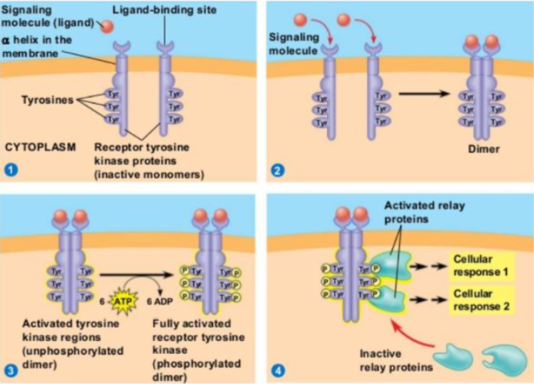
Receptor Tyrosine Kinase: A Kinase is an enzyme that transfers phosphate groups from energy molecules to other molecules. Both receptors need to have ligands so they can bind together and send signals. These signals can have to do with growth factors, such as cell division.
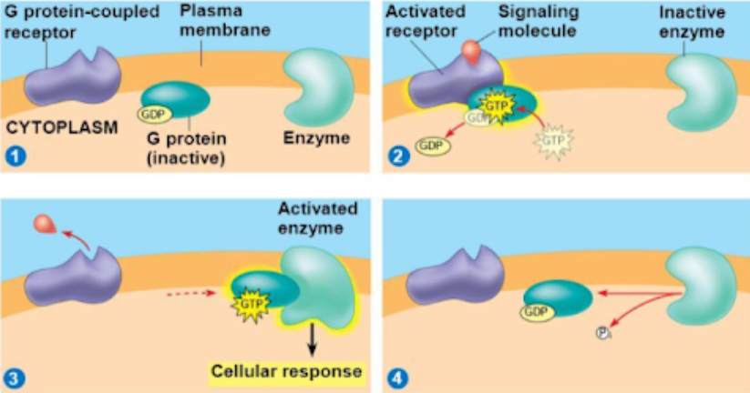
G Protein-Coupled Receptors: They are a common receptor composed of multiple proteins. When the ligand attaches to the receptor, a G-Protein is activated and a GTP attaches to it. Then the activated G-Protein moves across the membrane until it attaches to a membrane-bound enzyme, which then sends a signal inside of the cell. Cell Signaling uses these receptors as a cycle, as long as the ligand is attached.
GTP = Guanosine Triphosphate
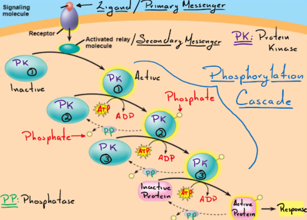
Transduction: A change in the receptor sets off a series of signaling events in a chain reaction, leading to a response. The many transduction pathways amplify the signal. The molecules that relay the signal are often proteins, but they can also be ions and phospholipids. The phosphorylation and dephosphorylation of molecules assist in the transmission of the cell.
Kinase: an enzyme that phosphorylates
Phosphatase: an enzyme that dephosphorylates
Phosphorylation: the attachment of a phosphate to a molecule
Dephosphorylation: the deattachment of a phosphate to a molecule
Secondary Messengers
They are small, non-protein molecules that pass along a signal initiated by the binding of a ligand (“first messenger”) to its receptor.
Examples of secondary messengers: Cyclic AMP (cAMP) and Inositol Phosphates
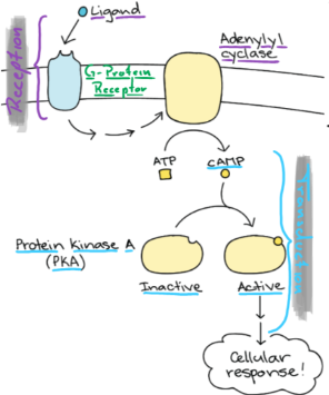
In response to signals, an enzyme called Adenylyl Cyclase is activated. Once activated, cAMP can activate an enzyme Protein Kinase to pass along a signal. These are mostly used in G-Protein Coupled Receptors.
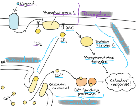
Ca 2+ ions: The Ca 2+ concentration is low in the cytoplasm and ion pumps in the membrane actively remove it. The Ca 2+ ions can be stored in the ER and these ions are used in Ligand Ion-Gated Channels.
Inositol Phosphates: Phosphatidylinositols (PIP2) are phospholipids that are found in the membrane for signaling. PIP2 can be broken down into DAG and IP3, which are used as secondary messengers. They are moihstly used in G-Protein Coupled Receptors.
Feedback Messengers
Homeostasis: The tendency to resist change in order to maintain a stable, relatively constant internal enviroment. Examples include body temperature and the concentration of ions, pH, sugar, and H2O in the bloodstream. It depends on the ability of your body to detect and oppose changes in your body by biological systems.
Negative and Positive Feedback mechanisms help your body to maintain internal balance.
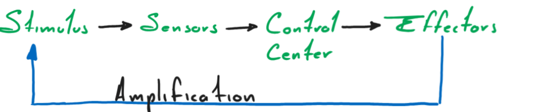
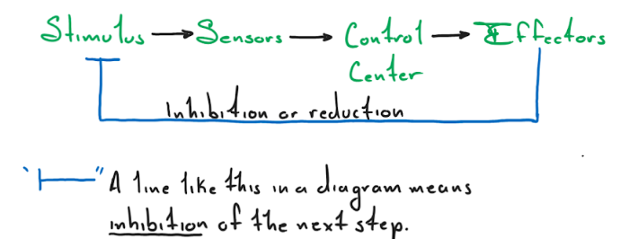
Every mechanism loop starts with a stimulus that is detected by a sensor. The sensor relays the signal to the control center, which activates the effectors to respond to the stimulus.
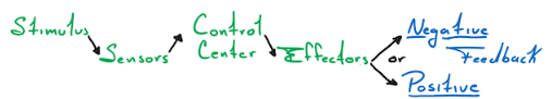
Negative Feedback: Homeostasis typically involves negative feedback that counteracts changes from internal balance. It inhibits or slows down the process that is moving your body out of homeostasis.
Positive Feedback: The less common mechanism inside the body. Positive Feedback uses it’s effectors to amplify the stimulus, which amplifies the effectors. This cycle can only be broken if something inside the cycle changes (Childbirth is an example of positive feedback)